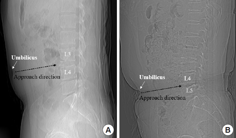
Fig. 1.

Preoperative sagittal scout computed tomography view used to identify the location of the umbilicus centered on the index levels. These images show examples of cases where the position of the patient’s umbilicus is parallel to the L3–4 levels (A) or parallel to the L4–5 levels (B).
