Abstract
Objectives
To evaluate the effects of different lingual retainers on periodontal health and stability of mandibular anterior teeth at the 1-year follow-up.
Materials and Methods
One hundred thirty-two patients were randomly allocated to four groups using different lingual retainers: group 1, 0.016 × 0.022-in dead-soft wire; group 2, 0.0215-in 5-strand stainless steel wire; group 3, 0.014 × 0.014-in computer-aided design/computer-aided manufacturing nitinol retainer (Memotain); group 4, connected bonding pads. Plaque, gingival, and calculus indexes were used to evaluate periodontal health, and Little's irregularity index, intercanine width, and arch length measurements were performed to evaluate stability. All measurements were performed at each time point (debonding and 3, 6, 9, and 12 months).
Results
The mean value of the gingival index obtained in group 3 was lower than the mean value for all other groups. The mean value of the calculus index was the lowest in group 3, and there was a significant difference between group 3 and groups 1 and 2. No differences were found among the groups in terms of plaque index, intercanine width, and arch length. The least irregularity was obtained in groups 2 and 3. There were no significant differences between these groups and groups 1 and 4.
Conclusions
Gingival inflammation and calculus accumulation were the lowest in group 3 (Memotain). The irregularity for Memotain and stainless steel retainers was less than or the other groups. However, no clinically significant worsening of periodontal health or relapse were seen in any groups after 1 year.
Keywords: CAD/CAM, Fixed retainer, Periodontal health, Stability
INTRODUCTION
Maintaining orthodontic treatment outcomes without relapse is an important issue for orthodontists. Although approximately 25% of displaced incisors could not be considered as being due to relapse of orthodontic treatment, loss of long-term stability is inevitable in many cases after fixed orthodontic treatment.1 In the literature, many studies have recommended the use of a removable retainer in the maxilla and a bonded retainer in the mandible.2–4 Long-term retention with fixed retainers is often advocated for the mandibular anterior region, where a high rate of relapse is observed.5 To date, however, no consensus exists on which type of fixed retainer to choose after orthodontic treatment.
Zachrisson2 claimed that 0.0215-in 5-strand stainless steel wires were the gold standard, and thinner wires showed more distortion. The disadvantage of this wire was that there were unexpected tooth movements when the wire was not fully passively adapted to the tooth surfaces.6 Dead-soft wires have been used to eliminate this problem because they can be easily adapted to tooth surfaces.7 In addition, a low probability of inadvertent third-order activation is another advantage of dead-soft wire. Nevertheless, these wires have high breakage rates.8
In recent years, retainers produced with computer-aided design/computer-aided manufacturing (CAD/CAM) systems have been used as an alternative to these retainers.9 Memotain is cut from a sheet of nickel-titanium, similar to the way in which scissors cut a piece of paper. This offers many advantages, such as greater fit accuracy, tighter interproximal adaptation, individually optimized placement, and resistance to microbial colonization compared with other options.10 Alternatively, connected bonding pads are produced in various sizes. These types of retainers have pads corresponding to each tooth. The manufacturer claims that this retainer provides maximum retention with the help of its pads.11 However, there is no study regarding connected bonding pad retainers in the literature.
Only a few studies have evaluated the effects of different types of lingual retainers in terms of periodontal health and stability.7,12–14 Gunay and Oz7 demonstrated that irregularity was higher in dead-soft than in multistrand stainless steel retainers. Störmann and Ehmer12 compared two different sizes of retainer wires made from stainless steel and found no significant differences in terms of irregularity and periodontal health. Knaup et al.13 concluded that Memotain showed better results than stainless steel retainers in terms of periodontal parameters in a short-term retrospective clinical study. In a recent study, Kartal et al.14 reported that Memotain and multistrand retainers showed similar periodontal outcomes. In addition, a systematic review emphasized the need for future studies to determine the effects of different lingual retainers on stability and periodontal health.15 The effects of Memotain and connected bonding pad retainers have not been evaluated in terms of stability during the retention period. Therefore, the aim of the current study was to investigate the effects of different lingual retainers on periodontal health and stability.
The primary aim of this study was to evaluate the impact of four different lingual retainers on periodontal health during a 1-year follow-up period. The secondary aim was to investigate the stability of treatment outcomes over this period. The null hypothesis tested in this trial was that there would be no significant difference in periodontal health and stability among patients with different retainer types.
MATERIALS AND METHODS
Trial Design and Ethical Approval
This was a single-center parallel-design prospective clinical trial with a 1:1 allocation ratio. Ethical approval was obtained from the Ethics Committee of Pamukkale University (06.03.2018/05). Written informed consent was obtained from the patients or their parents who agreed to participate in this study.
Participants, Eligibility Criteria, and Settings
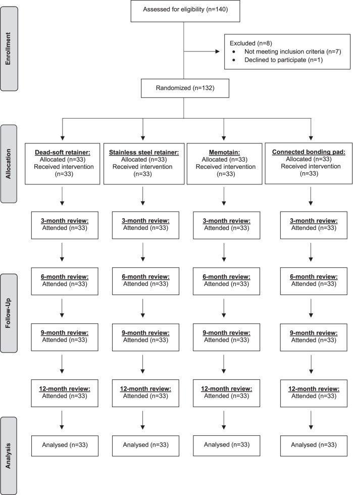
Before the debonding session, 132 patients (92 female, 40 male) selected from the orthodontic department of Pamukkale University were included based on the following criteria: (1) nonextraction treatment in the mandible, (2) moderate irregularity before treatment according to Little's irregularity index,16 (3) good oral hygiene (absence of visible plaque and redness in the gingiva), and (4) no caries. Patients were equally randomized to four groups (Figure 1).
Figure 1.
Study flow chart.
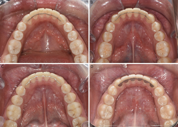
The study groups were as follows (Figure 2):
Figure 2.
Retainers applied: (A) group 1 (dead-soft wire), (B) group 2 (5-strand stainless steel wire), (C) group 3 (Memotain), and (D) group 4 (connected bonding pads).
Group 1: 0.016 × 0.022-in dead-soft wire (Bond-A-Braid, Reliance Orthodontic Products, Itasca, Ill, USA)
Group 2: 0.0215-in 5-strand stainless steel wire (Pentaflex, GC Orthodontics America Inc, Alsip, Ill, USA)
Group 3: 0.014 × 0.014-in computer-aided design/computer-aided manufacturing (CAD/CAM) nitinol retainer (Memotain, CA-Digital, Mettman, Germany)
Group 4: 0.012-in connected bonding pad retainer (Leone SpA, Firenze, Italy)
Interventions
All retainers were bonded directly by the same investigator (Dr Adanur-Atmaca). The same etching agent (Etch-Royale, Pulpdent, Watertown, Mass), adhesive primer (Transbond XT primer, 3M Unitek, Monrovia, Calif), and composite (Transbond LR, 3M Unitek) were used to bond all retainers. For all groups, vacuum-formed maxillary retainers were used concurrently.
All patients were taught how to clean their retainers. The patients were instructed to visit the clinic immediately in case of bond failure. All patients were followed up for 1 year after the retainers were bonded. All measurements were performed at the following time points by the same calibrated investigator (Dr Adanur-Atmaca): debonding session (T0), 3 months (T1), 6 months (T2), 9 months (T3), and 12 months (T4).
Outcomes
Periodontal measurements.
Plaque, gingival17 and calculus18 indexes were used to evaluate periodontal health. All scores were recorded on the lingual surfaces for lower anterior teeth. The mean for the six lower anterior teeth was calculated. Due to the dynamic nature of periodontal tissues, periodontal measurements could not be repeated to test intraexaminer reliability.
Stability measurements.
Little's irregularity index,16 intercanine width, and arch length measurements were performed with model analysis software (OrthoAnalyzer, 3Shape, Copenhagen, Denmark). Intercanine width and arch length were measured as described by Eslambolchi et al.19 Stability measurements were repeated on 44 randomly selected digital models to determine intraexaminer reliability 4 weeks later.
Sample Size
The sample size was calculated based on a previous study.7 Power analysis (G Power, version 3.0.10, Kiel, Germany) showed that 112 patients (28 patients for each group) would provide more than 80% power at a 95% confidence level with medium effect size (f = 0.35). Considering the possibility of patient dropouts (15%), 5 more patients were included in each group.
Randomization
Random numbers were assigned by the online randomization program to four groups. The numbers were placed in opaque envelopes, and one envelope was selected by each patient.
Blinding
Blinding of clinicians was not possible in this study because the outcome assessor and clinician were the same person.
Statistical Analysis
Data were analyzed with SPSS (version 24; IBM, Armonk, NY) software. The Kolmogorov-Smirnov test was used to determine normality. All measurements were examined according to group and time using generalized linear models (Wald χ2). After examining interactions with the main effects of group and time, multiple comparisons were performed with the Bonferroni correction for significant effects. The intraclass correlation coefficient (ICC) was used to determine intraexaminer reliability. The statistical significance level was set as P < .05.
RESULTS
Baseline Data
The groups were similar in terms of age, sex, and features of the original malocclusion (Table 1).
Table 1.
Baseline Data of the Samplea
| Overall Sample (n = 132) |
Dead-Soft Retainer Sample (n = 33) |
Stainless Steel Retainer Sample (n = 33) |
Memotain Sample (n = 33) |
Connected Bonding Pad Sample (n = 33) |
P Valueb |
|
| Age (y), median IQR | 16.0 (3.8) | 16 (3.5) | 15 (3) | 16 (3.5) | 16 (3.0) | NS |
| Sex, n (%) | ||||||
| Male | 40 (30.3) | 5 (15.2) | 11 (33.3) | 13 (39.4) | 11 (33.3) | NS |
| Female | 92 (69.7) | 28 (84.8) | 22 (66.7) | 20 (60.6) | 22 (66.7) | |
| Incisor classification, n (%) | ||||||
| Class I | 31 (23.5) | 8 (24.2) | 10 (30.3) | 7 (21.2) | 6 (18.2) | NS |
| Class II Division 1 | 55 (41.7) | 14 (42.5) | 13 (39.4) | 13 (39.4) | 15 (45.4) | NS |
| Class II Division 2 | 20 (15.1) | 4 (12.1) | 5 (15.2) | 5 (15.2) | 6 (18.2) | NS |
| Class III | 26 (19.7) | 7 (21.2) | 5 (15.2) | 8 (24.2) | 6 (18.2) | NS |
| Skeletal pattern, n (%) | ||||||
| Skeletal I | 43 (32.6) | 11 (33.3) | 12 (36.4) | 10 (30.3) | 10 (30.3) | NS |
| Skeletal II | 64 (48.5) | 16 (48.5) | 16 (48.5) | 15 (45.5) | 17 (51.5) | NS |
| Skeletal III | 25 (18.9) | 6 (18.2) | 5 (15.2) | 8 (24.2) | 6 (18.2) | NS |
| Irregularity (mm), median IQR | 4.6 (2.2) | 4.6 (2.1) | 4.8 (2.8) | 4.6 (1.7) | 4.3 (2.3) | NS |
IQR indicates interquartile range; NS, nonsignificant.
P value for comparison of group means by Kruskal-Wallis test or differences in proportions by χ2 test.
Periodontal Measurements
Periodontal measurements are shown in Tables 2 and 3. The main effect of time on the plaque index was statistically significant (P < .001). It was determined that the mean plaque index value obtained at T0 was lower than the values obtained at other times.
Table 2.
Comparison of Plaque, Gingival, and Calculus Index Values According to Group and Timea
| Plaque Index |
Gingival Index |
Calculus Index |
||||||||||
| Wald χ2 |
df |
P |
Partial Eta Square |
Wald χ2 |
df |
P |
Partial Eta Square |
Wald χ2 |
df |
P |
Partial Eta Square |
|
| (Intercept) | 694.061 | 1 | <.001 | 0.513 | 593.633 | 1 | <.001 | 0.474 | 378.199 | 1 | <.001 | 0.365 |
| Group | 6.902 | 3 | .075 | 0.010 | 15.234 | 3 | .002 | 0.023 | 11.179 | 3 | .011 | 0.017 |
| Time | 51.100 | 4 | <.001 | 0.072 | 49.772 | 4 | <.001 | 0.070 | 117.196 | 4 | <.001 | 0.151 |
| Group*Time | 7.588 | 12 | .816 | 0.011 | 24.383 | 12 | .018 | 0.036 | 7.418 | 12 | .829 | 0.011 |
df indicates degrees of freedom; bold P values indicate statistical significance.
Table 3.
Descriptive Statistics and Multiple Comparison Results of Plaque, Gingival, and Calculus Index Values According to Group and Timea
| Groups |
Time |
Plaque Index |
Gingival Index |
Calculus Index |
| Group 1 | T0 | 0.088 ± 0.203 | 0.103 ± 0.141C | 0.000 ± 0.000 |
| T1 | 0.168 ± 0.159 | 0.376 ± 0.232AB | 0.076 ± 0.09 | |
| T2 | 0.246 ± 0.193 | 0.468 ± 0.329A | 0.102 ± 0.102 | |
| T3 | 0.269 ± 0.243 | 0.216 ± 0.202ABC | 0.128 ± 0.107 | |
| T4 | 0.227 ± 0.146 | 0.288 ± 0.232ABC | 0.136 ± 0.116 | |
| Total | 0.200 ± 0.200 | 0.290 ± 0.265X | 0.088 ± 0.104YZ | |
| Group 2 | T0 | 0.066 ± 0.109 | 0.090 ± 0.149C | 0.000 ± 0.000 |
| T1 | 0.194 ± 0.165 | 0.385 ± 0.264 AB | 0.112 ± 0.107 | |
| T2 | 0.257 ± 0.201 | 0.418 ± 0.365AB | 0.102 ± 0.134 | |
| T3 | 0.260 ± 0.185 | 0.440 ± 0.412AB | 0.134 ± 0.150 | |
| T4 | 0.226 ± 0.178 | 0.327 ± 0.357ABC | 0.154 ± 0.175 | |
| Total | 0.200 ± 0.183 | 0.331 ± 0.343X | 0.100 ± 0.137Y | |
| Group 3 | T0 | 0.108 ± 0.180 | 0.111 ± 0.204C | 0.000 ± 0.000 |
| T1 | 0.203 ± 0.194 | 0.214 ± 0.249ABC | 0.048 ± 0.089 | |
| T2 | 0.229 ± 0.220 | 0.243 ± 0.346ABC | 0.090 ± 0.109 | |
| T3 | 0.213 ± 0.257 | 0.225 ± 0.361ABC | 0.081 ± 0.114 | |
| T4 | 0.210 ± 0.257 | 0.248 ± 0.368ABC | 0.098 ± 0.121 | |
| Total | 0.193 ± 0.225 | 0.208 ± 0.313Y | 0.063 ± 0.103X | |
| Group 4 | T0 | 0.146 ± 0.194 | 0.180 ± 0.234BC | 0.000 ± 0.000 |
| T1 | 0.244 ± 0.199 | 0.318 ± 0.266ABC | 0.042 ± 0.082 | |
| T2 | 0.222 ± 0.237 | 0.250 ± 0.253ABC | 0.088 ± 0.127 | |
| T3 | 0.293 ± 0.260 | 0.376 ± 0.395AB | 0.110 ± 0.142 | |
| T4 | 0.321 ± 0.280 | 0.328 ± 0.404ABC | 0.130 ± 0.153 | |
| Total | 0.245 ± 0.241 | 0.290 ± 0.323X | 0.074 ± 0.123XZ | |
| Total | T0 | 0.102 ± 0.176m | 0.121 ± 0.187k | 0.000 ± 0.000n |
| T1 | 0.202 ± 0.180l | 0.323 ± 0.259l | 0.070 ± 0.095m | |
| T2 | 0.239 ± 0.212kl | 0.345 ± 0.338l | 0.095 ± 0.118lm | |
| T3 | 0.259 ± 0.238k | 0.313 ± 0.361l | 0.113 ± 0.130kl | |
| T4 | 0.246 ± 0.224kl | 0.298 ± 0.344l | 0.130 ± 0.143k | |
| Total | 0.209 ± 0.214 | 0.280 ± 0.315 | 0.082 ± 0.118 |
Values are mean ± standard deviation, k–m: There is no difference between times with the same letter in terms of plaque, gingival, and calculus indexes, A–C: There is no difference between group and time interactions with the same letter in terms of gingival index, X–Z: There is no difference between groups with the same letter in terms of each parameter. Group 1, dead-soft retainer; group 2, stainless steel retainer; group 3, Memotain; group 4, connected bonding pad.
The main effect of the group (retainer type) on the gingival index values was found to be statistically significant (P = .002). The mean value of group 3 was lower than that of the other groups. There was no significant difference among groups 1, 2, and 4. The main effect of time was found to be statistically significant (P < .001). The mean gingival index values remained stable after T1. The main effect of the group and time interaction was statistically significant (P = .018). The value obtained at T0 in group 1 was significantly lower than at T1 and T2, and the value obtained at T0 in group 2 was significantly lower than at T1, T2, and T3. Despite the differences, all values were in the range 0.1–1.0 (mild gingivitis), so these changes may not be considered clinically significant.
The main effect of the group on the calculus index values was statistically significant (P = .011). The lowest mean value was in group 3, and there was a significant difference between group 3 and groups 1 and 2. It was observed that the main effect of time on the mean calculus index values was significant (P < .001). The mean calculus index value obtained at T0 was lower than that obtained at other times.
Stability Measurements
Intraexaminer reliability demonstrated excellent agreement associated with Little's irregularity index (ICC = 0.984; 95% confidence interval [CI] = 0.967–0.992), intercanine width (ICC = 0.955; 95% CI = 0.909–0.978), and arch length (ICC = 0.909; 95% CI = 0.816–0.955).
Stability measurements are shown in Tables 4 and 5. The main effects of group and time on the Little's irregularity index values were found to be statistically significant (P < .001). There was no significant difference between group 1 and group 4, and the highest mean values were obtained in these groups (0.336 mm and 0.38 mm, respectively). Similarly, there was no difference between group 2 and group 3, and the lowest mean values were obtained in these groups (0.122 mm and 0.075 mm, respectively). Although there was a difference between the groups, no clinically significant increase in irregularity was observed in any group after 1 year. The main effect of the group and time interaction on Little's irregularity index values was statistically significant (P = .005). The mean values obtained at T0 in group 1 and group 4 were different from the mean values obtained at T2, T3, and T4.
Table 4.
Comparison of Little's Irregularity Index, Intercanine Width and Arch Length Values According to Group and Timea
| Little's Irregularity Index |
Intercanine Width |
Arch Length |
||||||||||
| Wald χ2 |
df |
P |
Partial Eta Square |
Wald χ2 |
df |
P |
Partial Eta Square |
Wald χ2 |
df |
P |
Partial Eta Square |
|
| (Intercept) | 297.258 | 1 | <.001 | 0.311 | 329920.096 | 1 | <.001 | 0.998 | 351672.096 | 1 | <.001 | 0.998 |
| Group | 99.066 | 3 | <.001 | 0.131 | 4.214 | 3 | .239 | 0.006 | 4.932 | 3 | .177 | 0.007 |
| Time | 67.304 | 4 | <.001 | 0.093 | 4.635 | 4 | .327 | 0.007 | 0.009 | 4 | 1.000 | 0.000 |
| Group*Time | 28.166 | 12 | .005 | 0.041 | 2.352 | 12 | .999 | 0.004 | 0.528 | 12 | 1.000 | 0.001 |
df indicates degrees of freedom; bold P values indicate statistical significance.
Table 5.
Descriptive Statistics and Multiple Comparison Results of Little's Irregularity Index, Intercanine Width and Arch Length According to Group and Time
| Groups |
Time |
Little's Irregularity Index |
Intercanine Width |
Arch Length |
| Group 1 | T0 | 0.041 ± 0.103E | 26.309 ± 1.151 | 58.573 ± 1.892 |
| T1 | 0.268 ± 0.328ABCDE | 26.04 ± 1.206 | 58.47 ± 1.925 | |
| T2 | 0.376 ± 0.448ABCD | 25.989 ± 1.192 | 58.467 ± 1.934 | |
| T3 | 0.451 ± 0.531ABC | 25.945 ± 1.196 | 58.47 ± 1.931 | |
| T4 | 0.555 ± 0.732AB | 25.922 ± 1.177 | 58.445 ± 1.973 | |
| Total | 0.336 ± 0.501X | 26.041 ± 1.178 | 58.485 ± 1.908 | |
| Group 2 | T0 | 0.028 ± 0.069E | 26.326 ± 1.248 | 58.733 ± 2.74 |
| T1 | 0.079 ± 0.164DE | 26.304 ± 1.275 | 58.661 ± 2.732 | |
| T2 | 0.147 ± 0.239CDE | 26.268 ± 1.266 | 58.633 ± 2.733 | |
| T3 | 0.164 ± 0.251CDE | 26.262 ± 1.262 | 58.624 ± 2.727 | |
| T4 | 0.192 ± 0.267CDE | 26.256 ± 1.258 | 58.621 ± 2.724 | |
| Total | 0.122 ± 0.217Y | 26.283 ± 1.247 | 58.655 ± 2.698 | |
| Group 3 | T0 | 0.032 ± 0.092E | 26.154 ± 0.977 | 58.958 ± 3.103 |
| T1 | 0.068 ± 0.152E | 26.133 ± 1.009 | 58.915 ± 3.07 | |
| T2 | 0.091 ± 0.16DE | 26.022 ± 1.076 | 58.897 ± 3.065 | |
| T3 | 0.093 ± 0.164DE | 26.06 ± 1.048 | 58.897 ± 3.065 | |
| T4 | 0.093 ± 0.164DE | 26.06 ± 1.048 | 58.891 ± 3.061 | |
| Total | 0.075 ± 0.149Y | 26.086 ± 1.021 | 58.912 ± 3.035 | |
| Group 4 | T0 | 0.097 ± 0.235DE | 26.391 ± 1.248 | 58.821 ± 2.354 |
| T1 | 0.256 ± 0.273BCDE | 26.216 ± 1.211 | 59.015 ± 2.505 | |
| T2 | 0.437 ± 0.499ABC | 26.015 ± 1.272 | 59.079 ± 2.518 | |
| T3 | 0.545 ± 0.532AB | 25.918 ± 1.267 | 59.173 ± 2.497 | |
| T4 | 0.568 ± 0.53A | 25.906 ± 1.267 | 59.17 ± 2.485 | |
| Total | 0.38 ± 0.465X | 26.089 ± 1.252 | 59.052 ± 2.446 | |
| Total | T0 | 0.049 ± 0.142k | 26.295 ± 1.151 | 58.771 ± 2.536 |
| T1 | 0.168 ± 0.256l | 26.173 ± 1.17 | 58.765 ± 2.571 | |
| T2 | 0.263 ± 0.389m | 26.074 ± 1.196 | 58.769 ± 2.576 | |
| T3 | 0.312 ± 0.442mn | 26.046 ± 1.191 | 58.791 ± 2.572 | |
| T4 | 0.351 ± 0.516n | 26.036 ± 1.186 | 58.782 ± 2.576 | |
| Total | 0.228 ± 0.388 | 26.125 ± 1.179 | 58.776 ± 2.559 |
Values are mean ± SD, k-n: There is no difference between times with the same letter in terms of Little's irregularity index, A-E: There is no difference between group and time interactions with the same letter in terms of Little's irregularity index, X-Y: There is no difference between groups with the same letter in terms of Little's irregularity index. Group 1, Dead-soft retainer; Group 2, Stainless steel retainer; Group 3, Memotain; Group 4, Connected bonding pad.
The main effects of group, time, and their interactions on mean values of arch length and intercanine width were not statistically significant.
DISCUSSION
Periodontal Measurements
There are concerns that lingual retainers adversely affect periodontal health on long-term follow-up. However, no study has evaluated the effects on periodontal health of dead-soft retainers that have been commonly used in clinical practice. On the other hand, the effect of multistrand stainless steel retainers on periodontal status has been investigated in previous studies.13,20–24 Increases in plaque and calculus were related to the time that the retainers were in place rather than on the type of wire.21
Al-Moghrabi et al.20 evaluated the periodontal health of patients with 0.0175-in stainless steel retainers and observed increases in the plaque and gingival indexes. Although the follow-up period was extremely long, the calculus index value was zero at the end of the study. However, the calculus index values were slightly higher in the current study, although the patients were encouraged at each appointment to perform standard oral hygiene.
Pandis et al.22 concluded that patients with 0.0195-in stainless steel retainers demonstrated no significant changes in terms of gingival and plaque indexes between short- and long-term follow-up periods. However, the calculus index increased significantly in the long-term follow-up, and changes were related to the increased availability of retentive areas for microbial colonization. Taking this into consideration, it can be suggested that a 1-year period may be too short to evaluate the accumulation of remarkable levels of calculus.
Gökçe and Kaya23 investigated the effects of different thicknesses of stainless steel wires (0.0215-in and 0.0175-in) on periodontal status. They reported that plaque and gingival index values changed slightly, but there was no difference between the groups. Storey et al.24 found a mild increase in periodontal parameters at a 1-year follow-up when a thinner (0.0195-in) stainless steel wire was used. Compatible results were found in the current study, although a thicker wire was used. Based on these findings, it can be claimed that wire thickness did not have a significant effect on periodontal health.
In another study by Knaup et al.,13 plaque and gingival index scores and bleeding on probing were higher in patients with 0.0175-in stainless steel retainers than in those with Memotain retainers. Less bacterial adhesion was explained as being due to the smooth and electropolished surface of the Memotain. However, the investigators did not record the index scores before the application of the retainer.13 Therefore, it could be incorrect to say that Memotain was better than stainless steel by evaluating the index scores only at the end of a 6-month follow-up period. Recording the scores before and after any procedures and comparing these scores would give more accurate results in terms of periodontal evaluation. Supporting this view, the investigators reported that periodontal health remained stable in the mandibular anterior region with Memotain and 0.0215-in stainless steel retainers.13
Stability Measurements
Gunay and Oz7 compared the efficacy of 0.0195-in dead-soft and 0.0175-in stainless steel wire retainers and found greater increases in the dead-soft (1.97 mm) and stainless steel wire groups (0.82 mm) in terms of irregularity. These differences could be explained by bond failure rates in the dead-soft (18.9%) and stainless steel wires (13.2%). No failures were observed in any patients with the dead-soft and stainless steel wires during the current study. Another explanation was that a more rigid 0.0215-in stainless steel wire caused less deformation and unexpected tooth movement, as mentioned in a previous study.2
Forde et al.4 found an increase in irregularity (0.77 mm) when a 0.0195-in 3-strand stainless steel wire was used for retention. This difference may have resulted from the higher rate of failure observed (50%). Dahl and Zachrisson25 reported that 0.0215-in 5-strand wire had fewer fractures and loosening than thinner or 3-strand wires of the same thickness. Additionally, Zachrisson26 reported that thinner wires demonstrated more distortion, and 0.0215-in multistrand dead-soft or heat-treated wires were unsafe for maintaining anterior leveling. However, Renkema et al.27 evaluated the long-term effectiveness of a 0.0195-in 3-strand wire and found satisfactory mandibular anterior alignment in most patients.
Memotain was developed as an alternative to multistrand stainless steel retainers.9 Aycan and Goymen28 reported no deformation in Memotain, while significant deformation was observed in the dead-soft wire. This was attributed to the shape memory feature of this retainer provided by its nickel-titanium content. Similarly, Memotain was not deformed as a result of the forces experienced in clinical conditions, which may be the reason that no relapse occurred in patients with Memotain retainers during the current study.
Changes in the amount of irregularity may also affect the changes in arch length and intercanine width.7 In this study, arch length and intercanine width remained stable during the 1-year period, in agreement with previous studies.7,20,27,29 Although different sizes of wires were used, Gunay and Oz7 reported nearly identical results. Additionally, Egli et al.29 reported that posttreatment changes were not clinically significant in patients wearing stainless steel retainers during a longer follow-up period.
The major limitation of this trial was the unblinded operator because the outcome assessor and clinician were the same person. Additionally, this trial evaluated periodontal status and stability during the first year of retention. Taking the longer follow-up into consideration, the findings of this study may change.
CONCLUSIONS
Gingival inflammation and calculus accumulation were the least in patients with Memotain retainers.
The irregularity in patients with Memotain and stainless steel retainers was less than in the other groups.
However, no clinically significant worsening of periodontal health and relapse was seen in any groups after 1 year.
ACKNOWLEDGMENTS
We would like to thank Hande Senol for her statistical support. This project was supported by Pamukkale University (No. 2018DISF005).
REFERENCES
- 1.Abdulraheem S, Schütz-Fransson U, Bjerklin K. Teeth movement 12 years after orthodontic treatment with and without retainer: relapse or usual changes? Eur J Orthod. 2020;42:52–59. doi: 10.1093/ejo/cjz020. [DOI] [PubMed] [Google Scholar]
- 2.Zachrisson BU. Multistranded wire bonded retainers: from start to success. Am J Orthod Dentofacial Orthop. 2015;148:724–727. doi: 10.1016/j.ajodo.2015.07.015. [DOI] [PubMed] [Google Scholar]
- 3.Alexander RG. Retention: looking back over a 50-year practice career. Semin Orthod. 2017;23:215–220. [Google Scholar]
- 4.Forde K, Storey M, Littlewood SJ, Scott P, Luther F, Kang J. Bonded versus vacuum-formed retainers: a randomized controlled trial. Part 1: stability, retainer survival, and patient satisfaction outcomes after 12 months. Eur J Orthod. 2018;40:387–398. doi: 10.1093/ejo/cjx058. [DOI] [PubMed] [Google Scholar]
- 5.Schütz-Fransson U, Lindsten R, Bjerklin K, Bondemark L. Mandibular incisor alignment in untreated subjects compared with long-term changes after orthodontic treatment with or without retainers. Am J Orthod Dentofacial Orthop. 2019;155:234–242. doi: 10.1016/j.ajodo.2018.03.025. [DOI] [PubMed] [Google Scholar]
- 6.Katsaros C, Livas C, Renkema AM. Unexpected complications of bonded mandibular lingual retainers. Am J Orthod Dentofacial Orthop. 2007;132:838–841. doi: 10.1016/j.ajodo.2007.07.011. [DOI] [PubMed] [Google Scholar]
- 7.Gunay F, Oz AA. Clinical effectiveness of 2 orthodontic retainer wires on mandibular arch retention. Am J Orthod Dentofacial Orthop. 2018;153:232–238. doi: 10.1016/j.ajodo.2017.06.019. [DOI] [PubMed] [Google Scholar]
- 8.Shaughnessy TG, Proffit WR, Samara SA. Inadvertent tooth movement with fixed lingual retainers. Am J Orthod Dentofacial Orthop. 2016;149:277–286. doi: 10.1016/j.ajodo.2015.10.015. [DOI] [PubMed] [Google Scholar]
- 9.Schumacher P. CAD/CAM-fabricated lingual retainers made of nitinol. Dent Tribune. 20152020 https://www.dental-tribune.com/clinical/cadcam-fabricated-lingual-retainers-made-of-nitinol/ Accessed April 29.
- 10.Kravitz ND, Grauer D, Schumacher P, Jo Y-M. Memotain: a CAD/CAM nickel-titanium lingual retainer. Am J Orthod Dentofacial Orthop. 2017;151:812–815. doi: 10.1016/j.ajodo.2016.11.021. [DOI] [PubMed] [Google Scholar]
- 11.SpA Leone. Direct bonding retainer. Leone Orthodontic Catalogue. 2020 https://www.leone.it/english/services/download/Cat_Orthodontic_Eng-Sez_F.pdf Accessed April 29.
- 12.Störmann I, Ehmer U. A prospective randomized study of different retainer types. J Orofac Orthop. 2002;63:42–50. doi: 10.1007/s00056-002-0040-6. [DOI] [PubMed] [Google Scholar]
- 13.Knaup I, Wagner Y, Wego J, Fritz U, Jäger A, Wolf M. Potential impact of lingual retainers on oral health: comparison between conventional twistflex retainers and CAD/CAM fabricated nitinol retainers: a clinical in vitro and in vivo investigation. J Orofac Orthop. 2019;80:88–96. doi: 10.1007/s00056-019-00169-7. [DOI] [PubMed] [Google Scholar]
- 14.Kartal Y, Kaya B, Polat-Özsoy Ö. Comparative evaluation of periodontal effects and survival rates of Memotain and five-stranded bonded retainers: a prospective short-term study. J Orofac Orthop. 2021;82:32–41. doi: 10.1007/s00056-020-00243-5. [DOI] [PubMed] [Google Scholar]
- 15.Littlewood SJ, Millett DT, Doubleday B, Bearn DR, Worthington HV. Retention procedures for stabilising tooth position after treatment with orthodontic braces. Cochrane Database Syst Rev. 2016. 2016:CD002283. [DOI] [PubMed]
- 16.Little RM. The irregularity index: a quantitative score of mandibular anterior alignment. Am J Orthod. 1975;68:554–563. doi: 10.1016/0002-9416(75)90086-x. [DOI] [PubMed] [Google Scholar]
- 17.Löe H. The Gingival Index, the Plaque Index and the Retention Index systems. J Periodontol. 1967;38:610–616. doi: 10.1902/jop.1967.38.6.610. [DOI] [PubMed] [Google Scholar]
- 18.Greene JC, Vermillion JR. The oral hygiene index: a method for classifying oral hygiene status. J Am Dent Assoc. 1960;61:172–179. [Google Scholar]
- 19.Eslambolchi S, Woodside DG, Rossouw PE. A descriptive study of mandibular incisor alignment in untreated subjects. Am J Orthod Dentofacial Orthop. 2008;133:343–353. doi: 10.1016/j.ajodo.2006.04.038. [DOI] [PubMed] [Google Scholar]
- 20.Al-Moghrabi D, Johal A, O'Rourke N, et al. Effects of fixed vs removable orthodontic retainers on stability and periodontal health: 4-year follow-up of a randomized controlled trial. Am J Orthod Dentofacial Orthop. 2018;154:167–174.e1. doi: 10.1016/j.ajodo.2018.01.007. [DOI] [PubMed] [Google Scholar]
- 21.Artun J. Caries and periodontal reactions associated with long-term use of different types of bonded lingual retainers. Am J Orthod. 1984;86:112–118. doi: 10.1016/0002-9416(84)90302-6. [DOI] [PubMed] [Google Scholar]
- 22.Pandis N, Vlahopoulos K, Madianos P, Eliades T. Long-term periodontal status of patients with mandibular lingual fixed retention. Eur J Orthod. 2007;29:471–476. doi: 10.1093/ejo/cjm042. [DOI] [PubMed] [Google Scholar]
- 23.Gökçe B, Kaya B. Periodontal effects and survival rates of different mandibular retainers: comparison of bonding technique and wire thickness. Eur J Orthod. 2019;41:591–600. doi: 10.1093/ejo/cjz060. [DOI] [PubMed] [Google Scholar]
- 24.Storey M, Forde K, Littlewood SJ, Scott P, Luther F, Kang J. Bonded versus vacuum-formed retainers: a randomized controlled trial. Part 2: periodontal health outcomes after 12 months. Eur J Orthod. 2018;40:399–408. doi: 10.1093/ejo/cjx059. [DOI] [PubMed] [Google Scholar]
- 25.Dahl EH, Zachrisson BU. Long-term experience with direct-bonded lingual retainers. J Clin Orthod. 1991;25:619–630. [PubMed] [Google Scholar]
- 26.Zachrisson BU. Long-term experience with direct bonded retainers: update and clinical advice. J Clin Orthod. 2007;41:728–737. [PubMed] [Google Scholar]
- 27.Renkema AM, Renkema A, Bronkhorst E, Katsaros C. Long-term effectiveness of canine-to-canine bonded flexible spiral wire lingual retainers. Am J Orthod Dentofacial Orthop. 2011;139:614–621. doi: 10.1016/j.ajodo.2009.06.041. [DOI] [PubMed] [Google Scholar]
- 28.Aycan M, Goymen M. Comparison of the different retention appliances produced using CAD / CAM and conventional methods and different surface roughening methods. Lasers Med Sci. 2019;34:287–296. doi: 10.1007/s10103-018-2585-7. [DOI] [PubMed] [Google Scholar]
- 29.Egli F, Bovali E, Kiliaridis S, Cornelis MA. Indirect vs direct bonding of mandibular fixed retainers in orthodontic patients: comparison of retainer failures and posttreatment stability. A 2-year follow-up of a single-center randomized controlled trial. Am J Orthod Dentofacial Orthop. 2017;151:15–27. doi: 10.1016/j.ajodo.2016.09.009. [DOI] [PubMed] [Google Scholar]


