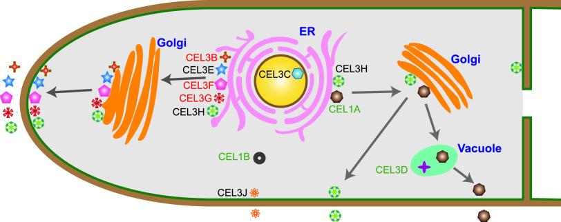
FIG 11.
Cellular distribution and protein secretion pathways of β-glucosidases CEL3B, CEL3E, CEL3F, CEL3H, CEL3J, CEL1A, CEL3C, CEL1B, CEL3G, and CEL3D in T. reesei. CEL3B, CEL3E, CEL3F, CEL3H, CEL3J, CEL1A, and CEL3G were extracellular β-glucosidases. CEL3B, CEL3E, CEL3F, and CEL3G were secreted through the tip-directed conventional ER-Golgi secretion pathway. CEL3H was located on the cell membrane and endomembrane and was secreted through tip- and non-tip-directed conventional ER-Golgi secretion pathways. CEL1A was in vacuole and secreted via the vacuole-mediated conventional ER-Golgi secretion pathway. CEL3J was distributed on the cell membrane and endomembrane and secreted via an unconventional protein pathway bypassing the ER and Golgi. The intracellular β-glucosidases CEL3C and CEL3D were in the nucleus and vacuole, respectively. CEL1B was observed in the cytoplasm with extremely low expression. The names of β-glucosidases are marked with different colors according to the impact of their overexpression on cellulase expression, which are categorized into three types—CEL1A, CEL1B, and CEL3D, marked in green, whose overexpression repressed all the cellulase activities, including pNPGase, pNPCase, and CMCase; CEL3B, CEL3F, and CEL3G, marked in red, whose overexpression decreased the pNPCase activity, increased the pNPGase activity, and did not reduce the CMCase activity; and CEL3C, CEL3E, CEL3H, and CEL3J, highlighted in black, whose overexpression decreased the pNPCase activity, and left the pNPGase activity unchanged, without compromising the CMCase activity.

