FIG 1.
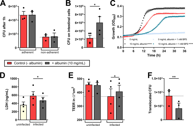
Influence of albumin on C. albicans pathogenicity mechanisms. (A) Adherent and nonadherent CFU of C. albicans to intestinal epithelial cells after 1 h in the presence or absence of 5 mg/ml albumin (HSA) in the lower compartment. (B) Fungal CFU grown on the intestinal epithelial cells were quantified after 18 h of infection with C. albicans in the presence or absence of 5 mg/ml albumin in the lower compartment. (C) Proliferation of C. albicans in the presence of 0 or 10 mg/ml albumin and of 0 or 1 mM BPS in RPMI was measured by determining the OD600 at 37°C for 36 h. (D to F) After 18 h of infection, the LDH release of intestinal epithelial cells in ng/ml was quantified from a 96-well plate (D), and the TEER (E) and CFU translocated through the intestinal epithelial layer (F) were quantified from the transwell assay. Bars represent mean values and the SEM of n = 3 (A, C, and F) or n = 4 (B, D, and E) independent experiments. Significances were calculated using a paired, parametric, two-tailed Student t test and compared to the control (= 0 mg/ml albumin or for panel B; additionally, 0 mg/ml + BPS versus 10 mg/ml + BPS) (*, P ≤ 0.05; **, P ≤ 0.01; ****, P ≤ 0.0001).

