FIG 2.
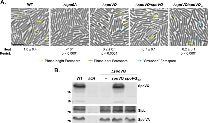
Loss of SpoVQ results in morphological and functional defects in spore formation. (A) Phase-contrast microscopy images of sporulating cultures of wild-type 630Δerm-p (WT, pyrE restored) and the indicated strains ∼20 h after sporulation induction. ΔspoVQ was complemented with either wild-type spoVQ or spoVQΔ32, the latter of which encodes an N-terminal truncation of SpoVQ’s transmembrane domain. Arrows mark mature phase-bright forepores (yellow), immature phase-dark forespores (green), and “smushed” forespores (blue), which appeared flattened relative to the oblong phase-dark or phase-bright forespores highlighted. Heat resistance efficiencies were calculated from 20- to 24-h sporulating cultures. The efficiencies represent the average ratio of heat-resistant spore CFU to total cells for a given strain relative to the WT based on a minimum of three biological replicates; the standard deviation is shown. The limit of detection of the assay is 10−6. Scale bar represents 5 μm. (B) Western blot analyses of strains shown in panel A using anti-SpoVQ, anti-SipL (20), and anti-SpoIVA antibodies.

