Abstract
RNA turnover is essential in all domains of life. The endonuclease RNase Y (rny) is one of the key components involved in RNA metabolism of the model organism Bacillus subtilis. Essentiality of RNase Y has been a matter of discussion, since deletion of the rny gene is possible, but leads to severe phenotypic effects. In this work, we demonstrate that the rny mutant strain rapidly evolves suppressor mutations to at least partially alleviate these defects. All suppressor mutants had acquired a duplication of an about 60 kb long genomic region encompassing genes for all three core subunits of the RNA polymerase—α, β, β′. When the duplication of the RNA polymerase genes was prevented by relocation of the rpoA gene in the B. subtilis genome, all suppressor mutants carried distinct single point mutations in evolutionary conserved regions of genes coding either for the β or β’ subunits of the RNA polymerase that were not tolerated by wild type bacteria. In vitro transcription assays with the mutated polymerase variants showed a severe decrease in transcription efficiency. Altogether, our results suggest a tight cooperation between RNase Y and the RNA polymerase to establish an optimal RNA homeostasis in B. subtilis cells.
INTRODUCTION
Among all organisms, bacteria are the ones multiplying most rapidly. Under optimal conditions, the model bacteria Escherichia coli and Bacillus subtilis have generation times of 20 to 30 min. On the other hand, bacteria are exposed to a variety of changing environmental conditions, and due to their small size, the impact of environmental changes is particularly severe for bacterial cells. To adapt to these potentially rapidly changing conditions, bacteria have evolved a huge arsenal of systems to sense and respond to the environment. Especially in the competition between microorganisms, it is crucial that these responses are both rapid and productive. However, while regulatory events may be very rapid, there is an element of retardation in the system, and this is the stability of mRNA and protein molecules. If the continued activity of a protein may become harmful to the bacteria, it is important not only to prevent expression of the corresponding gene but also to take two important measures: (i) switch off the protein's activity and (ii) degrade the mRNA to exclude further production of the protein. The inactivation or even degradation of proteins is well documented in the model bacteria. For example, in both E. coli and B. subtilis the uptake of toxic ammonium is limited by a regulatory interaction of the ammonium transporter with GlnK, a regulatory protein of the PII family (1,2). Similarly, the uptake of potentially toxic potassium can be prevented by inhibition of potassium transporters at high environmental potassium concentrations, either by the second messenger cyclic di-AMP or by interaction with a dedicated modified signal transduction protein, PtsN (3–5). To prevent the accumulation of potentially harmful mRNAs, bacteria rely on a very fast mRNA turnover. Indeed, in E. coli and B. subtilis >80% of all transcripts have average half-lives of less than 8 min, as compared to about 30 min and 10 h in yeast or human cells, respectively (6–9). Thus, the mRNA turnover is much faster than the generation time. The high mRNA turnover rate in bacteria contributes to the fast adaptation even in rapidly growing cells. The rapid mRNA turnover is therefore a major factor to resolve the apparent growth speed-adaptation trade-off.
RNases are the key elements to achieve the rapid mRNA turnover in bacteria. Theses enzymes can degrade bulk mRNA in a rather unspecific manner, just depending on the accessibility of the RNA molecules as well as perform highly specific cleavages that serve to process an RNA molecule to its mature form. In all organisms, RNA degradation involves an interplay of endo- and exoribonucleases as well as other proteins such as RNA helicases that resolve secondary structures (10–13). Often, these proteins form a complex called the RNA degradosome. In E. coli, the RNA degradosome is organized around the essential endoribonuclease RNase E (14,15). RNase E consists of two parts, the N-terminal endoribonuclease domain that harbors the enzymatic activity and the C-terminal macromolecular interaction domain that serves as the scaffold for the degradosome components and is responsible for the binding of RNase E to the cell membrane (15,16). As mentioned above, RNase E is essential for viability of the bacteria. An analysis of the contributions of the two parts of RNase E to its essentiality revealed that the enzymatically active N-terminal domain is essential whereas the C-terminal interaction domain is dispensable (17). This suggests that the endoribonucleolytic attack on mRNA molecules is the essential function of RNase E, whereas the interaction with other degradosome components is not required for viability. This conclusion is supported by the fact, that the other components of the E. coli degradosome are also dispensable (14).
RNase E is widespread in proteobacteria, cyanobacteria, and actinobacteria, but absent from many firmicutes, ϵ-proteobacteria, or from bacteria of the Deinococcus-Thermus class. However, an efficient RNA-degrading machinery is important also for these bacteria to allow both rapid growth and adaptation. Indeed, these bacteria possess a different endoribonuclease, RNase Y (18,19). A depletion of RNase Y results in a 2-fold increase of the average mRNA half-life in B. subtilis (19). Similar to RNase E, RNase Y is a membrane protein, and it is capable of interacting with several proteins involved in RNA degradation. Among these proteins are the 5′‐to‐3′ exoribonunclease RNase J1, polynucleotide phosphorylase, the RNA helicase CshA, the glycolytic proteins enolase and phosphofructokinase, and a protein complex composed of YaaT, YlbF and YmcA (18–23). Many of these interactions are likely to be transient as judged from the distinct localization of RNase Y and its interaction partners in the cell membrane and in the cytoplasm, respectively (24).
We are interested in the identification of the essential cellular components that are required for the viability of B. subtilis cells with the aim to construct strains that harbor only the minimal set of genes to fulfill the essential cellular functions (25–27). For B. subtilis, RNase Y and RNase J1 were originally described as being essential (18,19,28–30). Interestingly, these two RNases are also present in the most genome-reduced independently viable organism, Mycoplasma mycoides JCVI-syn3.0 (31). Both RNase J1 and RNase Y are involved in the processing and degradation of a large number of RNA molecules in B. subtilis (32–36). However, more recent studies demonstrated the possibility to delete the rnjA and rny genes, encoding the two RNases (37,38) and the dispensability of RNase Y was confirmed in a global approach to inactivate all genes of B. subtilis (39).
Comprehensive knowledge on essential genes and functions is the key to construct viable minimal genomes. By definition, essential genes cannot be individually deleted in a wild type genetic background under standard growth conditions (25). In this study, we have addressed the essentiality of RNase Y in B. subtilis. While the rny gene could indeed be deleted, this was accompanied by the rapid acquisition of suppressor mutations that affect the transcription apparatus. We demonstrate that a strongly reduced transcription activity is required to allow stable growth of B. subtilis in the absence of RNase Y. Our results suggest that the accumulation of mRNA that cannot be degraded is the growth-limiting factor in strains lacking RNase Y.
MATERIALS AND METHODS
Bacterial strains, plasmids and growth conditions
All B. subtilis strains used in this study are listed in Table 1. All strains are derived from the laboratory strain 168 (trpC2). B. subtilis and E. coli cells were grown in Lysogeny Broth (LB medium) (40). LB plates were prepared by addition of 17 g Bacto agar/l (Difco) to LB (40). The plasmids are listed in Table 2. Oligonucleotides are listed in Supplementary Table S1.
Table 1.
B. subtilis strains used in this study
| Strain | Genotypea | Source or reference |
|---|---|---|
| 168 | trpC2 | Laboratory collection |
| CCB441 | W168 Δrny::spc | 37 |
| BSB1 | Wild type | 92 |
| LK633 | MO1099 rpoE::aphA3 amyE::mls | 62 |
| LK1098 | ΔrpoE::aphA3 | LK633 → BSB1 |
| BP351 | trpC2 ΔgreA::cat | F. Commichau |
| GP2501b | trpC2 Δrny::spc | CCB441 → 168 |
| GP2503b | trpC2 Δrny::spc greA (C374T – Ser125Leu) (rrnW-rrnI)2 | Evolution of GP2501 at 22°C |
| GP2504 | trpC2 Δrny::spc greA (G169T – Glu57Stop) | Evolution of GP2501 at 22°C |
| GP2518b | trpC2 ΔgreA::cat Δrny::spc (rrnW-rrnI)2 | Evolution of GP2628 on LB agar at 37°C |
| GP2524 | trpC2 Δrny::ermC | This work |
| GP2525 | trpC2 greA-3xflag spc | pGP2542 → 168 |
| GP2529 | trpC2 Δrny::ermC greA-3xflag spc | GP2524 → GP2525 |
| GP2538 | trpC2 Δrny::ermC greA (Insertion A406)-3xflag spc | Evolution of GP2529 at 22°C |
| GP2539 | trpC2 Δrny::ermC greA (Deletion A66)-3xflag spc | Evolution of GP2529 at 22°C |
| GP2542 | trpC2 ΔrecA::spc | 44 |
| GP2614 | trpC2 ΔcspD::aphA3 | This work |
| GP2615 | trpC2 ΔcspD::aphA3 Δrny::spc | GP2501 → GP2614 |
| GP2628b | trpC2 ΔgreA::cat Δrny::spc | BP351 + GP2501 → 168 |
| GP2636b | trpC2 Δrny::spc cspD (G23A – Trp8Stop) (rrnW-rrnI)2 | Evolution of GP2501 on LB agar at 37°C |
| GP2637b | trpC2 Δrny::spc adeR (T163A – Tyr55Asn) rpoE-Δ199–208 Δskin (rrnW-rrnI)2 | Evolution of GP2501 on LB agar at 22°C |
| GP2678 | trpC2 Δrny::spc RBS of cspD(GGAGGA → GGAAGA) | Evolution of GP2501 on LB agar at 37°C |
| GP2901 | trpC2 rae1 (insertion T33) | This work |
| GP2902 | trpC2 dgk-rpoA-cat-yaaH | This work |
| GP2903 | trpC2 dgk-rpoA-cat-yaaH ΔrpoA::aphA3 | This work |
| GP2904 | trpC2 dgk-rpoA-cat-yaaH ΔrpoA::aphA3 Δrny::spc | GP2501 → GP2903 |
| GP2907 | trpC2 raeI Palf4- gfp-ermC sigH | This work |
| GP2909 | trpC2 dgk-rpoA-cat-yaaH ΔrpoA::aphA3 (rae1 Palf4- gfp-ermC sigH) | GP2907 → GP2903 |
| GP2910 | trpC2 dgk-rpoA-cat-yaaH ΔrpoA::aphA3 (rae1 Palf4- gfp-ermC sigH) Δrny::spc | GP2501 → 2909 |
| GP2912b | trpC2 dgk-rpoA-cat-yaaH ΔrpoA::aphA3 Δrny::spc rpoC (G263A – Arg88His) Δskin trnSL-Val1 (bp55T → C) | Evolution of GP2904 on LB agar at 37°C |
| GP2913b | trpC2 dgk-rpoA-cat-yaaH ΔrpoA::aphA3 (rae1 Palf4- gfp-ermC sigH) Δrny::spc rpoB (G3160T – Gly1054Cys) Δskin | Evolution of GP2910 on LB agar at 37°C |
| GP2915 | trpC2 dgk-rpoA-cat-yaaH ΔrpoA::aphA3 (rae1 Palf4- gfp-ermC sigH) Δrny::spc rpoC (G134A – Gly45Asp) | Evolution of GP2910 on LB agar at 37°C |
| GP3210 | trpC2 Δrny::spc rpoE (Insertion A88) | Evolution of GP2501 on LB agar at 22°C |
| GP3211b | trpC2 Δrny::spc (rrnW-rrnI)2 | Evolution of GP2501 at 37°C |
| GP3216 | trpC2 ΔrpoE::aphA3 | LK1098 → 168 |
| GP3217 | trpC2 ΔrpoE::aphA3 Δrny::spc | GP2501 → GP3216 |
| GP3220 | trpC2 purT-rpoB(partial)-cat-mpr | This work |
| GP3288 | trpC2 purT- rpoB-rpoC-spc-mpr | This work |
| GP3289 | trpC2 dgk-rpoA-cat::yaaH purT-rpoB-rpoC-spc-mpr | GP2903 → GP3288 |
| GP3295 | trpC2 dgk-rpoA-cat::yaaH purT-rpoB-rpoC-spc-mpr Δrny::ermC | GP2524 → GP3289 |
| GP3296 | trpC2 dgk-rpoA-cat::yaaH purT-rpoB-rpoC-spc-mpr ΔcspD::aphA3 Δrny::ermC | GP2524 + GP2614 → GP3289 |
| GP3297 | trpC2 dgk-rpoA-cat::yaaH purT-rpoB-rpoC-spc-mpr ΔrpoE::aphA3 Δrny::ermC | GP2524 + GP3216 → GP3289 |
aFor strains with suppressing point mutations the mutations are indicated using the one- and three letter code for nucleotide and amino acid substitutions, respectively.
bThese strains were analyzed by whole genome sequencing.
Table 2.
Plasmids used in this study
| Plasmid | Relevant characteristics | Source or reference |
|---|---|---|
| pCD2 | For overexpression of B. subtilis σA | 53 |
| pJOE8999 | CRISPR-Cas9 vector | 49 |
| pBSURNAP | PT7 rpoA rpoZ rpoE rpoY rpoB-rpoC-8xHis | This work |
| pGP1331 | Allows construction of triple FLAG-tag fusions | 93 |
| pGP2181 | PT7 rpoA rpoZ rpoE rpoY rpoB-rpoC*-8xHis (RpoC-R88H) | This work |
| pGP2182 | PT7 rpoA rpoZ rpoE rpoY rpoB*-rpoC-8xHis (RpoB-G1054C) | This work |
| pGP2542 | pGP1331/ greA-3xflag spc | This work |
| pGP2825 | pJOE8999/ rpoC (G263A) | This work |
| pGP2826 | pJOE8999/rea1 (insertion T33) | This work |
| pRLG770 | promoter vector | 94 |
| pRLG7558 | pRLG770 with B. subtilis Pveg (-38/-1, +1G) | 73 |
| pRLG7596 | pRLG770 with B. subtilis rrnB P1 (-39/+1) | 73 |
| pLK502 | pRLG770 with B. subtilis PilvB (-262/-1, +1GG) | This work |
DNA manipulation and genome sequencing
B. subtilis was transformed with plasmids, genomic DNA or PCR products according to the two-step protocol (40,41). Transformants were selected on LB plates containing erythromycin (2 μg/ml) plus lincomycin (25 μg/ml), chloramphenicol (5 μg/ml), kanamycin (10 μg/ml), or spectinomycin (250 μg/ml). Competent cells of E. coli were prepared and transformed following the standard procedure (40) and selected on LB plates containing kanamycin (50 μg/ml). S7 Fusion DNA polymerase (Mobidiag, Espoo, Finland) was used as recommended by the manufacturer. DNA fragments were purified using the QIAquick PCR Purification Kit (Qiagen, Hilden, Germany). DNA sequences were determined by the dideoxy chain termination method (40). Chromosomal DNA from B. subtilis was isolated using the peqGOLD Bacterial DNA Kit (Peqlab, Erlangen, Germany). To identify the mutations in the suppressor mutant strains GP2503, GP2518, GP2636, GP2637, GP2912, GP2913 and GP3211 (see Table 1), the genomic DNA was subjected to whole-genome sequencing. Concentration and purity of the isolated DNA was first checked with a Nanodrop ND-1000 (PeqLab Erlangen, Germany) and the precise concentration was determined using the Qubit® dsDNA HS Assay Kit as recommended by the manufacturer (Life Technologies GmbH, Darmstadt, Germany). Illumina shotgun libraries were prepared using the Nextera XT DNA Sample Preparation Kit and subsequently sequenced on a MiSeq system with the reagent kit v3 with 600 cycles (Illumina, San Diego, CA, USA) as recommended by the manufacturer. The reads were mapped on the reference genome of B. subtilis 168 (GenBank accession number: NC_000964) (42). Mapping of the reads was performed using the Geneious software package (Biomatters Ltd., New Zealand) (43). Frequently occurring hitchhiker mutations (44) and silent mutations were omitted from the screen. The resulting genome sequences were compared to that of our in-house wild type strain. Single nucleotide polymorphisms were considered as significant when the total coverage depth exceeded 25 reads with a variant frequency of ≥90%. All identified mutations were verified by PCR amplification and Sanger sequencing. Copy numbers of amplified genomic regions were determined by dividing the mean coverage of the amplified regions by the mean coverage of the remaining genome as described previously (44,45).
Construction of deletion mutants
Deletion of the rny, rpoA, and cspD genes was achieved by transformation with PCR products constructed using oligonucleotides to amplify DNA fragments flanking the target genes and intervening antibiotic resistance cassettes as described previously (46–48). The identity of the modified genomic regions was verified by DNA sequencing.
Chromosomal relocation of the rpoA gene
To construct a strain in which the genes for the core subunits of RNA polymerase are genomically separated, we decided to place the rpoA gene between the dgk and yaaH genes, and then to delete the original copy of the gene. First, the rpoA gene was fused in a PCR reaction with its cognate promoter and a chloramphenicol resistance gene at the 5′ and 3′ ends, respectively. In addition, the amplified dgk and yaaH genes were fused to this construct to direct the integration of the construct to the dgk-yaaH locus. The fusion of PCR products was achieved by overlapping primers. The final product was then used to transform B. subtilis 168. Correct insertion was verified by PCR amplification and sequencing. The resulting strain was B. subtilis GP2902. In the second step, the original rpoA gene was replaced by a kanamycin resistance gene as described above, leading to strain GP2903.
Chromosomal duplication of the rpoBC operon
To construct a strain carrying a duplication of the rpoBC operon, we inserted the operon in two steps into the genome of B. subtilis 168. First, the promoter and 5′ part of the rpoB gene was fused in a PCR reaction with a chloramphenicol resistance gene at the 3′ end as well as with the amplified purT and mpr genes to direct the integration of the construct to the purT-mpr locus. The fusion of the four PCR products was achieved by overlapping primers. The final product was then used to transform B. subtilis 168. Correct insertion was verified by PCR amplification and sequencing. The resulting strain was B. subtilis GP3220. In the second step, the cat gene was replaced by the 3′ part of rpoB and the complete rpoC gene fused to a spectinomycin resistance gene using the previously introduced 5′ part of rpoB and mpr to guide the homologous recombination. The resulting strain was GP3288. The correctness of the insert was verified by DNA sequencing.
Genome editing
Introduction of genetic changes in genes for RNA polymerase subunit RpoC or the non-essential RNase Rae1 at their native locus was attempted using CRISPR editing as described (49). Briefly, oligonucleotides encoding a 20 nucleotide gRNA with flanking BsaI sites and a repair fragment carrying mutations of interest with flanking SfiI restriction sites were cloned sequentially into vector pJOE8999 (49). The resulting plasmids pGP2825 and pGP2826 were used to transform recipient B. subtilis strain 168 and cells were plated on 10 μg/ml kanamycin plates with 0.2% mannose. Transformation was carried out at 30°C since replication of pJOE8999 derivatives is temperature-sensitive. The transformants were patched on LB agar plates and incubated at the non-permissive temperature of 50°C. The loss of the vector was verified by the inability of the bacteria to grow on kanamycin plates. The presence of the desired mutation in rae1 or rpoC was checked via Sanger sequencing. While the desired mutation could be introduced into the rae1 gene, this was not the case for rpoC.
Construction of the expression vector pBSURNAP
To facilitate the purification of different variants of B. subtilis RNA polymerase, we expressed and purified the core subunits of the RNA polymerase and the sigma factor separately in E. coli. For the expression of the core subunits, we cloned the corresponding B. subtilis genes into the backbone of a pET28a derivative as follows. The pRMS4 vector (a pET28a derivative, 50) containing Mycobacterium smegmatis RNA polymerase core subunit genes was used as a template to create an analogous vector containing the genes rpoA, rpoZ, rpoE, rpoY and rpoBC. The construct was designed to allow removal/substitution of each gene via unique restriction sites (Supplementary Figure S1). DNA encoding rpoA, rpoZ, rpoE and rpoY genes was cloned as one single fragment (purchased as Gene Art Strings from Invitrogen) via XbaI and NotI restriction sites. The rpoB and rpoC genes were amplified by PCR using genomic DNA of B. subtilis 168 as a template and inserted into the plasmid via NotI and NcoI or NcoI and KpnI restriction sites, respectively. The rpoC gene was inserted with a sequence encoding a 8xHis tag on the 3′ end. The cloned construct was verified by DNA sequencing. The final vector, pBSURNAP, encodes a polycistronic transcript for expression of all six RNA polymerase core subunits. Expression is driven from an IPTG-inducible T7 RNAP-dependent promoter. Each gene is preceded by a Shine-Dalgarno sequence (AGGAG) except for rpoC. RpoB-RpoC are expressed as one fused protein connected by a short linker (nine amino acid residues) to decrease the possibility that E. coli subunits would mix with B. subtilis subunits as done previously for RNA polymerase from Mycobacterium bovis (51). The full sequence of pBSURNAP has been deposited in GenBank under Accession No. MT459825. The mutant alleles of rpoB and rpoC were amplified from the mutant strains GP2913 and GP2912 and introduced into pBSURNAP by replacing the wild type alleles as NotI/NcoI and NcoI/KpnI fragments, respectively. The resulting plasmids were pGP2181 (RpoC-R88H) and pGP2182 (RpoB-G1054C).
Purification of B. subtilis RNA polymerase from E. coli cells
For purification, E. coli BL21 carrying pBSURNAP or the plasmids specifying the mutant alleles was cultivated in LB medium containing kanamycin (50 μg/ml). Expression was induced by the addition of IPTG (final concentration 0.3 mM) to logarithmically growing cultures (OD600 between 0.6 and 0.8), and cultivation was continued for 3 h. Cells were harvested and the pellets from 1 l of culture medium were washed in 50 ml buffer P (300 mM NaCl, 50 mM Na2HPO4, 3 mM β-mercaptoethanol, 1 mM PMSF, 5% glycerol) and the pellets were resuspended in 30 ml of the same buffer. Cells were lysed using a HTU DIGI-F Press (18 000 p.s.i., 138 000 kPa, two passes, G. Heinemann, Germany). After lysis, the crude extracts were centrifuged at 41 000 × g for 30 min at 4°C, and the RNA polymerase was purified from the supernatant via the His-tagged RpoC as described (52). The RNA polymerase-containing fractions were pooled and further purified by size exclusion chromatography. For this purpose, the complex was applied onto a HiLoad 16/600 Superdex 200 column (GE Healthcare) in buffer P. The buffer was filtered (0.2 μm filters) prior to protein separation on an Äkta Purifier (GE Healthcare). The fractions containing RNA polymerase were pooled and dialyzed against RNA polymerase storage buffer (50 mM Tris–HCl, pH 8.0, 3 mM β-mercaptoethanol, 0.15 M NaCl, 50% glycerol, 1:1000). The purified RNA polymerase was stored at –20°C.
The housekeeping sigma factor σA was overproduced from plasmid pCD2 (53) and purified as described (54).
In vitro transcription assays
Multiple round transcription assays were performed as described previously (55), unless stated otherwise. Initiation competent RNA polymerase was reconstituted using the core enzyme and saturating concentration of σA in dilution buffer (50 mM Tris–HCl, pH 8.0, 0.1 M NaCl, 50% glycerol) for 10 min at 30°C. Assays were carried out in 10 μl with 64 nM RNA polymerase holoenzyme and 100 ng plasmid DNA templates in transcription buffer containing 40 mM Tris–HCl (pH 8.0), 10 mM MgCl2, 1 mM dithiothreitol (DTT), 0.1 mg/ml bovine serum albumin (BSA), 150 mM NaCl, and NTPs (200 μM ATP, 2,000 μM GTP, 200 μM CTP, 10 μM UTP plus 2 μM of radiolabeled [α-32P]-UTP). The samples were preheated for 10 min at 37°C. The reaction was started by the addition of RNA polymerase and allowed to proceed for 20 min (30 min in the case of iNTP-sensing experiments) at 37°C. Subsequently, the reaction was stopped by the addition of 10 μl of formamide stop solution (95% formamide, 20 mM EDTA, pH 8.0). The samples were loaded onto 7 M urea–7% polyacrylamide gels. The gels were dried and exposed to Fuji MS phosphor storage screens, scanned with a Molecular Imager FX (BIORAD) and analyzed with Quantity One program (BIORAD).
Transcriptome analysis
Cells were grown in LB medium at 37°C to an OD600 of 0.5 to 0.6. 5 ml samples of the cultures were added to 10 ml RNA-protect (Qiagen) and allowed to incubate for 5 min at room temperature, followed by centrifugation at 5000 × g for 10 min at 4°C. Pellets were quickly frozen in liquid nitrogen and stored at −80°C. A total of three independent biological replicates were included. The harvested pellets were resuspended in 800 μl RLT buffer (RNeasy Mini Kit, Qiagen) with β-mercaptoethanol (10 μl/ml) and cell lysis was performed using a laboratory ball mill. Subsequently 400 μl RLT buffer with β-mercaptoethanol (10 μl/ml) and 1200 μl 96% [v/v] ethanol were added. For RNA isolation, the RNeasy Mini Kit (Qiagen) was used as recommended by the manufacturer, but instead of RW1 buffer RWT buffer (Qiagen) was used to facilitate the isolation of RNAs smaller 200 nt. To determine the RNA integrity number (RIN) the isolated RNA was run on an Agilent Bioanalyzer 2100 using an Agilent RNA 6000 Nano Kit as recommended by the manufacturer (Agilent Technologies, Waldbronn, Germany). Remaining genomic DNA was removed by digesting with TURBO DNase (Invitrogen, ThermoFischer Scientific, Paisley, United Kingdom). The Pan-Prokaryozes riboPOOL kit v1 (siTOOLS BIOTECH, Planegg/Martinsried, Germany) was used to reduce the amount of rRNA-derived sequences. For sequencing, the strand-specific cDNA libraries were constructed with a NEBNext Ultra II directional RNA library preparation kit for Illumina (New England BioLabs, Frankfurt am Main, Germany). To assess quality and size of the libraries, samples were run on an Agilent Bioanalyzer 2100 using an Agilent High Sensitivity DNA Kit (Agilent Technologies, Waldbronn, Germany). Concentration of the libraries were determined using the Qubit® dsDNA HS Assay Kit as recommended by the manufacturer (Life Technologies GmbH, Darmstadt, Germany). Sequencing was performed by using the HiSeq4000 instrument (Illumina Inc., San Diego, CA, USA) using the HiSeq 3000/4000 SR Cluster Kit for cluster generation and the HiSeq 3000/4000 SBS Kit (50 cycles for sequencing in the single-end mode and running 1 × 50 cycles. Between 12.623.708 and 16.865.134 raw reads were generated for the samples. For quality filtering and removing of remaining adaptor sequences, Trimmomatic-0.39 (56) and a cutoff phred-33 score of 15 were used. The mapping of the remaining sequences was performed with the Bowtie (version 2) program (57) using the implemented end-to-end mode, which requires that the entire read aligns from one end to the other. First, surviving reads were mapped against a database consisting of tRNA and rRNA sequences of B. subtilis 168 and unaligned reads were subsequently mapped against the genome of B. subtilis 168. Differential expression analyses were performed with the BaySeq program (58). Genes with fold change in expression of ≥2.0 or ≤ –2.0, a likelihood value of ≥0.9, and an adjusted P value of ≤0.05 (the P value was corrected by the false discovery rate [FDR] on the basis of the Benjamini-Hochberg procedure) were considered differentially expressed. The raw reads have been deposited in the National Center for Biotechnology Information's (NCBI) Sequence Read Archive (SRA) under accession no. SRP274247. Functional and regulation information on the differentially expressed genes was obtained from the SubtiWiki database (59).
RESULTS
Inactivation of the rny gene leads to evolution of suppressor mutations affecting transcription
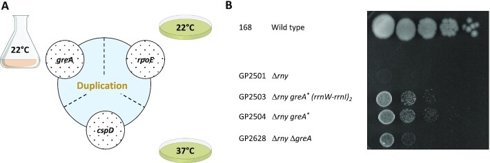
RNase Y had been considered to be essential (18,28); however, two studies reported that the rny gene could be deleted from the genome (37,39). The deletion leads to severe growth defects and morphological changes (37). In an attempt to get a better understanding of the importance of RNase Y for B. subtilis physiology, we deleted the rny gene in the genetic background of B. subtilis 168. The colonies of the resulting strain, GP2501, were small and lysed rapidly. Moreover, the cells grew very slowly at low temperatures (below 22°C). However, we observed the appearance of suppressor mutants after a few days. By analysis of such mutants we wished to gain a better understanding of the growth-limiting problem of the rny mutant. For this purpose, we isolated suppressor mutants in different experimental setups. First, the rny mutant GP2501 was adapted to growth in liquid LB medium at 22°C since the rny mutants had a severe growth defect at low temperatuRes. After the adaptation experiment, the culture was plated at 22°C, and two colonies were isolated for further investigation. In addition to the adaptation experiment in liquid medium, we also evolved suppressors on solid LB agar plates both at 22°C and 37°C. We isolated two mutants under each condition (see Figure 1A).
Figure 1.
Suppressors of rny show increased growth at 22°C. (A) Schematic depiction of different single nucleotide polymorphisms identified in the initial suppressor screen and their overlap with the duplication of ctsR-pdaB region. (B) Serial drop dilutions comparing growth of the wild type strain 168, the rny mutant GP2501, its greA suppressors (GP2503, greA (Ser125Leu) (rrnW-rrnI)2; GP2504, greA (Glu57Stop) and the rny greA double mutant GP2628 on LB-agar plate at 22°C. The greA suppressors are indicated with asterisks. The picture was taken after 2 days of incubation.
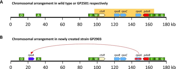
Growth of the isolated strains was verified (Figure 1B, see also Supplementary Figures S2 and S3), and for each selection scheme, one mutant was analysed by whole genome sequencing. In all cases, this confirmed the deletion of the rny gene and revealed the presence of additional mutations. Strikingly, there was one feature common for all the suppressors tested, regardless of the isolation condition, which was not present in the progenitor strain GP2501: It was an identical genomic duplication of the approximately 60 kb long ctsR-pdaB region. This genomic segment is flanked by clusters of ribosomal RNA operons. Upstream of the duplicated region are the rrnJ and rrnW operons, and downstream the rrnI, rrnH, and rrnG operons (see Figure 2A). This duplicated region contains 76 genes encoding proteins of various functions, among them proteolysis (ClpC), signal transduction (DisA), RNA modification (YacO, TruA), RNases (MrnC, Rae1), translation factors (EF-G, IF-1, EF-Tu), several ribosomal proteins, and proteins involved in transcription (NusG, RpoA, RpoB, RpoC, SigH). Strikingly, the genes for all three main subunits of the RNA polymerase—rpoA, rpoB and rpoC were present in the duplicated region. The observation, that this duplication was observed irrespective of the selective condition used to isolate suppressor mutants suggests that the duplication is relevant to overcome the poor growth associated with the loss of RNase Y. However, in addition, for each selection scheme we found additional mutations that affect genes involved in transcription.
Figure 2.
Genomic organization of the duplicated genomic region. (A) Schematic representation of the first 180 kb of the B. subtilis chromosome. The orange box indicates the duplicated region in the suppressors of rny strain GP2501. rRNA operons are depicted as green rectangles, RNA polymerase genes rpoA, rpoB, rpoC as blue arrows, the ctsR and pdaB genes are shown in yellow and red, respectively. (B) Chromosomal relocation of the rpoA gene. For the color code, see above; the relocated rpoA is shown as a purple arrow.
For the selection in liquid medium at 22°C, the suppressor mutant GP2503 had a point mutation that resulted in an amino acid substitution (S125L) in the greA gene encoding a transcription elongation factor (60). For the other suppressor mutant (GP2504) isolated under the same selective conditions, we sequenced the greA gene to test whether it had also acquired a mutation in this gene. Indeed, we found a different mutation in greA, resulting in the introduction of a premature stop codon after E56. Moreover, we evolved two additional suppressor mutants applying this adaptive scenario, and both contained frameshift mutations in greA that resulted in premature stop codons after amino acid 23 and 137 (GP2539 and GP2538, respectively; see Table 1).
The strain isolated on LB plates at 22°C (GP2637) had a deletion of the skin element, an amino acid substitution (Y55N) in the AdeR activator protein (61), and a short internal deletion in the rpoE gene encoding the δ subunit of RNA polymerase, which resulted in a frameshift after residue G66 (54,62). For the second mutant isolated at 22°C (GP3210), we re-sequenced the adeR and rpoE genes. While the adeR gene was identical to the wild type, we found an insertion of an adenine residue after position 87 of rpoE, resulting in a frameshift after 29 amino acids and premature stop codon after 38 amino acids. Therefore, the rpoE but not the adeR mutation is likely to be required for the suppressor phenotype.
The suppressor evolved at 37°C on LB plates (GP2636) contained a mutation resulting in the introduction of a premature stop at the eighth codon of the cspD gene encoding an RNA binding protein which has transcription antitermination activity in E. coli (63,64). Sanger sequencing of the second suppressor isolated under the same condition (GP2678) also identified a mutation affecting cspD, but this time in its ribosomal binding site (GGAGGA → GGAAGA).
Taken together, the duplication of the ctsR-pdaB genomic region was accompanied by specific additional suppressor mutations affecting transcription in every single suppressor mutant analysed. These mutations result in the inactivation of the greA gene in liquid medium at 22°C, whereas the selective pressure on agar plates at 22°C and 37°C was directed at the inactivation of the RNA polymerase subunit RpoE or the RNA binding protein CspD, respectively (see Figure 1A). It is therefore tempting to speculate that the inactivation of these genes combined with the ctsR-pdaB genomic duplication is causative for the suppression.
In order to test whether the inactivation of the greA, rpoE, or cspD genes alone is sufficient for the suppression of the rny mutant strain, we constructed the corresponding double mutants. As both rny and greA mutants are defective in genetic competence (39), the greA rny double mutant was obtained by transforming the wild type strain 168 with DNA molecules specifying both deletions simultaneously (see Table 1). For the greA and rpoE deletions, the double mutants did not phenocopy the original suppressor mutants, instead the gene deletions conferred only partial suppression (see Figure 1B for the rny greA double mutant GP2628, and Supplementary Figure S2 for the rny rpoE double mutant GP3217). In the case of the rny cspD double mutant GP2615, complete suppression was observed (see Supplementary Figure S3). However, we cannot exclude that the mutant had already acquired the duplication of the ctsR-pdaB genomic region. Thus, we conclude that the suppression depends on both, the duplication of the ctsR-pdaB region and the concomitant mutations that inactivate genes involved in transcription.
Transcriptome analysis of the rny mutant and a suppressor strain
As mentioned above, the deletion of greA allowed only partial suppression of the growth defect caused by the loss of RNase Y. However, the rny greA double mutant GP2628 eventually gave rise to a better suppressing mutant, GP2518. Whole genome sequencing of this strain revealed that in addition to the greA deletion it had only acquired the duplication of the ctsR-pdaB genomic region. Again, this highlights the relevance of the combination of the greA deletion and the ctsR-pdaB duplication for suppression.
To get insights into the global consequences of the suppressing mutations, we compared the transcriptomes of the wild type strain 168, the rny mutant GP2501, and the suppressor mutant GP2518 by RNA-Seq analysis. We identified 1102 genes (corresponding to about 25% of all genes of B. subtilis) with at least two-fold differential expression in the Δrny strain GP2501 as compared to the wild type 168. It should be noted that the number of differentially expressed genes is likely to be underestimated, since about 50% of all genes are not or only very poorly expressed during vegetative growth (27,65). The rny gene is encoded within an operon with the ymdB gene (66); however, there was no polar effect on the expression of ymdB, suggesting that the observed changes are a direct result of the loss of RNase Y.
From the dataset mentioned above, 587 and 515 genes were down- and upregulated, respectively, in the rny strain. The most severe difference (more than 100-fold decrease) was observed for the yxkC gene. This gene codes for protein of unknown function and is part of the σD regulon (67). Interestingly, 14 out of the 30 most strongly downregulated genes are σD dependent (see Supplementary Table S2). This may be the result of the reduced expression of the sigD gene itself. Since σD controls the expression of many genes responsible for motility as well as peptidoglycan autolysins (lytA, lytB, lytC, lytD and lytF) this reduced expression of target genes might cause the disordered cell wall of the rny deletion strain (37). Among the most strongly upregulated genes (see Supplementary Table S2), many are members of the general stress response factor σB regulon. Another set of upregulated genes is controlled by the sporulation specific sigma factors σF and σG, whose genes are also >4-fold upregulated. This is especially striking taking into an account that the rny mutant strain is not able to form spores (37).
Importantly, we wanted to test whether the suppressor mutant had restored a wild type-like expression of genes that were affected by the loss of RNase Y. We found 461 genes with differential expression between the suppressor mutant GP2518 and the rny mutant GP2501. Of these, however, only some were returned towards the expression levels of the wild type (176 genes, see Supplementary Table S3), while for others, the mRNA levels were even more distant from the wild type. In total 115 genes that were upregulated in the rny strain showed reduced expression in the suppressor mutant. On the other hand, also 61 genes which were downregulated in the rny mutant, had increased their expression again in the suppressor mutant GP2518 (see Supplementary Table S3). Among these genes with restored expression, four (murAA, tagA, tagB, ywpB) are essential, and only the expression of ywpB encoding an enzyme of fatty acid biosynthesis is 2.4-fold reduced in the rny mutant. This weak regulation suggests that fatty acid biosynthesis is not the growth-limiting factor for the rny mutant. In contrast, many of these genes with (partially) restored expression belong to prophage PBSX or are required for rather specific metabolic pathways. In conclusion, the evaluation of the genes which had their expression restored as a result of the suppressing mutations did not give a clear clue to the reason of suppression.
Genomic separation of the genes encoding the core subunits of RNA polymerase
As mentioned above, the region duplicated in all suppressor mutants contained genes encoding RNA modification enzymes, translation factors, ribosomal proteins, RNases, and proteins involved in transcription. MrnC and Rae1 are RNase Mini-III required for the maturation of 23S rRNA and ribosome-associated A site endoribonuclease, respectively (68,69). As our suppressor screen identified additional mutations related to transcription, we assumed that the translation-specific RNases encoded in this region might not be relevant for the suppression of the rny deletion. Therefore, we hypothesized that the duplication of the genes encoding the main three subunits of RNA polymerase made a major contribution to the selective advantage provided by the duplication.
To test the idea that simultaneous duplication of all three genes for the RNA polymerase core subunits is key for the suppression of the loss of RNase Y, we decided to interfere with this possibility. The duplicated region is located between two highly conserved rrn gene clusters which may facilitate the duplication event (see Figure 2A). Therefore, we attempted to separate the core RNA polymerase genes by relocating the rpoA gene out of this genomic region flanked by the rrn operons. We assumed that if RNA polymerase was indeed key to the original suppression, such a duplication would not be likely in the new background with relocated rpoA, since simultaneous duplication of all three RNA polymerase subunit genes would be disabled there. For this purpose, the rpoA gene kept under the control of its natural promoter PrpsJ was placed between the dgk and yaaH genes, and the original copy of rpoA was deleted (see Figure 2B, Materials and Methods for details). We then compared the growth of the wild type strain 168 and the strain with the relocated rpoA GP2903 using a drop-dilution assay. No differences were observed, thus excluding a possible negative impact of the rpoA relocation on B. subtilis physiology (see Supplemental Figure S4).
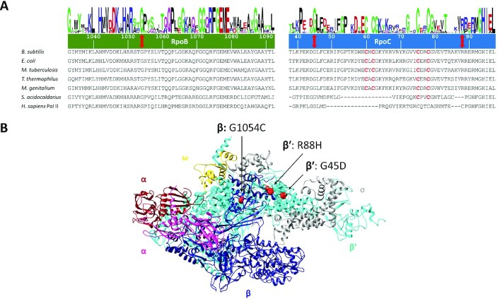
Strain GP2903 was then used to delete the rny gene, and to isolate suppressor mutants. Indeed, even with the genomically separated RNA polymerase genes, suppressor mutations appeared upon the deletion of the rny gene encoding RNase Y. There were three possibilities for the outcome of the experiment. First, the same genomic region as in the original suppressors might duplicate thus falsifying our hypothesis that the simultaneous duplication of all three genes encoding the core subunits of RNA polymerase is required for suppression. Second, both regions containing the rpoA and rpoBC genes might be duplicated. Third, in the new genetic background completely new suppressing mutations might evolve. Two of these suppressor mutants were subjected to whole genome sequencing. None of them had the duplication of the ctsR-pdaB region as in the original suppressors. Similarly, none of the mutants had the two regions containing the rpoA and the rpoBC genes duplicated. Instead, both mutants had point mutations in the RNA polymerase subunit genes that resulted in amino acid substitutions (GP2912: RpoC, R88H; GP2913: RpoB, G1054C; see Table 1). A mutation affecting RNA polymerase was also evolved in one strain (GP2915) not subjected to whole genome sequencing. In this case, the mutation resulted in an amino acid substitution (G45D) in RpoC.
An analysis of the localization of the amino acid substitutions in RpoB and RpoC revealed that they all affect highly conserved amino acid residues (see Figure 3A). G1054 of RpoB and G45 of RpoC are universally conserved in RNA polymerases in all domains of life, and R88 of RpoC is conserved in the bacterial proteins. This high conservation underlines the importance of these residues for RNA polymerase function. The mutations G45D and R88H in RpoC affect the N-terminal β’ zipper and the zinc-finger like motif of the β′ subunit, respectively, that are required for the processivity of the elongating RNA polymerase (70,71). G1054C in RpoB is located in the C-terminal domain of the β subunit that is involved in transcription termination (72). In the three-dimensional structure of RNA polymerase, these regions of the β and β′ subunits are located in close vicinity opposite to each other in the region of the RNA exit channel which guides newly transcribed RNA out of the enzyme (see Figure 3B, 71), and they are both in direct contact with DNA (70).
Figure 3.
Suppressor mutations in RNA polymerase localize to evolutionary conserved regions. (A) Multiple sequence alignment of RpoB and RpoC sequences from various species, the numbering of amino acid residues is based on the B. subtilis sequence. The positions of mutations are indicated with red double head arrows, conserved cysteines involved in Zn-finger formation are shown in red. Logos were created as described (95). Abbreviations: B. subtilis, Bacillus subtilis; E. coli, Escherichia coli; M. tuberculosis, Mycobacterium tuberculosis; T. thermophilus, Thermus thermophilus; M. genitalium, Mycoplasma genitalium; S. acidocaldarius, Sulfolobus acidocaldarius; H. sapiens, Homo sapiens. (B) Localization of the mutations (indicated as red spheres) in the RNA polymerase shown at their corresponding position in the structure of T. thermophilus (PDB ID: 1IW7; 96). The two α subunits are shown in dark red and violet, respectively, the ß subunit is shown in dark blue, ß’ in cyan, ω in gold and the σ subunit is shown in grey. The image was created using UCSF Chimera (97).
The fact that several independent mutations affecting RNA polymerase were obtained in the suppressor screen strongly supports the idea that RNA polymerase is key for the suppression. As the mutations affect highly conserved residues, they are likely to compromise the enzyme's activity. Based on the structural information, the mutations might weaken RNA polymerase-nucleic acid interactions and therefore, destabilize the transcription elongation complex which may result in increased premature termination and reduced RNA polymerase processivity. However, RNA polymerase is essential, therefore the mutations cannot inactivate the protein completely.
Establishing the rpoB and rpoC mutations in wild type background
Based on the essentiality of transcription, we expected that the mutations in rpoB and rpoC that we have identified in the suppressor screen with the rny mutant and genomically separated RNA polymerase genes might adjust some of the properties of RNA polymerase. To study the consequences of these mutations for the RNA polymerase and hence also for the physiology of B. subtilis, we decided to introduce one of them (RpoC-R88H) into the wild type background of B. subtilis 168. For this purpose, the CRISPR/Cas9 system designed for use in B. subtilis was employed (49). As a control, we used the same procedure to introduce a mutation in the rae1 gene, which is located nearby on the chromosome. Although this system readily allowed the introduction of a frameshift mutation (introduction of an extra T after 32 bp) in rae1 (strain GP2901), we failed to isolate genome-edited clones expressing the RpoC-R88H variant in multiple attempts. This failure to construct the RpoC-R88H variant in the wild type background suggests that the properties of the protein are altered in a way that is incompatible with the presence of an intact RNA degradation machine.
Mutated RNA polymerases have highly decreased activity in vitro
Since our attempts to study the effect of the mutations in vivo failed, we decided to test the properties of the mutant RNA polymerases using in vitro transcription. B. subtilis RNA polymerase is usually purified from a strain expressing His-tagged RpoC (52). However, the loss of competence of the rny mutant and the lethality of the rpoC mutation in the wild type background prevented the construction of a corresponding strain. To solve this problem, we used an approach to purify B. subtilis RNA polymerase from E. coli that had been applied successfully before for RNA polymerase of Mycobacterium smegmatis (50). Briefly, plasmid pBSURNAP containing genes rpoA, rpoB, rpoC, rpoE, rpoY and rpoZ for the RNA polymerase subunits under control of an IPTG inducible promoter was constructed in a way that each individual gene for a subunit could be cleaved out using unique restriction sites and replaced with its mutant counterpart, yielding pGP2181 (RpoC-R88H) and pGP2182 (RpoB-G1054C) (for details of the construction, see Materials and Methods). The variant RNA polymerases were expressed in E. coli BL21 and purified via affinity chromatography and subsequent size exclusion chromatography.
We purified the wild type and two mutant RNA polymerases (RpoC-R88H and RpoB-G1054C) and assessed their activity by in vitro transcription on three different templates, containing well-studied promoters of the veg and ilvB genes and the P1 promoter of the rrnB operon (73,74). In agreement with previous results on wild type RNA polymerase (75), this enzyme performed well on all three substrates. In contrast, the mutated variants of RNA polymerase exhibited a drastic decrease of transcription activity on all three promoters; for the RpoB-G1054C variant the transcripts were only barely detectable (Figure 4A).
Figure 4.
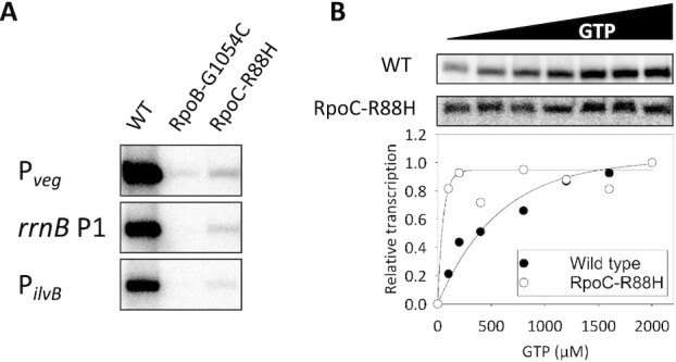
Comparison of transcriptional activity between RNA polymerase variants. (A) The RNA polymerase variants (64 nM) were reconstituted with saturating concentrations of σA (1:10). Holoenzymes were used to initiate transcription on three promoters as indicated. A representative image from three independent experiments is shown. (B) Transcription from the rrnB P1 promoter in dependence on increasing concentration of iNTP (GTP). The intensity of the transcripts generated by RNA polymerase containing RpoC-R88H was adjusted for better visibility. The relative activity of this mutant RNA polymerase was 2.5% of the wild type RNA polymerase at 2,000 μM GTP. Representative primary data are shown. The graph shows the averages of two replicates normalized for maximal transcription of each polymerase (set as 1).
On many promoters, including the P1 promoter of the rrnB operon, B. subtilis RNA polymerase is sensitive to the concentration of the first transcribed nucleotide both in vitro and in vivo (73). This prompted us to compare the response of the wild type and the RpoC-R88H variant RNA polymerases to different concentrations of GTP, the initiation NTP for the rrnB P1 transcript. As described before, transcription with the wild type enzyme increased gradually in response to the GTP concentration (73). In contrast, the mutated variant was saturated with a relatively low GTP concentration, suggesting that this important regulatory mechanism is not functional here (see Figure 4B).
Taken together, our results suggest that a reprogramming of the properties of RNA polymerase as indicated by a substantial reduction in RNA polymerase activity and its altered ability to be regulated by iNTPs allows the suppressor mutants to overcome the loss of RNase Y.
A pre-existing duplication of the genomic region containing rpoA and rpoBC is fixed in response to the deletion of rny
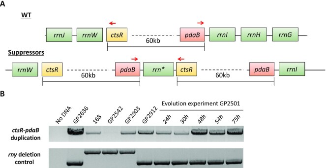
The screen for suppressor mutations that facilitate growth of strains lacking RNase Y yielded two classes of mutants: the first set harboured mutations in genes involved in transcription (greA, rpoE or cspD) in addition to a duplication of the chromosomal region encoding the core subunits of RNA polymerase. The second class had point mutations affecting the β or β’ subunits of RNA polymerase that result in strongly decreased transcription activity. At a first glance, these results seem to be conflicting. Considering RNA degradation as the function of RNase Y, it seemed plausible that the selective pressure caused by deletion of rny would result in alleviating the stress from mRNA accumulation. This seems to be the case in the second class of suppressors (see above), whereas the reason for the duplication seems to be less obvious. Importantly, this duplication was always accompanied by one of the other aforementioned mutations reducing transcription efficiency. In an attempt to determine the order of the evolutionary events in these suppressors we established a method to detect the presence of the duplication without whole genome sequencing. For this, we made use of a pair of oligonucleotides that binds to the pdaB and ctsR genes giving a product of about 10 kb, if the region is duplicated or amplified but no product in the absence of duplication or amplification (see Figure 5A). This PCR product was very prominent for the strain GP2636 that is known to carry the duplication. However, a band was also observed in the wild type strain 168, indicating that the duplication is present in a part of the population independent from the selective pressure exerted by the rny deletion (Figure 5B).
Figure 5.
Duplication of the ctsR-pdaB region in suppressors of the Δrny mutant GP2501. (A) Schematic representation of the ctsR-pdaB region and its duplication in suppressors of GP2501. In the suppressors, a chimeric rrn operon (shown as rrn*) is located between the pdaB and ctsR genes. The binding sites of the oligonucleotides used for the PCR detection of the duplication is indicated by red arrows. (B) Upper panel: The PCR product obtained by PCR using primers binding to pdaB and ctsR genes indicating presence of the duplication. Lower panel: The PCR product for the amplification of the rny region. Note the 5 μl of the PCR product were loaded in the upper panel, and 1 μl in the lower panel.
It is well-established that genomic duplications or amplifications occur frequently in bacterial populations, even in the absence of selective pressure (76). In Salmonella typhimurium, rrn operons have been shown to be a hotspot of gene duplications or amplifications (77). Since evolution of such a genomic duplication is dependent on homologous recombination, we performed the PCR also on the recA mutant GP2542, which is defective in homologous recombination and thus unable to amplify chromosomal regions (44,45). Indeed, in this case we did not obtain even a faint band. Interestingly, the genomic duplication can also be observed in cells having the core subunits of RNA polymerase at distinct genomic regions (GP2903). For the derived suppressor mutant GP2912 that carries a point mutation in rpoC, the band indicating the presence of the duplication was also detectable by PCR analysis although the duplication could not be detected by whole genome sequencing. This apparent discrepancy is most easily resolved by assuming that the duplication was present only in a small subpopulation (as observed for the wild type strain) and therefore only detectable by the very sensitive PCR assay.
Obviously, the different genomic and genetic backgrounds of the rny mutants generate distinct selective forces: While the duplication is not fixed in strains with separated rpo genes, it seems to become fixed in the suppressor mutants that have the rpo genes in one genomic region. To investigate the order of evolutionary events, we cultivated the rny mutant strain GP2501 for 75 h and monitored the status of the rpoA-rpoBC chromosomal region by PCR (see Figure 5B). The initial sample for the rny mutant GP2501 that was used for the experiment, already revealed the presence of the duplication in a small sub-population similar to the wild type strain. This supports the finding that the duplication is present irrespective of any selection. The band corresponding to the duplicated pdaB-ctsR region became more and more prominent in the course of the experiment, after 75 h it was comparable to the signal obtained with strain GP2636 that carries the duplication. As a control, we also amplified the genomic region of the rny gene. In the wild type strain, this PCR product has a size of 2.5 kb, whereas the replacement of rny by a spectinomycin resistance gene resulted in a product of 2 kb. Importantly, the intensity of this PCR product did not change during the course of the evolution experiment, thus confirming that the increased intensity of the product for the pdaB-ctsR region represents the spread of the duplication in the bacterial population. To verify the duplication and to check for the presence of accompanying mutations, we subjected genomic DNA of the strain obtained in this evolution experiment after 75 h (GP3211) to whole genome sequencing. The sequencing confirmed presence of the duplication, but did not reveal any additional suppressor mutation. Based on this result, we can assume that upon deletion of rny the bacteria first fixed the duplication of the pdaB-ctsR region and then, later, may acquire the point mutations affecting greA, rpoE, or cspD.
The duplication of genes encoding the subunits of RNA polymerase is not sufficient for suppression
In the investigation of suppressor mutants, we have found suppressor mutants that exhibited severely reduced RNA polymerase activity as well as suppressor mutants with increased copy number of core RNA polymerase subunit genes. In the latter mutants, one might expect that the increased copy number of RNA polymerase core subunit genes would result even in increased transcription, which seems to be in contradiction to the other set of suppressors. To address this obvious discrepancy, we decided to construct a strain carrying two copies of the genes encoding the RNA polymerase core subunits and compared the effect of rny deletion in strains with one or two copies of the rpoA, rpoB, and rpoC genes. For the duplication of the core subunit genes, we first assembled a second chromosomal copy of the rpoBC operon adjacent to the purT gene (see Material and Methods). Into the resulting strain GP3288, we inserted the second copy of the rpoA gene and then deleted the rny gene (see Table 1). Growth of the resulting strains was compared on LB plates. As shown in Figure 6, the duplication of the RNA polymerase core subunit genes did not overcome the severe growth defect of the rny deletion mutant. Even the additional deletion of cspD or rpoE, which were found to be inactivated in the original suppressor mutants that carry the duplication of the pdaB-ctsR region, did not restore growth (see Figure 6). This observation shows that the duplication of the genes for the RNA polymerase α, β, and β’ subunits is not sufficient for suppression of the rny deletion, and suggests that one or more additional genes that are encoded in the pdaB-ctsR region are involved in the suppression as well.
Figure 6.
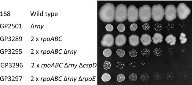
The duplication of the genes for core RNA polymerase is not sufficient to compensate for the loss of RNase Y. Serial drop dilutions comparing growth of the wild type strain 168, the rny mutant GP2501, and of the strains carrying the duplication of the rpoA, rpoB and rpoC genes on LB-agar plate at 37°C. The picture was taken after 2 days of incubation.
DISCUSSION
RNases E and Y are the main players in RNA degradation in E. coli and B. subtilis, respectively. Recently, it has been estimated that about 86% of all bacteria contain either RNase E or RNase Y (or, sometimes, both) supporting the broad relevance of these two enzymes (13). While RNase E of E. coli is essential (78), conflicting results concerning the essentiality of RNase Y have been published (18,28,29,37,39). In this study, we have examined the properties of B. subtilis mutants lacking RNase Y due to deletion of the corresponding rny gene. We observed that the rny mutant grew poorly, and rapidly acquired secondary mutations that suppressed, at least partially, the growth defect caused by the deletion of the rny gene. Thus, we conclude that RNase Y is in fact quasi-essential (31) for B. subtilis, since the mutant cannot be stably propagated on complex medium without acquiring suppressor mutations.
A lot of effort has been devoted to the understanding of the reason(s) of the (quasi)-essentiality of RNases E and Y for E. coli and B. subtilis, respectively. Initially, it was assumed that the essentiality is caused by the involvement of these RNases in one or more key essential processing event(s) that may affect the mRNAs of essential genes as has been found for B. subtilis RNase III and E. coli RNase P (33–35,79,80). However, such a target was never identified. Instead, different conclusions were drawn from suppressor studies with E. coli rne mutants lacking RNase E: some studies reported suppression by the inactivation or overexpression of distinct genes, such as deaD encoding a DEAD-box RNA helicase and ppsA encoding phosphoenolpyruvate synthetase, respectively (81,82). In addition, the processing and degradation of the essential stable RNAs, such as tRNAs and rRNAs was shown to be an essential function of RNase E (83). Yet another study suggested that mRNA turnover is the growth-limiting factor of the E. coli rne mutant (78). The results presented here lend strong support to the idea that the main task of RNase Y in B. subtilis is the control of intracellular mRNA concentration via the initiation of mRNA degradation. The transcriptome analysis with the rny mutant and a suppressor mutant revealed that only a limited number of genes shows restored expression in the suppressor mutant. Moreover, most of these genes are part of the prophage PBSX or encode very specific metabolic functions. In addition, irrespective of the conditions used in the different suppressor screens, we identified a coherent set of mutations that resulted in improved growth of the B. subtilis rny mutant. The initial mutants carry a duplication of the chromosomal region that contains the genes for the core subunits of RNA polymerase (RpoA, RpoB, RpoC) and point mutations in greA, rpoE, and cspD that all affect transcription. If this duplication was prevented by genomically separating the RNA polymerase genes, we found suppressor mutants affecting the core subunits of RNA polymerase which result in strongly compromised transcription activity. Taken together, these findings suggest that the (quasi)-essentiality of RNases E and Y is related to their general function in initiating mRNA turnover rather than to the processing of specific RNA species. This idea is further supported by three lines of evidence: First, mutations that mimic a stringent response and therefore reduce RNA polymerase activity suppressed the growth defect of a rne mutant. Second, artificial expression of RNase Y or of the ribonucleases RNase J1 or J2 from B. subtilis partially suppressed the E. coli strain lacking RNase E, but only under specific growth conditions (84,85), and third, RNase E can functionally replace RNase Y (86).
With the initiation of global mRNA degradation as the (quasi)-essential function of RNases E and Y in E. coli and B. subtilis, respectively, one might expect that the overexpression of other RNases might compensate for their loss. By analogy, such a compensation has been observed for the essential DNA topoisomerase I of B. subtilis, which could be replaced by overexpression of topoisomerase IV (44). However, in all the seven suppressor mutants analysed by whole genome sequencing (Table 1), we never observed a mutation affecting any of the known RNases of B. subtilis. Similarly, no such compensatory mutations resulting from overexpression of other cognate RNases have been found in suppressor screens for E. coli RNase E. While RNase Y does not have a paralog in B. subtilis, E. coli possesses the two related RNases E and G. However, not even the overexpression of RNase G allowed growth of an E. coli rne mutant (87,88) suggesting that RNase G has a much more narrow function than RNase E and that none of the other RNases in either bacterium is capable of initiating global mRNA degradation.
An interesting result of this study was the apparent contradiction between the isolation of suppressor mutants with increased copy number of core RNA polymerase subunit genes in one setup, intuitively suggesting increased transcription activity, and the isolation of mutants that exhibited severely reduced RNA polymerase activity in the other setup. We therefore tested with a theoretical model whether duplication of the core subunits leads to abortive incomplete complexes, as the composition of the RNA polymerase complex might be perturbed by the duplication of the core. The model confirmed that perturbing the stoichiometry of the transcription machinery may result in a strong reduction of the fraction of core RNA polymerases that assemble a functional complex. However, our experimental data demonstrate that duplication of the RNA polymerase core genes is not sufficient for suppression indicating that other factors encoded in the pdaB-ctsR chromosomal region are required to compensate for the loss of RNase Y.
In each organism, an optimal trade-off between RNA synthesis and degradation must be adjusted to allow optimal growth. Obviously, the loss of the major RNA decay-initiating enzyme will bring this adjustment out of equilibrium. This idea is supported by the observation that reduced RNA degradation in B. subtilis is accompanied by the acquisition of mutations that strongly reduce transcription activity of the RNA polymerase. In fact, the reduction of activity was so strong that it was not tolerated in a wild type strain with normal RNA degradation. This indicates that the suppressor mutants have reached a new stable equilibrium between RNA synthesis and degradation, which, however, is not optimal as judged from the reduced growth rates of the suppressor mutants as compared to the wild type strain. It has already been noticed that generation times and RNA stability are directly related (9,89). This implies that a stable genetic system requires a balance between transcription and RNA degradation to achieve a specific growth rate. In bacteria, rapid growth requires high transcription rates accompanied by rapid RNA degradation. The association between RNA polymerase and components of the RNA degrading machinery, as shown for B. subtilis and Mycobacterium tuberculosis might be a factor to achieve this coupling between RNA synthesis and degradation (90,91).
In conclusion, our study suggests that the initiation of mRNA degradation to keep the equilibrium between RNA synthesis and degradation is the function of RNase Y that makes it quasi-essential for B. subtilis. In addition to RNase Y, RNase J1 is also quasi-essential for this bacterium. In the future, it will be interesting to understand the reasons behind the critical role of this enzyme as well in order to get a more comprehensive picture of the physiology of RNA metabolism.
Supplementary Material
ACKNOWLEDGEMENTS
We are grateful to Alžbeta Rabatinová, Victoria Keidel, Janek Meißner and Fabian Commichau for providing strains and to Gabriele Beyer, Melanie Heinemann and Sarah Teresa Schüßle.r for technical support. We thank Jonas Jennrich for helpful discussions.
Author contributions: Design of the study: M.B., K.G., L.K. and J.S. Experimental work: M.B., S.W., P.F., K.G., D.K., S.K. and HŠ. Sequence analysis: M.B., A.P. and R.D. Wrote the paper: M.B., L.K. and J.S.
Contributor Information
Martin Benda, Department of General Microbiology, GZMB, Georg-August-University Göttingen, Göttingen, Germany.
Simon Woelfel, Department of General Microbiology, GZMB, Georg-August-University Göttingen, Göttingen, Germany.
Patrick Faßhauer, Department of General Microbiology, GZMB, Georg-August-University Göttingen, Göttingen, Germany.
Katrin Gunka, Department of General Microbiology, GZMB, Georg-August-University Göttingen, Göttingen, Germany.
Stefan Klumpp, Institute for the Dynamics of Complex Systems, Georg-August-University Göttingen, Göttingen, Germany.
Anja Poehlein, Department of Genomic and Applied Microbiology & Göttingen Genomics Laboratory, GZMB, Georg-August-University Göttingen, Göttingen, Germany.
Debora Kálalová, Laboratory of Microbial Genetics and Gene Expression, Institute of Microbiology of the Czech Academy of Sciences, Prague, Czech Republic.
Hana Šanderová, Laboratory of Microbial Genetics and Gene Expression, Institute of Microbiology of the Czech Academy of Sciences, Prague, Czech Republic.
Rolf Daniel, Department of Genomic and Applied Microbiology & Göttingen Genomics Laboratory, GZMB, Georg-August-University Göttingen, Göttingen, Germany.
Libor Krásný, Laboratory of Microbial Genetics and Gene Expression, Institute of Microbiology of the Czech Academy of Sciences, Prague, Czech Republic.
Jörg Stülke, Department of General Microbiology, GZMB, Georg-August-University Göttingen, Göttingen, Germany.
SUPPLEMENTARY DATA
Supplementary Data are available at NAR Online.
FUNDING
Deutsche Forschungsgemeinschaft [SFB860]; EU Horizons 2020 program [Rafts4Biotech, 720776 to J.S.]; Czech Science Foundation [20-12109S to L.K.]. Funding for open access charge: Deutsche Forschungsgemeinschaft [SFB860].
Conflict of interest statement. None declared.
REFERENCES
- 1. Coutts G., Thomas G., Blakey D., Merrick M.. Membrane sequestration of the signal transduction protein GlnK by the ammonium transporter AmtB. EMBO J. 2002; 21:536–545. [DOI] [PMC free article] [PubMed] [Google Scholar]
- 2. Detsch C., Stülke J.. Ammonium utilization in Bacillus subtilis: transport and regulatory functions of NrgA and NrgB. Microbiology. 2003; 149:3289–3297. [DOI] [PubMed] [Google Scholar]
- 3. Corrigan R.M., Campeotto I., Jeganathan T., Roelofs K.G., Lee V.T., Gründling A.. Systematic identification of conserved bacterial c-di-AMP receptor proteins. Proc. Natl. Acad. Sci. U.S.A. 2013; 110:9084–9089. [DOI] [PMC free article] [PubMed] [Google Scholar]
- 4. Gundlach J., Krüger L., Herzberg C., Turdiev A., Poehlein A., Tascón I., Weiß M., Hertel D., Daniel R., Hänelt I.et al.. Sustained sensing in potassium homeostasis: Cyclic di-AMP controls potassium uptake by KimA at the levels of expression and activity. J. Biol. Chem. 2019; 294:9605–9614. [DOI] [PMC free article] [PubMed] [Google Scholar]
- 5. Lee C.R., Cho S.H., Yoon M.J., Peterkofsky A, Seok Y.J.. Escherichia coli enzyme IIANtr regulates the K+ transporter TrkA. Proc. Natl. Acad. Sci. U.S.A. 2007; 104:4124–4129. [DOI] [PMC free article] [PubMed] [Google Scholar]
- 6. Geisberg J.V., Moqtaderi Z., Fan X., Ozsolak F., Struhl K.. Global analysis of mRNA isoform half-lives reveals stabilizing and destabilizing elements in yeast. Cell. 2014; 156:812–824. [DOI] [PMC free article] [PubMed] [Google Scholar]
- 7. Hambraeus G., von Wachenfeldt C., Hederstedt L.. Genome-wide survey of mRNA half-lives in Bacillus subtilis identifies extremely stable mRNAs. Mol. Gen. Genomics. 2003; 269:706–714. [DOI] [PubMed] [Google Scholar]
- 8. Bernstein JA., Lin P.H., Cohen S.N., Lin-Chao S.. Global analysis of Escherichia coli RNA degradosome function using DNA microarrays. Proc. Natl. Acad. Sci. U.S.A. 2004; 101:2758–2763. [DOI] [PMC free article] [PubMed] [Google Scholar]
- 9. Yang E., van Nimwegen E., Zavolan M., Rajewsky N., Schroeder M., Magnasco M., Darnell J.E. Decay rates of human mRNAs: correlation with functional characteristics and sequence attributes. Genome Res. 2003; 13:1863–1872. [DOI] [PMC free article] [PubMed] [Google Scholar]
- 10. Lehnik-Habrink M., Lewis R.J., Mäder U., Stülke J.. RNA degradation in Bacillus subtilis: an interplay of essential endo- and exoribonucleases. Mol. Microbiol. 2012; 84:1005–1017. [DOI] [PubMed] [Google Scholar]
- 11. Durand S., Tomasini A., Braun F., Condon C., Romby P.. sRNA and mRNA turnover in Gram-positive bacteria. FEMS Microbiol. Rev. 2015; 39:316–330. [DOI] [PubMed] [Google Scholar]
- 12. Redder P. Molecular and genetic interactions of the RNA degradation machineries in Firmicutes bacteria. Wiley Interdiscip. Rev. RNA. 2018; 9:e1460. [DOI] [PubMed] [Google Scholar]
- 13. Tejada-Arranz A., de Crécy-Lagard V., de Reuse H.. Bacterial RNA degradosomes: molecular machines under tight control. Trends Biochem. Sci. 2020; 45:42–57. [DOI] [PMC free article] [PubMed] [Google Scholar]
- 14. Carpousis A.J. The RNA degradosome of Escherichia coli: an mRNA-degrading machine assembled on RNase E. Annu. Rev. Microbiol. 2007; 61:71–87. [DOI] [PubMed] [Google Scholar]
- 15. Mackie G.A. RNase E: at the interface of bacterial RNA processing and decay. Nat. Rev. Microbiol. 2013; 11:45–57. [DOI] [PubMed] [Google Scholar]
- 16. Khemici V., Poljak L., Luisi B.F., Carpousis A.J.. The RNase E of Escherichia coli is a membrane-binding protein. Mol. Microbiol. 2008; 70:799–813. [DOI] [PMC free article] [PubMed] [Google Scholar]
- 17. Kido M., Yamanaka K., Mitani T., Niki H., Ogura T., Hiraga S.. RNase E polypeptides lacking a carboxyl-terminal half suppress a mukB mutation in Escherichia coli. J. Bacteriol. 1996; 178:3917–3925. [DOI] [PMC free article] [PubMed] [Google Scholar]
- 18. Commichau F.M., Rothe F.M., Herzberg C., Wagner E., Hellwig D., Lehnik-Habrink M., Hammer E., Völker U., Stülke J.. Novel activities of glycolytic enzymes in Bacillus subtilis: interactions with essential proteins involved in mRNA processing. Mol. Cell. Proteomics. 2009; 8:1350–1360. [DOI] [PMC free article] [PubMed] [Google Scholar]
- 19. Shahbabian K., Jamalli A., Zig L., Putzer H.. RNase Y, a novel endoribonuclease, initiates riboswitch turnover in Bacillus subtilis. EMBO J. 2009; 28:3523–3533. [DOI] [PMC free article] [PubMed] [Google Scholar]
- 20. Lehnik-Habrink M., Newman J., Rothe F.M., Solovyova A.S., Rodrigues C., Herzberg C., Commichau F.M., Lewis R.L., Stülke J.. RNase Y in Bacillus subtilis: a natively disordered protein that is the functional equivalent to RNase E from Escherichia coli. J. Bacteriol. 2011; 193:5431–5441. [DOI] [PMC free article] [PubMed] [Google Scholar]
- 21. Newman J.A., Hewitt L., Rodrigues C., Solovyova A.S., Harwood C.R., Lewis R.J.. Dissection of the network of interactions that links RNA processing with glycolysis in the Bacillus subtilis degradosome. J. Mol. Biol. 2012; 416:121–136. [DOI] [PubMed] [Google Scholar]
- 22. Salvo E., Alabi S., Liu B., Schlessinger A., Bechhofer D.H.. Interaction of Bacillus subtilis polynucleotide phosphorylase and RNase Y: structural mapping and effect on mRNA turnover. J. Biol. Chem. 2016; 291:6655–6663. [DOI] [PMC free article] [PubMed] [Google Scholar]
- 23. deLoughery A., Dengler V., Chai Y., Losick R. Biofilm formation by Bacillus subtilis requires an endoribonuclease-containing multisubunit complex that controls mRNA levels for the matrix gene repressor SinR. Mol. Microbiol. 2016; 99:425–437. [DOI] [PMC free article] [PubMed] [Google Scholar]
- 24. Cascante-Estepa N., Gunka K., Stülke J.. Localization of components of the RNA-degrading machine in Bacillus subtilis. Front. Microbiol. 2016; 7:1492. [DOI] [PMC free article] [PubMed] [Google Scholar]
- 25. Commichau F.M., Pietack N., Stülke J.. Essential genes in Bacillus subtilis: a re-evaluation after ten years. Mol. Biosyst. 2013; 9:1068–1075. [DOI] [PubMed] [Google Scholar]
- 26. Reuß D.R., Commichau F.M., Gundlach J., Zhu B., Stülke J.. The blueprint of a minimal cell: MiniBacillus. Microbiol. Mol. Biol. Rev. 2016; 80:955–987. [DOI] [PMC free article] [PubMed] [Google Scholar]
- 27. Reuß D.R., Altenbuchner J., Mäder U., Rath H., Ischebeck T., Sappa P.K., Thürmer A., Guérin C., Nicolas P., Steil L.et al.. Large-scale reduction of the Bacillus subtilis genome: consequences for the transcriptional network, resource allocation, and metabolism. Genome Res. 2017; 27:289–299. [DOI] [PMC free article] [PubMed] [Google Scholar]
- 28. Kobayashi K., Ehrlich S.D., Albertini A., Amati G., Andersen K.K., Arnaud M., Asai K., Ashikaga S., Aymerich S., Bessières P.et al.. Essential Bacillus subtilis genes. Proc. Natl. Acad. Sci. U.S.A. 2003; 100:4678–4683. [DOI] [PMC free article] [PubMed] [Google Scholar]
- 29. Hunt A., Rawlins J.P., Thomaides H.B., Errington J.. Functional analysis of 11 putative essential genes in Bacillus subtilis. Microbiology. 2006; 152:2895–2907. [DOI] [PubMed] [Google Scholar]
- 30. Mathy N., Bénard L., Pellegrini O., Daou R., Wen T., Condon C.. 5′-to-3′ exoribonuclease activity in bacteria: role of RNase J1 in rRNA maturation and 5′ stability of mRNA. Cell. 2007; 129:681–692. [DOI] [PubMed] [Google Scholar]
- 31. Hutchison C.A. 3rd, Chuang R.Y., Noskov V.N., Assad-Garcia N., Deerinck T.J., Ellisman M.H., Gill J., Kannan K., Karas B.J., Ma L.et al.. Design and synthesis of a minimal bacterial genome. Science. 2016; 351:aad6253. [DOI] [PubMed] [Google Scholar]
- 32. Mäder U., Zig L., Kretschmer J., Homuth G., Putzer H.. mRNA processing by RNases J1 and J2 affects Bacillus subtilis gene expression on a global scale. Mol. Microbiol. 2008; 70:183–196. [DOI] [PubMed] [Google Scholar]
- 33. Lehnik-Habrink M., Schaffer M., Mäder U., Diethmaier C., Herzberg C., Stülke J.. RNA processing in Bacillus subtilis: Identification of targets of the essential RNase Y. Mol. Microbiol. 2011; 81:1459–1473. [DOI] [PubMed] [Google Scholar]
- 34. Durand S., Gilet L., Bessières P., Nicolas P., Condon C.. Three essential ribonucleases – RNase Y, J1 and III – control the abundance of a majority of Bacillus subtilis mRNAs. PLoS Genet. 2012; 8:e1002520. [DOI] [PMC free article] [PubMed] [Google Scholar]
- 35. Laalami S., Bessières P., Rocca A., Zig L., Nicolas P., Putzer H.. Bacillus subtilis RNase Y activity in vivo analysed by tiling microarrays. PLoS One. 2013; 8:e54062. [DOI] [PMC free article] [PubMed] [Google Scholar]
- 36. deLougheri A., Lalanne J.B., Losick R., Li G.-W. Maturation of polycistronic mRNAs by the endoribonuclease RNase Y and its associated Y-complex in Bacillus subtilis. Proc. Natl. Acad. Sci. U.S.A. 2018; 115:E5585–E5594. [DOI] [PMC free article] [PubMed] [Google Scholar]
- 37. Figaro S., Durand S., Gilet L., Cayet N., Sachse M., Condon C.. Bacillus subtilis mutants with knockouts of the genes encoding ribonucleases RNase Y and RNase J1 are viable, with major defects in cell morphoplogy, sporulation, and competence. J. Bacteriol. 2013; 195:2340–2348. [DOI] [PMC free article] [PubMed] [Google Scholar]
- 38. Šiková M., Wiedermannova J., Převorovský M., Barvík I., Sudzinová P., Kofroňová O., Benada O., Šanderová H., Condon C., Krásný L.. The torpedo effect in Bacillus subtilis: RNase J1 resolves stalled transcription complexes. EMBO J. 2020; 39:e102500. [DOI] [PMC free article] [PubMed] [Google Scholar]
- 39. Koo B.M., Kritikos G., Farelli J.D., Todor H., Tong K., Kimsey H., Wapinski I., Galardini M., Cabal A., Peters J.M.et al.. Construction and analysis of two genome-scale deletion libraries for Bacillus subtilis. Cell Syst. 2017; 4:291–305. [DOI] [PMC free article] [PubMed] [Google Scholar]
- 40. Sambrook J., Fritsch E.F., Maniatis T.. Molecular Cloning: A Laboratory Manual. 1989; NY: Cold Spring Harbor Laboratory. [Google Scholar]
- 41. Kunst F., Rapoport G.. Salt stress is an environmental signal affecting degradative enzyme synthesis in Bacillus subtilis. J. Bacteriol. 1995; 177:2403–2407. [DOI] [PMC free article] [PubMed] [Google Scholar]
- 42. Barbe V., Cruveiller S., Kunst F., Lenoble P., Meurice G., Sekowska A., Vallenet D., Wang T., Moszer I., Médigue C., Danchin A.. From a consortium sequence to a unified sequence: the Bacillus subtilis 168 reference genome a decade later. Microbiology. 2009; 155:1758–1775. [DOI] [PMC free article] [PubMed] [Google Scholar]
- 43. Kearse M., Moir R., Wilson A., Stones-Havas S., Cheung M., Sturrock S., Buxton S., Cooper A., Markowitz S., Duran C.et al.. Geneious basic: an integrated and extendable desktop software platform for the organization and analysis of sequence data. Bioinformatics. 2012; 28:1647–1649. [DOI] [PMC free article] [PubMed] [Google Scholar]
- 44. Reuß D.R., Faßhauer P., Mroch P.J., Ul-Haq I., Koo B.M., Pöhlein A., Gross C.A., Daniel R., Brantl S., Stülke J.. Topoisomerase IV can functionally replace all type 1A topoisomerases in Bacillus subtilis. Nucleic Acids Res. 2019; 47:5231–5242. [DOI] [PMC free article] [PubMed] [Google Scholar]
- 45. Dormeyer M., Lübke A.L., Müller P., Lentes S., Reuß D.R., Thürmer A., Stülke J., Daniel R., Brantl S., Commichau F.M.. Hierarchical mutational events compensate for glutamate auxotrophy of a Bacillus subtilis gltC mutant. Environ. Microbiol. Rep. 2017; 9:279–289. [DOI] [PubMed] [Google Scholar]
- 46. Guérout-Fleury A.M., Shazand K., Frandsen N., Stragier P.. Antibiotic-resistance cassettes for Bacillus subtilis. Gene. 1995; 167:335–336. [DOI] [PubMed] [Google Scholar]
- 47. Youngman P. Hardwood C.R., Cutting S.M.. Use of transposons and integrational vectors for mutagenesis and construction of gene fusions in Bacillus subtilis. Mol. Biol. Methods for Bacillus. NY: Wiley; 221–266. [Google Scholar]
- 48. Wach A. PCR-synthesis of marker cassettes with long flanking homology regions for gene disruptions in S. cerevisiae. Yeast. 1996; 12:259–265. [DOI] [PubMed] [Google Scholar]
- 49. Altenbuchner J. Editing of the Bacillus subtilis genome by the CRISPR-Cas9 system. Appl. Environ. Microbiol. 2016; 82:5421–5427. [DOI] [PMC free article] [PubMed] [Google Scholar]
- 50. Kouba T., Pospíšil J., Hnilicová J., Šanderová H., Barvík I., Krásný L.. The core and holoenzyme forms of RNA polymerase from Mycobacterium smegmatis. J. Bacteriol. 2019; 201:e00583-18. [DOI] [PMC free article] [PubMed] [Google Scholar]
- 51. Czyz A., Mooney R.A., Iaconi A., Landick R.. Mycobacterial RNA polymerase requires a U-tract at intrinsic terminators and is aided by NusG at suboptimal terminators. mBio. 2014; 5:e00931-14. [DOI] [PMC free article] [PubMed] [Google Scholar]
- 52. Qi Y., Hulett F.M.. Pho-P and RNA polymerase sigma A holoenzyme are sufficient for transcription of Pho regulon promoters in Bacillus subtilis: PhoP-P activator sites within the coding region stimulate transcription in vitro. Mol. Microbiol. 1998; 28:1187–1197. [DOI] [PubMed] [Google Scholar]
- 53. Chang B.Y., Doi R.H.. Overproduction, purification, and characterization of Bacillus subtilis RNA polymerase sigma A factor. J. Bacteriol. 1990; 172:3257–3263. [DOI] [PMC free article] [PubMed] [Google Scholar]
- 54. Juang Y.L., Helmann J.D.. The delta subunit of Bacillus subtilis RNA polymerase. An allosteric effector of the initiation and core-recycling phases of transcription. J. Mol. Biol. 1994; 239:1–14. [DOI] [PubMed] [Google Scholar]
- 55. Wiedermannová J., Sudzinová P., Koval T., Rabatinová A., Šanderová H., Ramaniuk O., Rittich Š., Dohnálek J., Fu Z., Halada P.et al.. Characterization of HelD, an interacting partner of RNA polymerase from Bacillus subtilis. Nucleic Acids Res. 2014; 42:5151–5163. [DOI] [PMC free article] [PubMed] [Google Scholar]
- 56. Bolger A.M., Lohse M., Usadel B.. Trimmomatic: a flexible trimmer for Illumina sequence data. Bioinformatics. 2014; 30:2114–2120. [DOI] [PMC free article] [PubMed] [Google Scholar]
- 57. Langmead B., Salzberg S.. Fast gapped-read alignment with Bowtie 2. Nat. Methods. 2012; 9:357–359. [DOI] [PMC free article] [PubMed] [Google Scholar]
- 58. Mortazavi A., Williams B.A., McCue K., Schaeffer L., Wold B.. Mapping and quantifying mammalian transcriptomes by RNA-Seq. Nat. Methods. 2008; 5:621–628. [DOI] [PubMed] [Google Scholar]
- 59. Michna R.M., Commichau F.M., Tödter D., Zschiedrich C.P., Stülke J.. SubtiWiki – a database for the model organism Bacillus subtilis that links pathway, interaction and expression information. Nucleic Acids Res. 2014; 42:D692–D698. [DOI] [PMC free article] [PubMed] [Google Scholar]
- 60. Kusuya Y., Kurokawa K., Ishikawa S., Ogasawara N., Oshima T.. Transcription factor GreA contributes to resolving promoter-proximal pausing of RNA polymerase in Bacillus subtilis cells. J. Bacteriol. 2011; 193:3090–3099. [DOI] [PMC free article] [PubMed] [Google Scholar]
- 61. Lin T.-H., Wei G.-T., Su C.-C., Shaw G.-C.. AdeR, a PucR-type transcription factor, activates expression of L-alanine dehydrogenase and is required for sporulation of Bacillus subtilis. J. Bacteriol. 2012; 194:4995–5001. [DOI] [PMC free article] [PubMed] [Google Scholar]
- 62. Rabatinová A., Šanderová H., Matějčková J.J., Korelusová J., Sojka L., Barvik I., Popoušková V., Sklenář V., Židek L., Krásný L.. The δ subunit of RNA polymerase is required for rapid changes in gene expression and competitive fitness of the cell. J. Bacteriol. 2013; 195:2603–2611. [DOI] [PMC free article] [PubMed] [Google Scholar]
- 63. Graumann P., Wendrich T.M., Weber M.H.W., Schröder K., Marahiel M.A. A family of cold shock proteins in Bacillus subtilis is essential for cellular growth and for efficient protein synthesis at optimal and low temperature. Mol. Microbiol. 1997; 25:741–756. [DOI] [PubMed] [Google Scholar]
- 64. Bae W., Xia B., Inoye M., Severinov K.. Escherichia coli CspA-family RNA chaperones are transcription antiterminators. Proc. Natl. Acad. Sci. U.S.A. 2000; 97:7784–7789. [DOI] [PMC free article] [PubMed] [Google Scholar]
- 65. Rasmussen S., Nielsen H.B., Jarmer H.. The transcriptionally active regions in the genome of Bacillus subtilis. Mol. Microbiol. 2009; 73:1043–1057. [DOI] [PMC free article] [PubMed] [Google Scholar]
- 66. Diethmaier C., Pietack N., Gunka K., Wrede C., Lehnik-Habrink M., Herzberg C., Hübner S., Stülke J.. A novel factor controlling bistability in Bacillus subtilis: the YmdB protein affects flagellin expression and biofilm formation. J. Bacteriol. 2011; 193:5997–6007. [DOI] [PMC free article] [PubMed] [Google Scholar]
- 67. Serizawa M., Yamamoto H., Yamaguchi H., Fujita Y., Kobayashi K., Ogasawara N., Sekiguchi J.. Systematic analysis of SigD-regulated genes in Bacillus subtilis by DNA microarray and Northern blotting analyses. Gene. 2004; 329:125–136. [DOI] [PubMed] [Google Scholar]
- 68. Redko Y., Bechhofer D.H., Condon C.. Mini-III, an unusual member of the RNase III family of enzymes, catalyses 23S ribosomal RNA maturation in Bacillus subtilis. Mol. Microbiol. 2008; 68:1096–1106. [DOI] [PubMed] [Google Scholar]
- 69. Leroy M., Piton J., Gilet L., Pellegrini O., Proux C., Coppée J.-Y., Figaro S., Condon C.. Rae1/YacP, a new endoribonuclease involved in ribosome-dependent mRNA decay in Bacillus subtilis. EMBO J. 2017; 36:1167–1181. [DOI] [PMC free article] [PubMed] [Google Scholar]
- 70. Nudler E., Avetissova E., Markovtsov V., Goldfarb A.. Transcription processivity: protein-DNA interactions holding together the elongation complex. Science. 1996; 273:211–217. [DOI] [PubMed] [Google Scholar]
- 71. Nudler E. RNA polymerase active center: the molecular engine of transcription. Annu. Rev. Biochem. 2009; 78:335–361. [DOI] [PMC free article] [PubMed] [Google Scholar]
- 72. Clerget M., Jin D.J., Weisberg R.A.. A zinc-binding region in the beta’ subunit of RNA polymerase is involved in antitermination of early transcription of phage HK022. J. Mol. Biol. 1995; 248:768–780. [DOI] [PubMed] [Google Scholar]
- 73. Krásný L., Gourse R.L.. An alternative strategy for bacterial ribosome synthesis: Bacillus subtilis RNA polymerase rRNA transcription regulation. EMBO J. 2004; 23:4473–4483. [DOI] [PMC free article] [PubMed] [Google Scholar]
- 74. Krásný L., Tiserová H., Jonák J., Rejman D., Šanderová H.. The identity of the transcription +1 position is crucial for changes in gene expression in response to amino acid starvation in Bacillus subtilis. Mol. Microbiol. 2008; 69:42–54. [DOI] [PubMed] [Google Scholar]
- 75. Sojka L., Kouba T., Barvík I., Šanderová H., Maderová Z., Jonák J., Krásný L.. Rapid changes in gene expression: DNA determinants of promoter regulation by the concentration of the transcription initiating NTP in Bacillus subtilis. Nucleic Acids Res. 2011; 39:4598–4611. [DOI] [PMC free article] [PubMed] [Google Scholar]
- 76. Andersson D.I., Hughes D.. Gene amplification and adaptive evolution in bacteria. Annu. Rev. Genet. 2009; 43:167–195. [DOI] [PubMed] [Google Scholar]
- 77. Anderson P., Roth J.. Spontaneous tandem genetic duplications in Salmonella typhimurium arise by unequal recombination between rRNA (rrn) cistrons. Proc. Natl. Acad. Sci. U.S.A. 1981; 78:3113–3117. [DOI] [PMC free article] [PubMed] [Google Scholar]
- 78. Hammarlöf D.L., Bergman J.M., Garmendia E., Hughes D.. Turnover of mRNAs is one of the essential functions of RNase E. Mol. Microbiol. 2015; 98:34–45. [DOI] [PubMed] [Google Scholar]
- 79. Mohanty B.K., Agrawal A., Kushner S.R.. Generation of pre-tRNAs from polycistronic operons is the essential function of RNase P in Escherichia coli. Nucleic Acids Res. 2020; 48:2564–2578. [DOI] [PMC free article] [PubMed] [Google Scholar]
- 80. Durand S., Gilet L., Condon C.. The essential function of B. subtilis RNase III is to silence foreign toxin genes. PLoS Genet. 2012; 8:e1003181. [DOI] [PMC free article] [PubMed] [Google Scholar]
- 81. Tamura M., Kers J.A., Cohen S. Second-site suppression of RNase E essentiality by mutation of the deaD RNA helicase in Escherichia coli. J. Bacteriol. 2012; 194:1919–1926. [DOI] [PMC free article] [PubMed] [Google Scholar]
- 82. Tamura M., Honda N., Fujimoto H., Cohen S.N., Kato A.. PpsA-mediated alternative pathway to complement RNase E essentiality in Escherichia coli. Arch. Microbiol. 2016; 198:409–421. [DOI] [PubMed] [Google Scholar]
- 83. Sulthana S., Basturea G.N., Deutscher M.P.. Elucidation of pathways of ribosomal RNA degradation: an essential role for RNase E. RNA. 2016; 22:1163–1171. [DOI] [PMC free article] [PubMed] [Google Scholar]
- 84. Himabindu P., Anupama K.. Decreased expression of stable RNA can alleviate the lethality associated with RNase E deficiency in Escherichia coli. J. Bacteriol. 2017; 199:e00724-16. [DOI] [PMC free article] [PubMed] [Google Scholar]
- 85. Tamura M., Kageyama D., Honda N., Fujimoto H., Kato A.. Enzymatic activity necessary to restore the lethality due to Escherichia coli RNase E deficiency is distributed among bacteria lacking RNase E homologues. PLoS One. 2017; 12:e0177915. [DOI] [PMC free article] [PubMed] [Google Scholar]
- 86. Laalami S., Cavaiuolo M., Roque S., Chagneau C., Putzer H.. Escherichia coli RNase E can efficiently replace RNase Y in Bacillus subtilis. Nucleic Acids Res. 2021; 49:4643–4654. [DOI] [PMC free article] [PubMed] [Google Scholar]
- 87. Deana A., Belasco J.G.. The function of RNase G in Escherichia coli is constrained by its amino and carboxyl termini. Mol. Microbiol. 2004; 51:1205–1217. [DOI] [PubMed] [Google Scholar]
- 88. Chung D.-H., Min Z., Wang B.-C., Kushner S.R. Single amino acid changes in the predicted RNase H domain of Escherichia coli RNase G lead to complementation of RNase E deletion mutants. RNA. 2010; 16:1371–1385. [DOI] [PMC free article] [PubMed] [Google Scholar]
- 89. Rustad T.R., Minch K.J., Brabant W., Winkler J.K., Reiss D.J., Baliga N.S., Sherman D.R.. Global analysis of mRNA stability in Mycobacterium tuberculosis. Nucleic Acids Res. 2013; 41:509–517. [DOI] [PMC free article] [PubMed] [Google Scholar]
- 90. Delumeau O., Lecointe F., Muntel J., Guillot A., Guédon E., Monnet V., Hecker M., Becher D., Polard P., Noirot P.. The dynamic partnership of RNA polymerase in Bacillus subtilis. Proteomics. 2011; 11:2992–3001. [DOI] [PubMed] [Google Scholar]
- 91. Płociński P., Macios M., Houghton J., Niemiec E., Płocińska R., Brzostek A., Słomka M., Dziadek J., Young D., Dziembowski A.. Proteomic and transcriptomic experiments reveal an essential role of RNA degradosome complexes in shaping the transcriptome of Mycobacterium tuberculosis. Nucleic Acids Res. 2019; 47:5892–5905. [DOI] [PMC free article] [PubMed] [Google Scholar]
- 92. Nicolas P., Mäder U., Dervyn E., Rochat T., Leduc A., Pigeonneau N., Bidnenko E., Marchadier E., Hoebeke M., Aymerich S.et al.. Condition-dependent transcriptome reveals high-level regulatory architecture in Bacillus subtilis. Science. 2012; 335:1103–1106. [DOI] [PubMed] [Google Scholar]
- 93. Lehnik-Habrink M., Pförtner H., Rempeters L., Pietack N., Herzberg C., Stülke J.. The RNA degradosome in Bacillus subtilis: Identification of CshA as the major RNA helicase in the multi-protein complex. Mol. Microbiol. 2010; 77:958–971. [DOI] [PubMed] [Google Scholar]
- 94. Ross W., Thompson J.F., Newlands J.T., Gourse R.L.. E. coli Fis protein activates ribosomal RNA transcription in vitro and in vivo. EMBO J. 1990; 9:3733–3742. [DOI] [PMC free article] [PubMed] [Google Scholar]
- 95. Crooks G.E., Hon G., Chandonia J.-M., Brenner S.E.. WebLogo: a sequence logo generator. Genome Res. 2004; 14:1188–1190. [DOI] [PMC free article] [PubMed] [Google Scholar]
- 96. Vassylyev D.G., Sekine S., Laptenko O., Lee J., Vassylyeva M.N., Borukhov S.. Crystal structure of a bacterial RNA polymerase holoenzme at 2.6 Å resolution. Nature. 2002; 417:712–719. [DOI] [PubMed] [Google Scholar]
- 97. Pettersen E.F., Goddard T.D., Huang C.C., Couch G.S., Greenblatt D.M., Meng E.C., Ferrin T.E.. UCSF Chimera – a visualization system for exploratory research and analysis. J. Comput. Chem. 2004; 25:1605–1612. [DOI] [PubMed] [Google Scholar]
Associated Data
This section collects any data citations, data availability statements, or supplementary materials included in this article.


