Abstract
Purpose
To determine the role of scavenger receptor expressed by endothelial cell-1 (SREC-Ⅰ) in vitro and in a mouse model of Aspergillus fumigatus keratitis.
Methods
SREC-Ⅰ mRNA and protein expression were tested in both normal and A fumigatus stimulated human corneal epithelial cells (HCECs). Immunofluorescence was used to detect SREC-Ⅰ expression in human corneas with or without A fumigatus infection. HCECs were incubated with SREC-Ⅰ small interfering RNA, then the mRNA levels of LOX-1, IL-1β, and TNF-α were detected after A fumigatus stimulation. A mouse fungal keratitis (FK) model was established and SREC-Ⅰ mRNA and protein expression were detected by RT-PCR, Western blot and immunofluorescence. The severity of FK was evaluated by clinical score. CLCX1, LOX-1, IL-1β, and TNF-α mRNA expression levels were tested before and after anti–SREC-Ⅰ treatment.
Results
SREC-Ⅰ expressed in normal and A fumigatus treated HCECs and human corneal epithelium. In vitro experiment showed that SREC-Ⅰ mRNA and protein levels were significantly increased after A fumigatus stimulation. SREC-Ⅰ small interfering RNA treatment inhibited the expressions of LOX-1, IL-1β, and TNF-α in HCECs. The expressions of CLCX1, LOX-1, IL-1β, and TNF-α were elevated in mice with A fumigatus keratitis, which could be decreased by SREC-Ⅰ–neutralizing antibody treatment.
Conclusions
SREC-Ⅰ is a key mediator in inflammatory response induced by A fumigatus keratitis. SREC-Ⅰ blockade could be a potential therapeutic approach for FK.
Keywords: Aspergillus fumigatus, corneal epithelial cells, innate immunity, SREC-Ⅰ
Fungal keratitis (FK) is a major cause of visual impairment and blindness globally, associated with agriculture-related ocular trauma, overuse of contact lenses, and postoperative corneal infection.1,2 The incidence of FK ranges from 6% and 56% in various regions of the world.3 Mycotic keratitis is expected to be more common in the tropical and subtropic locations due to the hot, humid climate and the agriculture-based occupation,3 in which Aspergillus species caused keratitis is one of the most common FK. However, there have been no new treatments since natamycin was introduced in 1960s.2 Research on the pathogenesis and immune mechanisms of FK has become a hot spot in recent years, aiming to provide new targets for FK treatment. The innate immune system provides the first line of defense to recognize and resist pathogens against fungal infection, and the pattern recognition receptors play a vital role in innate immunity,4 including Toll-like receptors (TLRs), C-type lectin-like receptors, and the scavenger receptor family.5–8
Scavenger receptor expressed by endothelial cell-Ⅰ (SREC-Ⅰ) is a member of scavenger receptor family F, known as SCARF-Ⅰ.9 It is an 86-kDa protein with an extended extracellular domain and first cloned from human umbilical vein endothelial cells.10–11 SREC-Ⅰ is expressed by multiple types of cells, such as endothelial cells, epithelial cells, dendritic cells, and macrophages,12–14 but the expression in corneal cells is largely unknown. It is reported that SREC-Ⅰ could recognize modified self-ligands such as acetylated low-density lipoprotein, heat shock proteins, and apoptotic bodies,15–18 as well as several exogenous ligands from microbial pathogens like lipopolysaccharide and lipoteichoic acid to mediate endocytosis and phagocytosis,19–20 thus participating in the host's innate immunity.21
Moreover, studies have shown that SREC-Ⅰ could recognize and bind to β-glucans present on the cell surface of Cryptococcus neoformans and Candida albicans, then mediate the cytokine production in association with TLR2.22 However, the expression and function of SREC-Ⅰ during A fumigatus keratitis have yet to be determined. Our study investigated the role of SREC-Ⅰ in innate immunity to A fumigatus keratitis in vitro and in vivo. Results indicated that SREC-Ⅰ expressed in HCECs and human/mice corneal epithelial cells, and SREC-Ⅰ mRNA and protein levels were upregulated after A fumigatus stimulation. SREC-Ⅰ inhibition could decrease the expression of inflammatory factors, including IL-1β, TNF-α, and LOX-1, as well as the trending factor CXCL1 that is elevated in A fumigatus keratitis. Thus, our study suggested that SREC-Ⅰ works as an important member of innate immunity during A fumigatus keratitis, which could be a potential therapeutic or an additional target for FK.
Methods
Clinical Specimens
Six patients (four females and two males, 40–50 years old) with FK underwent penetrating keratoplasty at the Department of Ophthalmology (The Affiliated Hospital of Qingdao University) were included. The patients enrolled in the research were diagnosed clinically by staining of corneal scrapings, fungal culture (verified A fumigatus growth), or confocal microscopy at 25 to 30 days before the keratoplasty. All the samples were extracted during the keratoplasty. In total, six healthy corneal tissue samples from the remaining peripheral tissues of donor corneas and six patients (six eyes) with A fumigatus infection after corneal transplantation were used for immunofluorescence analysis. All patients gave their informed consent for inclusion before they participated in the study. Research adhered to the tenets of the Declaration of Helsinki. The agreement of patients was obtained and the informed consents were signed for specimen management. The experiment had the approval of the hospital's ethics committee.7
Aspergillus fumigatus Culture
A fumigatus standard strain 3.0772 (China General Microbiological Culture Collection Center, Beijing, China) was cultured in Sabouraud liquid medium at 37 °C, 110 rpm for 2 days. Then, the harvested mycelia of A fumigatus was washed three times by sterile PBS, then the supernatant was discarded without inactivation (for animals) or inactivated at 4 °C overnight in 70% alcohol (for human corneal epithelial cells [HCECs]).7,23 The density of the fungal mycelia was read in a blood cell counting board, and diluted to the final concentration of 1 × 10⁸ CFU/mL. The inactivated A fumigatus mycelia was stored at −20 °C.
Cell Culture and A fumigatus Stimulation
HCECs (ATCC CRL-11135, kindly provided by Ocular Surface Laboratory of Zhongshan Ophthalmic Center, Guangzhou, Guangdong, China) were cultured in DMEM with 10% fetal bovine serum (Gibco, Shanghai, China), 0.075% growth factor fibroblast growth factor 2 (Gibco), 0.075% insulin (Solarbio, Beijing, China), 1% penicillin G (Gibco), and streptomycin sulfate (Solarbio) in a humidified 5% CO2 incubator at 37 °C.7 HCECs suspensions of 1 × 10⁵/mL were seeded onto 12- or 6-well tissue culture plates until 80% of the cells were attached.24 Then the cells were rinsed by serum-free DMEM and treated with A fumigatus hyphae (to the final concentration of 5 × 10⁶ CFU/mL) for 0, 4, 8, 12, 16, 24, and 36 hours. HCECs were harvested for RT-PCR and Western blot. The mRNA levels of SREC-Ⅰ in HCECs were detected by RT-PCR, and the SREC-Ⅰ protein levels of HCECs were detected by Western blot.
SREC-Ⅰ Small Interfering RNA (siRNA) Treatment of HCECs
HCECs were incubated with SREC-Ⅰ siRNA (final concentration 200 nM; RiboBio, Guangzhou, Guangdong, China) or scrambled control siRNA (final concentration 200 nM; RiboBio) with transfection reagent (Ribo FECT CP Transfection Kit, Reagent No. C10511-05) at 1 day before infection. Then the cells were treated with inactivated A fumigatus for 8, 24, and 36 hours.25 The cells were harvested for RT-PCR to estimate the mRNA expression of SREC-I, IL-1β, TNF-α, and LOX-1 at 8 hours, and for Western blot to detect SREC-I protein level at 36 hours. The supernatants were collected for ELISA to evaluate the release of IL-1β and TNF-α protein at 24 hours.
Animals and Corneal Infection
Female 8-week-old C57BL/6 mice were purchased from SPF Biotechnology Co., Ltd. (Beijing, China) and weighed between 20 and 25 g. The animals were treated in accordance with the guidelines provided in the ARVO Statement for the Use of Animals in Ophthalmic and Vision Research. The method to obtain mice FK models was in accordance with Zhan et al.26 Mice were anesthetized with 8% chloral hydrate, placed beneath a stereoscopic microscope at ×25 magnification, and treated with intrastromal injections using a sterile microliter syringe (10 µL; Hamilton Corp., Bonaduz, GR, Switzerland). Conidia were harvested by rinsing the A fumigatus malt agar slants with PBS containing 0.1% Tween 20 (Sigma-Aldrich Corp., St. Louis, MO). Conidia suspension was prepared by repeated resuspending, centrifuging (12000g for 5 minutes), and washing using PBS (to the final concentration of 2.6 × 10⁷ CFU/mL). 2.5 µL of A fumigatus conidia suspension was injected into the midstromal level in the center of the right cornea and the left eyes were chosen as a sham control injected with PBS. Mice corneas were harvested for RT-PCR and Western blot at 0.5, 1.0, and 2.0 days after the experimental model was established. Eyeballs were removed at 48 hours for immunofluorescence analysis.
SREC-Ⅰ Neutralizing Antibody Treatment of Animals
Rabbit anti-mouse SREC-Ⅰ neutralizing antibody (20 µg/10 µL; Affinity Biosciences, Changzhou, China) or control IgG (20 µg/10 µL; Affinity Biosciences) was given subconjunctivally into the right eyes of mice (n = 6/group/time) 1 day before infection. One hour post infection (p.i.), an additional anti-SREC-Ⅰ antibody (200 µg/100 µL) was injected intraperitoneally (i.p.), and controls were similarly injected with IgG. Mice corneas at 12 hours p.i. were harvested for RT-PCR, and 2 days p.i. were harvested for Western blot to evaluate the expression level of SREC-I. The severity of keratitis was evaluated at 1, 3, and 5 days p.i. by clinical score that was the sum of the three aspects of cornea including opacity density, opacity area, and surface regularity, each of which has a grade of 0 to 4.27 Meanwhile, ranging from 0 to 12, the severity of keratitis was divided into normal (0), mild (1–5), moderate (6–9), and severe (10–12).26,28 Mice corneas at 12 and 48 hours p.i. were harvested for RT-PCR to evaluate IL-1β, TNF-α, LOX-1, and CXCL1 mRNA levels, and for ELISA to test the protein expression of IL-1β and TNF-α. Eyeballs were removed at 48 hours for hematoxylin and eosin staining.
Immunofluorescence
Immunofluorescence method was determined as reported elsewhere.29 Tissue samples were embedded in OCT compound (Tissue-Tek; Miles, Elkhart, IN), and frozen in liquid nitrogen, then cut into 8-µm-thick serial tissue sections and stored at 37 °C for 6 hours. After a 5-minute fixation in acetone, sections were blocked using normal goat serum (Bioss, Beijing, China) diluted 1:100 with PBS for 30 minutes, then incubated with rabbit anti-human SREC-Ⅰ (Abcam, Cambridge, MA) diluted 1:100 or rabbit anti-mouse SREC-Ⅰ (Bioss) diluted 1:100 overnight at 4 °C.30 This process was followed by FITC-conjugated goat anti-rabbit secondary antibody (1:200; Elabscience, Wuhan, China) for another hour. Cell nuclei were stained with DAPI (Bioss). The fluorescent micrographs were taken with a Zeiss Axiovert microscope at an original magnification of ×40.
Real-Time PCR
Total RNA of cells or corneas were extracted by the RNA iso plus reagent (TaKaRa, Dalian, China), and the RNA was quantified by the NanoDrop ND-1000 Spectrophotometry (Thermo Fisher Scientific). The first strand cDNA was synthesized by a two-step method using PrimeScript RT reagent Kit from total RNA. Then cDNA was used for PCR in 20-µL reaction volume following the manufacturer's instructions. The PCR program for the reactions was described as before.31 The ΔΔCT method was used for analysis of target gene products. Data are expressed as fold of increase in mRNA expression. The primer pair sequences are listed in the Table.
Table.
Nucleotide Sequence of Human and Mouse Primers for RT-PCR Amplification
| Gene | Primer | Nucleotide Sequence | GenBank |
|---|---|---|---|
| β-Actin(human) | FR | 5′-GAT TAC TGC TCT GGC TCC TAG C-3′5′-GAC TCA TCG TAC TCC TGC TTG C-3′ | NM_007393.3 |
| SREC-Ⅰ(human) | FR | 5′- GGT GAC AGC TTC ATC GAG CC-3′5′- AAC CTC ATC TGC CTC TCC AGA C-3′ | NM_145350.3 |
| LOX-1(human) | FR | 5′- GAG TGA ACA TAT CCA TCA TC-3′5′- TGG AGA CAT ATG AAT CTC AA-3′ | NM_002543.4 |
| IL-1β(human) | FR | 5′-GCT GAT GGC CCT AAA CAG ATG AA-3′5′-TCC ATG GCC ACA ACA ACT GAC-3′ | NM_008361.3 |
| TNF-α(human) | FR | 5′-TGC TTG TTC CTC AGC CTC TT-3′5′-CAG AGG GCT GAT TAG AGA GAG GT-3′ | NM_013693.2 |
| β-Actin(mouse) | FR | 5′-GAT TAC TGC TCT GGC TCC TAG C-3′5′-GAC TCA TCG TAC TCC TGC TTG C-3′ | NM_007393.3 |
| SREC-Ⅰ(mouse) | FR | 5′- TGG GAC TAG AGC TGG TGT TCT-3′5′- CAG ATG GGG ATG GTG CAT TCT-3′ | NM_001004157.2 |
| IL-1β(mouse) | FR | 5′-CGC AGC AGC ACA TCA ACA AGA GC-3′5′-TGT CCT CAT CCT GGA AGG TCC ACG-3′ | NM_008361.4 |
| TNF-α(mouse) | FR | 5′-ACC CTC ACA CTC AGA TCA TCT T-3′5′-GGT TGT CTT TGA GAT CCA TGC-3′ | NM_013693.3 |
| LOX-1(mouse) | FR | 5′- AGG TCC TTG TCC ACA AGA CTG G-3′5′- ACG CCC CTG GTC TTA AAG AAT TG-3′ | NM_138648.2 |
| CXCL1(mouse) | FR | 5′- TCA CCT CAA GAA CAT CCA GAG C-3′5′- ACT TGG GGA CAC CTT TTA GCA T-3′ | NM_008176.3 |
F, forward; R, reverse.
Western Blot
The protein separation procedure of C57BL/6 corneas and HCECs has been interpreted previously.32 The protein concentration was measured using BCA Protein Assay Kit (Solarbio). Then total protein was separated by SDS-PAGE electrophoresis and transferred onto PVDF membranes (Solarbio). The membranes were blocked with Western blocking buffer (Beyotime, Jiangsu, China) at 37 °C for 2 hours and then were incubated with primary antibody against β-actin (1:1000; Elabscience) or GAPDH (1:1000; Abcam) and primary antibody against SREC-Ⅰ (1:2000; Abcam) overnight at 4 °C. After washing in PBS/Tween three times, the membranes were incubated with corresponding peroxidase-conjugated secondary antibody (1:5000, Beyotime) at 37 °C for 1 hour.30 Finally, the blots were visualized by chemiluminescence (Thermo Fisher Scientific).
ELISA
Double-sandwich ELISA for human IL-1β and TNF-α was performed following the manufacturer's instructions (Elabscience) to detect the concentration of IL-1β and TNF-α proteins in HCECs from different treatments. The supernatant of HCECs was collected and centrifuged for 5 minutes at 1000 g at 4 °C, and 100 µL of each sample was assayed in duplicate to evaluate the protein levels of IL-1β and TNF-α. The optical density (optical density value) was measured at 450 nm. Sensitivities were 4.69 pg/mL (TNF-α) and 4.69 pg/mL (IL-1β).
Hematoxylin and Eosin Staining
Mice eyeballs (n = 3/group/time) were fixed in 4% paraformaldehyde at 4 °C for 3 days, embedded in paraffin, and sliced into 5-µm-thick serial tissue sections. The sections were stained with hematoxylin and eosin successively. After dehydration by alcohol, the sections were fixed and photographed under a light microscopy at an original magnification of ×40.
Statistical Analysis
All data were presented as mean ± SD from independent experiments. RT-PCR, ELISA, and Western blot were analyzed by one-way ANOVA to make comparison among three or more groups. An unpaired, two-tailed Student t-test was used to identify the difference between each two groups. All experiments were performed at least three times to ensure practicability. A P value of less than 0.05 was considered significant.
Results
SREC-Ⅰ Expression Increased in HCECs and Human Corneal Epithelium With A fumigatus
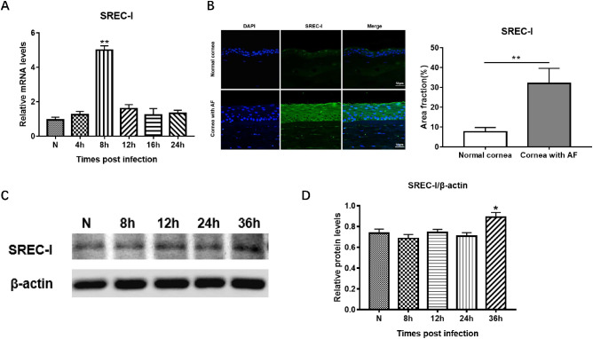
We demonstrated that SREC-Ⅰ is expressed in both normal uninfected and A fumigatus–treated HCECs and human corneal epithelium (Figs. 1A–D). The relative SREC-Ⅰ mRNA level was significantly higher in A fumigatus–stimulated HCECs at 8 hours than in the normal group (Fig. 1A; **P < 0.001). The localization and expression of SREC-Ⅰ were examined in corneal tissues of patients with A fumigatus keratitis by immunofluorescence staining. After A fumigatus infection, the corneas were noticeably thickened (Fig. 1B). SREC–positive cells in infected corneal tissues were detected with strong green fluorescence, although little immunoreactivity of SREC-Ⅰ was detected in healthy corneal tissue (Fig. 1B; **P < 0.001). In addition, we tested the protein level of SREC-Ⅰ in A fumigatus stimulated HCECs by Western blot. The results indicated that the SREC-Ⅰ protein level was increased in HCECs at 36 hours after A fumigatus stimulation compared with normal controls (Figs. 1C, D; *P < 0.05).
Figure 1.
Expression of SREC-Ⅰ in normal and A fumigatus–treated HCECs and corneas. SREC-Ⅰ mRNA level was significantly higher in A fumigatus–stimulated HCECs at 8 hours comparing with control (A). Immunofluorescence staining for SREC-Ⅰ (green) in normal and A fumigatus–infected human corneas, and quantitative analysis (n = 6/group/time) (t-test). Scale bar = 50 µm. (B) SREC-Ⅰ protein expression level in A fumigatus–stimulated HCECs at 0, 8, 12, 24 and 36 hours (C) and quantitative analysis (D). Values represents means ± SD. *P < 0.05, **P < 0.001 compared with untreated cells by one-way ANOVA test.
SREC-Ⅰ siRNA Inhibited the Expression of Inflammatory Factors in HCECs
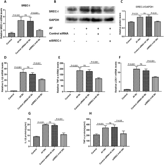
Previous study demonstrated that IL-1β, TNF-α, and LOX-1 mRNA and protein levels were significantly upregulated in HCECs after A fumigatus stimulation.33–34 We next sought to determine whether the expression of cytokines induced in A fumigatus–stimulated HCECs depends on SREC-Ⅰ expression. SREC-I siRNA effectively downregulated the mRNA (Fig. 2A; P < 0.001) and protein levels (Figs. 2B, C; P < 0.01) of SREC-I in HCECs. In addition, SREC-Ⅰ siRNA significantly inhibited the mRNA expressions of IL-1β (Fig. 2D; P < 0.001), TNF-α (Fig. 2E; P < 0.001), and LOX-1 (Fig. 2F; P < 0.001) at 8 hours compared with control siRNA treated HCECs after A fumigatus stimulation. Moreover, the ELISA results showed SREC-Ⅰ siRNA significantly inhibited IL-1β (Fig. 2G; P < 0.001) and TNF-α (Fig. 2H; P < 0.05) protein levels at 24 hours when compared with controls. These findings indicate that SREC-Ⅰ may be a key mediator involved in the A fumigatus–induced inflammatory response.
Figure 2.
SREC-Ⅰ siRNA inhibited the expression of inflammatory factors in HCECs. The expression of SREC-I mRNA at 8 hours (A) and protein at 36 hours (B, C) was significantly downregulated in HCECs after SREC-I siRNA transfection. SREC-Ⅰ siRNA significantly inhibited the mRNA expression of IL-1β (D), TNF-α (E) and LOX-1 (F) in HCECs at 8 hours after A fumigatus treatment, and inhibited the protein secretion of IL-1β (G) and TNF-α (H) at 24 hours compared with control. ns, no significance.
SREC-Ⅰ Expression Increased in A fumigatus–Infected Mice Corneal Epithelium
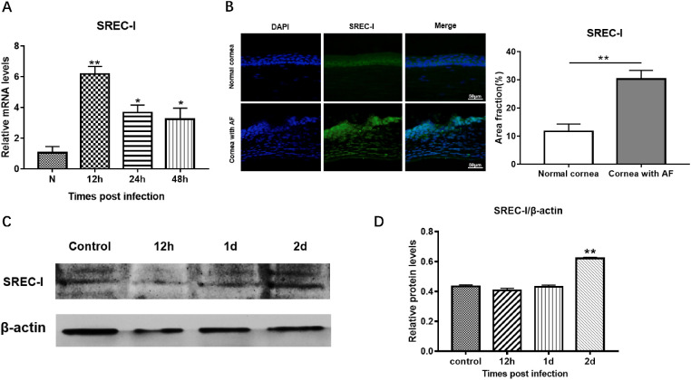
We next tested SREC-Ⅰ expression in both healthy and A fumigatus infected corneal epithelium of mice. Real-time PCR demonstrated that the SREC-Ⅰ mRNA expression significantly increased in corneal epithelium of A fumigatus-infected mice at 12, 24, and 48 hours and peaked at 12 hours (Fig. 3A; *P < 0.01, **P < 0.001). The upregulated expression of SREC-Ⅰ was confirmed by immunofluorescence staining at protein level, in which SREC-Ⅰ protein expression markedly increased in infected corneas of mice compared with normal corneas (Fig. 3B; **P < 0.001). Higher SREC-Ⅰ protein level was also detected in mice corneal epithelium at 2 days after A fumigatus infection (Figs. 3C, D; **P < 0.01).
Figure 3.
Expression of SREC-Ⅰ in A fumigatus infected mice cornea. SREC-Ⅰ mRNA expression increased in corneal epithelium of A fumigatus infected mice at 12, 24, and 48 hours p.i. and peaked at 12 hours (A). Immunofluorescence staining for SREC-Ⅰ (green) in normal and A fumigatus infected corneas at 2 days p.i., and quantitative analysis. (n = 3/group) (t-test). Scale bar = 50 µm. (B). Western blot for SREC-Ⅰ protein expression in A fumigatus–infected mouse corneal epithelium at 12 hours, 1 day and 2 days p.i. (C) and quantitative analysis (n = 6/group/time) (t-test) (D). Values represents means ± SD. **P < 0.001, * P < 0.01 compared with untreated cells by one-way ANOVA test.
SREC-Ⅰ–Neutralizing Antibody Attenuated A fumigatus FK Progression In Vivo
To further investigate the role of SREC-Ⅰ in A fumigatus–induced FK, SREC-Ⅰ antibody was applied to block SREC-Ⅰ in mice with A fumigatus FK. After SREC-Ⅰ neutralization, the mRNA (Fig. 4A) and protein (Figs. 4B, C) expression levels of SREC-Ⅰ were significantly downregulated. Clinical score in the anti–SREC-Ⅰ blockade group was significantly lower than the control group at 3 and 5 days p.i., although there was no significant difference observed at 1 day p.i. (Fig. 4D). The corneal photographs taken at 5 days p.i. illustrated that corneas in SREC-Ⅰ antibody neutralized mice (Fig. 4F) were more transparent and with markedly decreased ulcer area than control group (Fig. 4E). Hematoxylin and eosin staining showed SREC-I blockade significantly decreased the number of inflammatory cells in A fumigatus–infected mice cornea (Figs. 4G, H). These results demonstrate that SREC-Ⅰ blockade could decrease corneal ulcer area and attenuate A fumigatus FK progression in vivo.
Figure 4.
SREC-Ⅰ neutralizing antibody treatment of A fumigatus–infected mice cornea. SREC-Ⅰ mRNA (A) and protein (B, C) levels were significantly downregulated after anti-SREC-Ⅰ treatment. Clinical scores of anti–SREC-Ⅰ treatment group were significantly lower than the control group at 3 and 5 days p.i., with no difference between groups at 1 day p.i. (D). Representative slit lamp photographs of anti–SREC-Ⅰ (F) and control IgG-treated mice cornea (E). Hematoxylin and eosin staining showed normal cornea (G1), A fumigatus–infected cornea (G2), control IgG treated cornea (G3) and anti–SREC-Ⅰ antibody–treated cornea (G4). Quantitative analysis for the number of inflammatory cells in HE staining (H) (n = 6/group/time). Original magnification ×400. ns, no significance.
Blockade of SREC-Ⅰ Inhibits the Production of Proinflammatory Mediators in an A fumigatus Keratitis Mouse Model
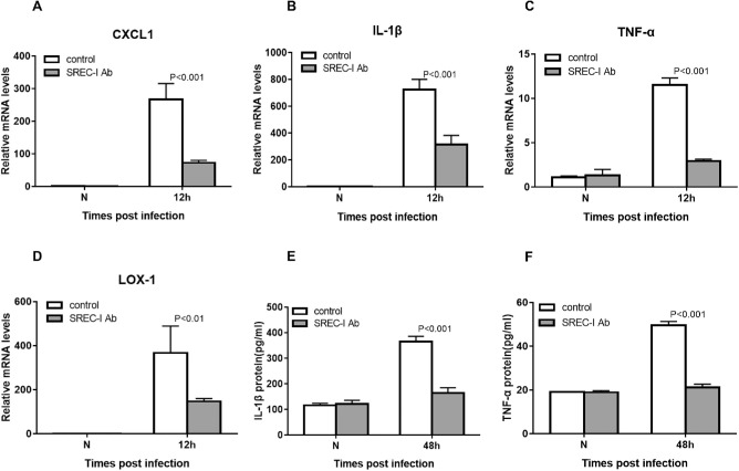
Compared with uninfected controls, the mRNA expressions of several proinflammatory mediators such as CXCL1, LOX-1, IL-1β, and TNF-α were elevated in A fumigatus infected mice corneas at 12 hours p.i., and SREC-Ⅰ antibody significantly restrained the mRNA expression levels of CXCL1 (Fig. 5A), IL-1β (Fig. 5B), TNF-α (Fig. 5C), and LOX-1(Fig. 5D) at 12 hours p.i. In addition, protein levels of IL-1β (Fig. 5E) and TNF-α (Fig. 5F) were downregulated at 48 hours p.i. These data indicate that SREC-Ⅰ blockade could suppress inflammatory response induced by A fumigatus.
Figure 5.
SREC-Ⅰ–neutralizing antibody significantly restrained the mRNA expression levels of CXCL1 (A), IL-1β (B), TNF-α (C) and LOX-1 (D) in mice corneas at 12 hours p.i., and inhibited the protein levels of IL-1β (E) and TNF-α (F) in mice corneas at 48 hours p.i. (n = 6/group/time).
Discussion
SREC-Ⅰ is becoming a key player in the immune response.12 Previous studies have found that SREC-Ⅰ is expressed in endothelial cells, macrophages, dendritic cells, and epithelial cells.13–14,35–36 Our research first showed that SREC-Ⅰ is expressed in normal human corneas and HCECs. After A fumigatus stimulation, the SREC-Ⅰ mRNA expression was significantly upregulated at 8 hours, whereas the protein expression was later than gene expression. These data indicated that SREC-Ⅰ may play an important role in FK. Previous studies have reported that SREC-Ⅰ can recognize β-glucans presented on the cell surface of Cryptococcus neoformans and Candida albicans, then trigger the production of inflammatory chemokines and cytokines such as IL-1β, CXCL2, and CXCL1 in association with TLR2,22 which suggested that SREC-Ⅰ may play a proinflammatory role in the immune response upon fungal infection. In Ayesha Murshid et al’ study,37 SREC-Ⅰ promoted TLR4-induced signal transduction through nuclear factor-κB and MAPK pathways, leading to enhanced inflammatory cytokine IL-6 and TNF-α release in RAW 264.7 cells. To determine the role of SREC-Ⅰ in the corneal epithelium during A fumigatus stimulation, SREC-Ⅰ siRNA was applied to inhibit the expression of SREC-Ⅰ in HCECs. Our data showed that SREC-Ⅰ inhibition significantly decreased the upregulated production of IL-1β, TNF-α, and LOX-1 induced by A fumigatus treatment. These findings are consistent with previous studies that SREC-I knockdown (siRNA) decreased the levels of lipopolysaccharide-mediated proinflammatory cytokines IL-6 and TNF-α secretion.37 Thus, SREC-Ⅰ may act as an important member of innate immunity during FK, and its activation promotes the release of proinflammatory factors that in turn facilitate inflammation. Inhibiting the activity of SREC-Ⅰ at an appropriate level could be beneficial for controlling inflammation-related diseases.
Our previous studies have shown several inflammation-related factors were upregulated in A fumigatus infected mice, such as LOX-1, TNF-α, and IL-6.38–40 In this study, high expression of SREC-Ⅰ mRNA level was also detected in mouse corneal epithelium after A fumigatus infection and peaked at 12 hours, whereas the protein expression increased at 48 hours p.i., which was remarkably suppressed by an SREC-Ⅰ–neutralizing antibody. In agreement with the decreased protein expression of SREC-Ⅰ, the SREC-Ⅰ mRNA level was also downregulated after anti–SREC-Ⅰ antibody treatment in mice. We consider it may be associated with the attenuated level of inflammatory response in mice cornea. Simultaneously, the corneal inflammatory state in SREC-Ⅰ antibody-neutralized mice was significantly inhibited compared with controls. Fewer inflammatory cells and lower LOX-1, CXCL1, TNF-α, and IL-1β expression levels were observed after anti–SREC-Ⅰ antibody treatment. These data indicate that SREC-I blockade could inhibit A fumigatus–induced inflammatory response in vivo. As a member of scavenger receptor, SREC-Ⅰ and the other C-type lectin, scavenger receptor LOX-1 can induce the production of IL-6 and IL-8 through recognizing the outer membrane protein A from Klebsiella pneumonia cooperated with TLR2.41 The role of SREC-Ⅰ as an interacting receptor in inflammatory diseases has been reported many times.41–43 Future study may focus on the interactions between SREC-Ⅰ and other pattern recognition receptors like LOX-1 and TLRs to explore other mechanisms of how SREC-Ⅰ is induced and functions in response to A fumigatus infection.
Our study showed that SREC-Ⅰ is expressed in both HCECs and human/mice corneal epithelium. In A fumigatus–stimulated HCECs, SREC-Ⅰ mRNA and protein expression levels were significantly increased, and SREC-Ⅰ siRNA inhibited the expression of IL-1β, TNF-α, and LOX-1. In mice with A fumigatus keratitis, the expression of SREC-Ⅰ was upregulated, and SREC-Ⅰ blockade not only inhibited the expression of proinflammatory factors including LOX-1, CXCL1, TNF-α, and IL-1β, but also attenuated A fumigatus keratitis progression. These data suggest that SREC-Ⅰ plays an important proinflammatory role during FK, providing a potential therapeutic or an additional target for FK.
Acknowledgments
Supported by the National Natural Science Foundation of China (No. 81870632), and the Youth Project of Natural Science Foundation of Shandong Province (No. ZR2019BH004).
Disclosure: R. Zhang, None; X. Peng, None; J. Lin, None; Y. Zhang, None; L. Zhan, None; X. Tian, None; J. Yin, None; G. Zhao, None
References
- 1. Huang JF, Zhong J, Chen GP, et al.. A hydrogel-based hybrid theranostic contact lens for fungal keratitis. ACS Nano. 2016; 10(7): 6464–6473. [DOI] [PubMed] [Google Scholar]
- 2. Austin A, Lietman T, Rose-Nussbaumer J, et al.. Update on the management of infectious keratitis. Ophthalmology. 2017; 124(11): 1678–1689. [DOI] [PMC free article] [PubMed] [Google Scholar]
- 3. Gopinathan U, Garg P, Fernandes M, et al.. The epidemiological features and laboratory results of fungal keratitis: a 10-year review at a referral eye care center in South India. Cornea. 2002; 21(6): 555–559. [DOI] [PubMed] [Google Scholar]
- 4. Li N, Zhao GQ.. Mechanism of the immune response to keratomycosis. Zhonghua Yanke Zazhi. 2011; 47(4): 378–381. [PubMed] [Google Scholar]
- 5. Kaur A, Kumar V, Singh S, et al.. Toll-like receptor-associated keratitis and strategies for its management. 3 Biotech. 2015; 5(5): 611–619. [DOI] [PMC free article] [PubMed] [Google Scholar]
- 6. Briard B, Karki R, Malireddi RKS, et al.. Fungal ligands released by innate immune effectors promote inflammasome activation during Aspergillus fumigatus infection. Nat Microbiol. 2019; 4(2): 316–327. [DOI] [PMC free article] [PubMed] [Google Scholar]
- 7. Li C, Zhao G, Che C, et al.. The role of LOX-1 in innate immunity to Aspergillus fumigatus in corneal epithelial cells. Invest Ophthalmol Vis Sci. 2015; 56(6): 3593–3603. [DOI] [PubMed] [Google Scholar]
- 8. Xu J, Flaczyk A, Neal LM, et al.. Scavenger receptor MARCO orchestrates early defenses and contributes to fungal containment during cryptococcal infection. J Immunol. 2017; 198(9): 3548–3557. [DOI] [PMC free article] [PubMed] [Google Scholar]
- 9. Berwin B, Delneste Y, Lovingood RV, et al.. SREC-I, a type F scavenger receptor, is an endocytic receptor for calreticulin. J Biol Chem. 2004; 279(49): 51250–51257. [DOI] [PubMed] [Google Scholar]
- 10. Adachi H, Tsujimoto M, Arai H, et al.. Expression cloning of a novel scavenger receptor from human endothelial cells. J Biol Chem. 1997; 272(50): 31217–31220. [DOI] [PubMed] [Google Scholar]
- 11. Ishii J, Adachi H, Aoki J, et al.. SREC-II, a new member of the scavenger receptor type F family, trans-interacts with SREC-I through its extracellular domain. J Biol Chem. 2002; 277(42): 39696–70. [DOI] [PubMed] [Google Scholar]
- 12. Murshid A, Borges TJ, Calderwood SK.. Emerging roles for scavenger receptor SREC-I in immunity. Cytokine. 2015; 75(2): 256–260. [DOI] [PMC free article] [PubMed] [Google Scholar]
- 13. Baur S, Rautenberg M, Faulstich M, et al.. A nasal epithelial receptor for Staphylococcus aureus WTA governs adhesion to epithelial cells and modulates nasal colonization. PLoS Pathog. 2014; 10(5): e1004089. [DOI] [PMC free article] [PubMed] [Google Scholar]
- 14. Rechner C, Kühlewein C, Müller A, et al.. Host glycoprotein Gp96 and scavenger receptor SREC interact with PorB of disseminating Neisseria gonorrhoeae in an epithelial invasion pathway. Cell Host Microbe. 2007; 2(6): 393–403. [DOI] [PubMed] [Google Scholar]
- 15. Taha EA, Ono K, Eguchi T, et, al. Roles of extracellular HSPs as biomarkers in immune surveillance and immune evasion. Int J Mol Sci. 2019; 20(18): 4588, 10.3390/ijms20184588. [DOI] [PMC free article] [PubMed] [Google Scholar]
- 16. Apostolov EO, Shah SV, Ray D, et al.. Scavenger receptors of endothelial cells mediate the uptake and cellular proatherogenic effects of carbamylated LDL. Arterioscler Thromb Vasc Biol. 2009; 29(10): 1622–30. [DOI] [PMC free article] [PubMed] [Google Scholar]
- 17. Sano M, Korekane H, Ohtsubo K, et al.. N-glycans of SREC-I (scavenger receptor expressed by endothelial cells): essential role for ligand binding, trafficking and stability. Glycobiology. 2012; 22(5): 714–724. [DOI] [PubMed] [Google Scholar]
- 18. Murshid A, Gong J, Calderwood SK.. Hsp90-peptide complexes stimulate antigen presentation through the class II pathway after binding scavenger receptor SREC-I. Immunobiology. 2014; 219(12): 924–931. [DOI] [PMC free article] [PubMed] [Google Scholar]
- 19. Schade J, Weidenmaier C.. Cell wall glycopolymers of Firmicutes and their role as nonprotein adhesins. FEBS Lett. 2016; 590(21): 3758–3771. [DOI] [PubMed] [Google Scholar]
- 20. Gordon S. Pattern recognition receptors: doubling up for the innate immune response. Cell. 2002; 111(7): 927–930. [DOI] [PubMed] [Google Scholar]
- 21. Patten DA. SCARF1: a multifaceted, yet largely understudied, scavenger receptor. Inflamm Res. 2018; 67(8): 627–632. [DOI] [PMC free article] [PubMed] [Google Scholar]
- 22. Means TK, Mylonakis E, Tampakakis E, et al.. Evolutionarily conserved recognition and innate immunity to fungal pathogens by the scavenger receptors SCARF1 and CD36. J Exp Med. 2009; 206(3): 637–653. [DOI] [PMC free article] [PubMed] [Google Scholar]
- 23. Yin M, Li C, Peng XD, et al.. Expression and role of calcitonin gene-related peptide in mouse Aspergillus fumigatus keratitis. Int J Ophthalmol. 2019; 12(5): 697–704. [DOI] [PMC free article] [PubMed] [Google Scholar]
- 24. Gu L, Lin J, Wang Q, et al.. Dimethyl itaconate protects against fungal keratitis by activating the Nrf2/HO-1 signaling pathway. Immunol Cell Biol. 2020; 98(3): 229–241. [DOI] [PMC free article] [PubMed] [Google Scholar]
- 25. Fan Y, Li C, Peng X, et al.. Perillaldehyde ameliorates Aspergillus fumigatus keratitis by activating the Nrf2/HO-1 signaling pathway and inhibiting dectin-1-mediated inflammation. Invest Ophthalmol Vis Sci. 2020; 61(6): 51. [DOI] [PMC free article] [PubMed] [Google Scholar]
- 26. Zhan L, Peng X, Lin J, et al.. Honokiol reduces fungal load, Toll-like receptor-2, and inflammatory cytokines in Aspergillus fumigatus keratitis. Invest Ophthalmol Vis Sci. 2020; 61(4): 48. [DOI] [PMC free article] [PubMed] [Google Scholar]
- 27. Peng X, Ekanayaka SA, McClellan SA, et al.. Characterization of three ocular clinical isolates of P. aeruginosa: viability, biofilm formation, adherence, infectivity, and effects of glycyrrhizin. Pathogens. 2017; 6(4): 52. [DOI] [PMC free article] [PubMed] [Google Scholar]
- 28. Zhu GQ, Zhao GQ, Li C, et al.. Regulation of LOX-1 on adhesion molecules and neutrophil infiltration in mouse Aspergillus fumigatus keratitis. Int J Ophthalmol. 2020; 13(6): 870–878. [DOI] [PMC free article] [PubMed] [Google Scholar]
- 29. Peng XD, Zhao GQ, Lin J, et al.. Interaction of mannose binding lectin and other pattern recognition receptors in human corneal epithelial cells during Aspergillus fumigatus infection. Int Immunopharmacol. 2018; 63: 161–169. [DOI] [PubMed] [Google Scholar]
- 30. Niu Y, Zhao G, Li C, et al.. Aspergillus fumigatus increased PAR-2 expression and elevated proinflammatory cytokines expression through the pathway of PAR-2/ERK1/2 in cornea. Invest Ophthalmol Vis Sci. 2018; 59(1): 166–175. [DOI] [PubMed] [Google Scholar]
- 31. Peng XD, Zhao GQ, Lin J, et al.. Fungus induces the release of IL-8 in human corneal epithelial cells, via Dectin-1-mediated protein kinase C pathways. Int J Ophthalmol. 2015; 8(3): 441–447. [DOI] [PMC free article] [PubMed] [Google Scholar]
- 32. You J, Lin J, Zhou YF, et al.. Role of the IL-33/ST2/p38 signaling pathway in the immune response of corneal epithelial cells to Aspergillus fumigatus infection. Int J Ophthalmol. 2019; 12(4): 549–556. [DOI] [PMC free article] [PubMed] [Google Scholar]
- 33. Zhao G, Hu M, Li C, et al.. Osteopontin contributes to effective neutrophil recruitment, IL-1β production and apoptosis in Aspergillus fumigatus keratitis. Immunol Cell Biol. 2018; 96(4): 401–412. [DOI] [PubMed] [Google Scholar]
- 34. Wang Q, Zhao G, Lin J, et al.. Role of the mannose receptor during Aspergillus fumigatus infection and interaction with dectin-1 in corneal epithelial cells. Cornea. 2016; 35(2): 267–273. [DOI] [PubMed] [Google Scholar]
- 35. Murshid A, Borges TJ, Bonorino C, et al.. Immunological outcomes mediated upon binding of heat shock proteins to scavenger receptors SCARF1 and LOX-1, and endocytosis by mononuclear phagocytes. Front Immunol. 2019; 10: 3035. [DOI] [PMC free article] [PubMed] [Google Scholar]
- 36. Piccolo P, Vetrini F, Mithbaokar P, et al.. SR-A and SREC-I are Kupffer and endothelial cell receptors for helper-dependent adenoviral vectors. Mol Ther. 2013; 21(4): 767–774. [DOI] [PMC free article] [PubMed] [Google Scholar]
- 37. Murshid A, Gong J, Prince T, et al.. Scavenger receptor SREC-I mediated entry of TLR4 into lipid microdomains and triggered inflammatory cytokine release in RAW 264.7 cells upon LPS activation. PLoS One. 2015; 10(4): e0122529. [DOI] [PMC free article] [PubMed] [Google Scholar]
- 38. He K, Yue LH, Zhao GQ, et al.. The role of LOX-1 on innate immunity against Aspergillus keratitis in mice. Int J Ophthalmol. 2016; 9(9): 1245–1250. [DOI] [PMC free article] [PubMed] [Google Scholar]
- 39. Li C, Liu YY, Zhao GQ, et al.. Role of vasoactive intestinal peptide in Aspergillus fumigatus-infected cornea. Int J Ophthalmol. 2018; 11(2): 183–188. [DOI] [PMC free article] [PubMed] [Google Scholar]
- 40. Jiang N, Zhang L, Zhao G, et al.. Indoleamine 2,3-dioxygenase regulates macrophage recruitment, polarization and phagocytosis in Aspergillus fumigatus keratitis. Invest Ophthalmol Vis Sci. 2020; 61(8): 28. [DOI] [PMC free article] [PubMed] [Google Scholar]
- 41. Jeannin P, Bottazzi B, Sironi M, et al.. Complexity and complementarity of outer membrane protein A recognition by cellular and humoral innate immunity receptors. Immunity. 2005; 22(5): 551–560. [DOI] [PubMed] [Google Scholar]
- 42. Murshid A, Borges TJ, Lang BJ, et al.. The scavenger receptor SREC-c Cooperates with Toll-like receptors to trigger inflammatory innate immune responses. Front Immunol. 2016; 7: 226. [DOI] [PMC free article] [PubMed] [Google Scholar]
- 43. Murshid A, Gong J, Ahmad R, et al.. Scavenger receptor SREC-I promotes double stranded RNA-mediated TLR3 activation in human monocytes. Immunobiology. 2015; 220(6): 823–832. [DOI] [PMC free article] [PubMed] [Google Scholar]


