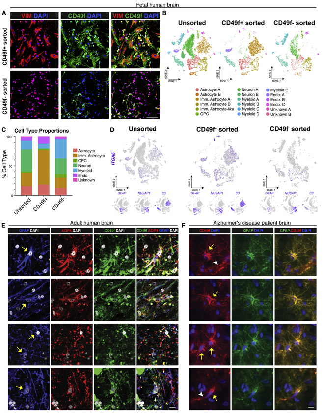
Figure 4: CD49f-sort from human fetal brain enriches for astrocytes, and CD49f is localized in astrocytes in slides from human adult brains.
See also Figure S7 and Supplemental Table 1.
a) Representative immunofluorescence images showing vimentin (red), CD49f (green), and DAPI (blue) in CD49f+ and CD49f− sorted cells from human fetal brain tissue. Scale bar, 100μm.
b) tSNE plots of single-cell RNA-Seq data from unsorted (left, n = 11,817), CD49f+ (middle, n = 6,069), and CD49f sorted (right, n = 4,409) cells from fetal brain tissue. In total, 18 clusters were identified. All data are from an 18-week-old human fetus (n=1).
c) Quantification of cell type proportions from unsorted, CD49f+, and CD49f− sorted cells from fetal brain tissue. CD49f+ cells are highly enriched in astrocytes and immature astrocytes.
d) tSNE feature plots highlighting in purple cells that express CD49f (ITGA6), mature astrocyte (GFAP), and immature astrocyte (NUSAP1, C3) transcripts from unsorted, CD49f+, and CD49f− sorted cells from fetal brain tissue.
e) Representative immunofluorescence images showing GFAP+ (blue), AQP4+ (red), and CD49f+ (green) cells with DAPI nuclei (grey) in cryosections from the subventricular zone of an adult brain from healthy individual. Yellow arrows indicate cells that are CD49f+, AQP4+, and GFAP+. Scale bar, 10μm.
f) Representative immunofluorescence images showing CD49f+ (red) and GFAP+ (green) cells with DAPI nuclei (blue) in cryosections from the prefrontal cortex of an Alzheimer’s disease patient. Yellow arrows indicate cells that are CD49f+ and GFAP+. White arrowheads indicate CD49f+ endothelial cells. Scale bar, 10μm.
Abbreviations: Endo.=endothelial cell; Imm.=immature; OPC=oligodendrocyte progenitor cell.

