PLOS ONE logoLink to PLOS ONE
. 2021 Jul 14;16(7):e0252040. doi: 10.1371/journal.pone.0252040

Feasibility and safety of targeted focal microwave ablation of the index tumor in patients with low to intermediate risk prostate cancer: Results of the FOSTINE trial

Nicolas Barry Delongchamps 1,2,*, Alexandre Schull 1, Julien Anract 1,2, Jean-Paul Abecassis 3, Marc Zerbib 1, Mathilde Sibony 4, Léa Jilet 5, Hendy Abdoul 5, Vincent Goffin 2, Michaël Peyromaure 1,2
Editor: Neal Shore6
PMCID: PMC8279354  PMID: 34260598

Abstract

Objective

To assess the feasibility, safety and precision of organ-based tracking (OBT)-fusion targeted focal microwave ablation (FMA), in patients with low to intermediate risk prostate cancer.

Patients and method

Ten patients with a visible index tumor of Gleason score ≤3+4, largest diameter <20mm were included. Transrectal OBT-fusion targeted FMA was performed using an 18G needle. Primary endpoint was the evidence of complete overlap of the index tumor by ablation zone necrosis on MRI 7 days after ablation. Urinary and sexual function were assessed with IPSS, IIEF5 and MSHQ-EjD-SF. Oncological outcomes were assessed with PSA at 2 and 6 months, and re-biopsy at 6 months.

Results

Median [IQR] age was 64.5 [61–72] years and baseline PSA was 5 [4.3–8.1] ng/mL. Seven (70%) and 3 (30%) patients had a low and intermediate risk cancer, respectively. Median largest tumor axis was of 11 [9.0–15.0] mm. Median duration of procedure was of 82 [44–170] min. No patient reported any pain or rectal bleeding, and all 10 patients were discharged the next day. Seven days after ablation, total necrosis of the index tumor on MRI was obtained in eight (80% [95%CI 55%-100%]) patients. One patient was treated with radical prostatectomy. Re-biopsy at 6 months in the other 9 did not show evidence of cancer in 4 patients. IPSS, IIEF-5 and MSHQ-EjD-SF were not statistically different between baseline and 6 months follow up.

Conclusions

OBT-fusion targeted FMA was feasible, precise, and safe in patients with low to intermediate risk localized prostate cancer.

1. Introduction

Although prostate cancer is frequently a multifocal disease, the index tumor is believed to have the most malignant potential among other smaller lesions (secondary tumors) within the prostate. Tumor volume and Gleason score are predictive of disease recurrence after radical treatments [1, 2]. These observations led to the option of Focal therapy (FT) of the index tumor, aiming to decrease the risk of cancer progression, while preserving genitourinary function. A number of sources of energy have been employed so far, and different sizes of ablation have been proposed, including tumor-only, zonal or hemi-gland ablation [3].

Interest in FT has recently been renewed owing to improved biopsy and imaging techniques, allowing a more comprehensive management of the index tumor. The ability of magnetic resonance imaging (MRI) to detect significant cancer foci [49], together with the reliability of organ-based tracking (OBT) MRI-ultrasound fusion [1012], now allows clinicians to detect and target the index tumor precisely: Performing an elastic fusion between MRI and ultrasound images can create a precise three-dimensional mapping of the prostate, accurately showing the index tumor. OBT enables physicians to guide and to distribute, in a real-time fashion, the biopsy cores in three-dimensions. Another major value of OBT resides in its capacity to memorize and then recall the location of interest in the prostate, from biopsy to FT, and then during follow-up, providing quality control.

Dodd et al. demonstrated microwave energy resulted in coagulation necrosis with a low “heat-sink” effect, while also reporting the volume of necrosis obtained seemed to be predictable and repeatable [13].

We hypothesized that combining the advantages provided by OBT-targeting and microwave therapy would allow a safe and precise FT of index tumor-only. We thus undertook a prospective trial to assess the feasibility, safety and precision of OBT-fusion targeted focal microwave ablation (FMA), in patients with localized PCa of low to intermediate risk of progression.

2. Patients and method

This trial was approved by the French national committee for ethics (CPP, ref: Am7730-2-3439) and registered on ClinicalTrials.gov (NCT03023345). All patients gave their written consent before inclusion.

Between September 2017 and 2018, 10 eligible patients were included. The inclusion criteria were: (1) age between 45 and 76 years; (2) prostate cancer with a visible index tumor on MRI, confirmed on targeted biopsy, and (3) signed informed consent. Exclusion criteria were: (1) PSA level >15ng/ml, (2) severe low urinary tract symptoms defined by IPSS>18, (3) index tumor largest diameter >20mm, (4) lower distance between index tumor and rectum wall <5mm, (5) evidence of extra-capsular extension or seminal vesicle invasion on MRI, (6) evidence of Gleason grade 4 on systematic biopsies, (7) more than 50% of Gleason grade 4 on targeted biopsy, (8) contraindication to general anesthesia, and (9) untreated bacteriuria less than 2 days before surgery. No protocol violations were identified.

The primary objective was to demonstrate the feasibility and precision of targeted FMA under OBT registration. The primary endpoint was the evidence of complete overlap of the index tumor by ablation zone necrosis on MRI performed 7 days after ablation.

Secondary endpoints were: (1) intra and postoperative outcomes, including safety and pain evaluation, (2) urinary and sexual outcomes, assessed with IPSS, IIEF5 and MSHQ-EjD-SF self-questionnaires, respectively (3) oncological outcomes, assessed with PSA at 2 and 6 months, as well as prostate re-biopsy, at 6 months.

All patients received a preoperative rectal preparation (enema) and prophylactic antibiotic using oral fluoroquinolones. Procedures were performed under general anesthesia. No urethral catheterization was performed. The ultrasound probe was inserted transrectally and held with a mechanical arm (Steady Pro™, Koelis, Meylan, France). Ultrasound-MRI image fusion was performed with OBT-registration using Trinity™ station (Koelis, Meylan, France) [14]. Microwave thermal ablation was provided by the TATO generator (Biomedical Srl, Firenze, Italy) using a single 18G needle inserted transrectally. Duration and power of microwave application were set according to a pre-clinical predictive ablation chart. Patients were discharged the next day.

Multiparametric prostate MRI (mpMRI), including T2 and diffusion weighted imaging, as well as dynamic contrast-enhanced imaging, was performed 7 days after ablation and at 6 months. The primary endpoint was evaluated by a first radiologist by comparing baseline prostate MRI with day-7 MRI. The extent of necrosis 7 days after ablation was evaluated with T1- contrast enhanced weighted sequences. In the absence of complete necrosis covering of the index tumor, the proportion of necrosis extent was visually estimated, according to the surface leaved untreated on each MRI image. A second radiologist evaluated the primary endpoint, blinded to patient characteristics. Six months after the procedure, three targeted biopsies were performed in the treated zone, as well as 10 to 12-core systematic biopsies.

A phase II study design was used to estimate the number of necessary subjects. We considered that complete necrosis of the index tumor was desirable in 99% of patients. We also considered that if less than 60% of patients showed complete necrosis of the index tumor, the treatment under investigation would be considered inefficient. With a two-tailed test, a risk alpha of 0.1 and beta of 0.1, a total number of 10 patients was considered sufficient.

Continuous data were reported as means (sd) or medians [IQR] (for non-normal data), while categorical data were summarized as counts and percentages. To estimate the proportion of patients with complete necrosis of the index tumor 7 days after ablation, a two-sided 95% confidence interval was calculated. A Wilcoxon signed-rank test was used to compare the data from the questionnaires, with a risk alpha of 0.05. All analyses were conducted using SAS software 9.4.

3. Results

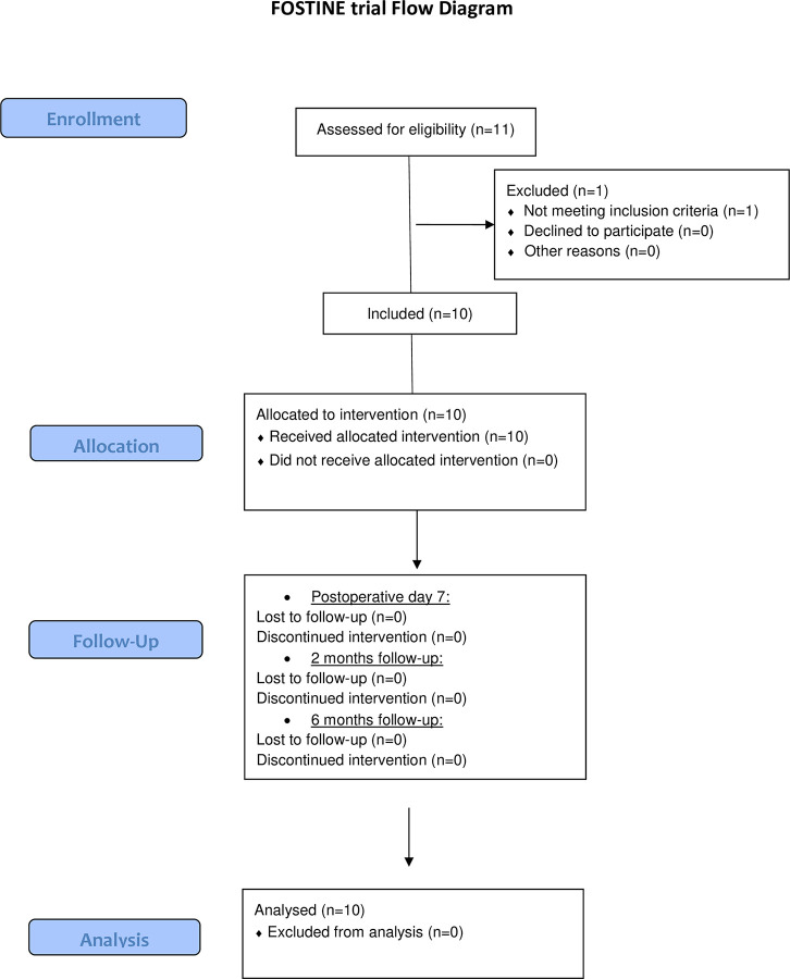
Eleven patients were enrolled. One patient did not meet eligibility criteria, and only 10 patients were finally included and treated according to the pre-established protocol (Fig 1). Median [IQR] age was 64 [61–72], median PSA was of 5 [4.3–8.1] ng/mL, and median prostate volume of 50 [40–55] mL. Seven patients had low and 3 patients had intermediate D’Amico risk cancer, respectively. Three patients had multifocal disease, with evidence of non MRI-visible secondary tumor foci of Gleason 3+3 cancer on systematic biopsies. The 7 other patients had no evidence of cancer on systematic biopsies. Index tumor characteristics are reported in Table 1 and Fig 2. Baseline median [IQR] IPSS, IIEF-5, and MSHQ-EjD were of 8.5 [413], 18 [1323], and 10 [713], respectively.

Fig 1. Flow chart.

Fig 1

Table 1. Summarizes the index tumor characteristics of the 10 patients included.

Variable Median Interquartile range
Maximum diameter on mpMRI (mm) 11.5 9.0–15.0
MCCL of targeted biopsies (mm) 7.0 6.0–7.0
Variable N (total = 10) %
Gleason pattern
    3+3 8 80.0
    3+4 2 20.0
Location
    Apex 3 30.0
    Median zone 3 30.0
    Base 4 40.0

MCCL: Maximum Cancer Core Length.

Fig 2. Index tumor location at baseline in the 10 patients before targeted FMA.
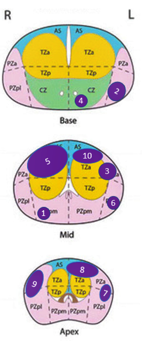
Fig 2

Schematic view of index tumor location. Index tumors have been represented with a 1:1 scale on a prostate with 4 cm height (antero-posterior axis), 6 cm width (lateral axis).

3.1. Intra and post-operative outcomes

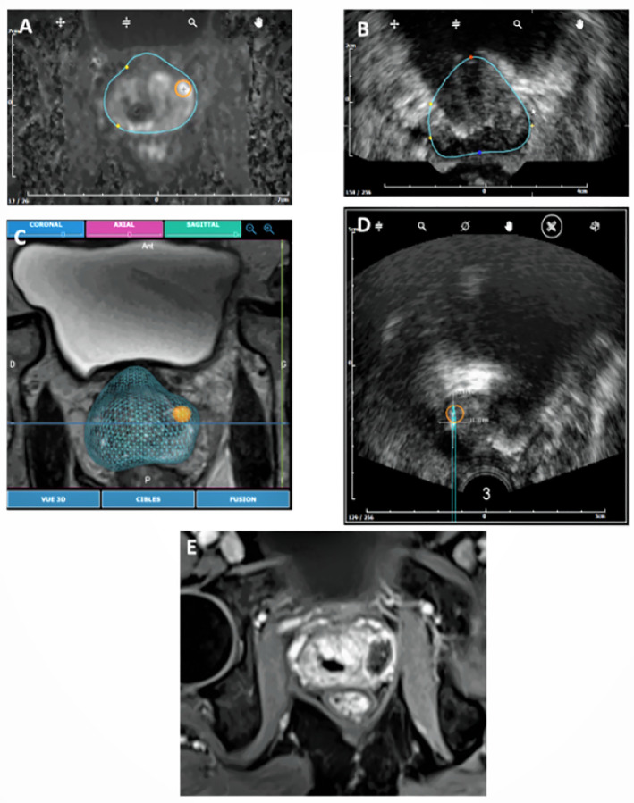
Median duration of the surgical procedure was of 82 [44–170] min, including a median time of 16 [823] and 2 [26] min for MRI-ultrasound fusion and targeting, and microwave application, respectively (Fig 3). Microwave ablation was set at 15W for 3 min, 15W for 2 min, and 10W for 2 min, in 3 (30%), 5 (50%), and 2 (20%) patients, respectively. In 2 patients, 2 consecutive 3 min applications of 15W were performed.

Fig 3.

Fig 3

Elastic fusion and ablation process in patient N°3: MRI prostate contours were delineated and the target (index tumor) was defined on the station (A). Prostate contours were then delineated on ultrasound images (B), and elastic MRI/Ultrasound fusion was performed (C). This allowed an OBT-Fusion registration of the microwave applicator and measurement on ultrasound of the expected ablation (D), based on ex-vivo predictive ablation charts. The site of ablation was then visualized 7 days after ablation on Dynamic Contrast Enhanced MRI (E).

Postoperatively, all patients recovered spontaneous micturition within the next few hours. No patient reported any pain or rectal bleeding, and all 10 patients were discharged at day 1, according to protocol. None of the patients received any antibiotics after surgery. Outcomes were uneventful, and no adverse events were reported during the 6-month follow-up.

3.2. Primary endpoint

Seven days after ablation, total necrosis of the index tumor on MRI was visually confirmed in eight (80% [95%CI 55%-100%]) patients (Fig 4). Median [Q1-Q3] largest dimension of necrosis was of 17.5 [1622] mm. Table 2 compares, for the 8 patients who had only one microwave application, the largest dimension of necrosis measured on MRI with that of our predictive ablation chart.

Fig 4. Assessment of index tumor coverage by necrosis 7 days after ablation in patients 1, 2, 4 and 6: Preoperative MRI the index tumor (arrow).
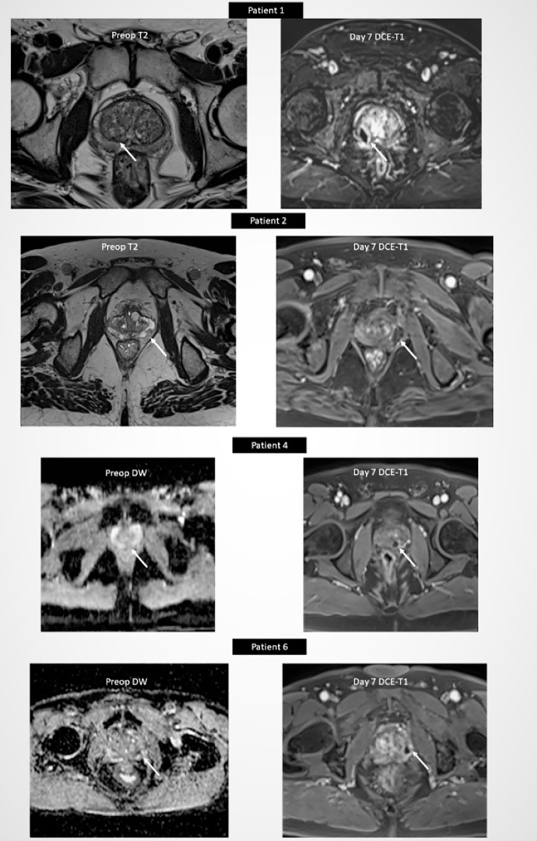
Fig 4

Postoperative DCE-T1 MRI showing total necrosis of index tumor (arrow).

Table 2. Compares the largest dimension of necrosis to that of preclinical ablation charts.

N° Patient Ablation power (in W) Ablation time (in min) Largest dimension of ablation on preclinical charts (in mm) Largest dimension of necrosis on day 7 DCE MRI (in mm) Difference (in mm)
1 10 2 17,00 17,74 0,74
2 15 2 20,60 18,65 -1,95
3 15 2 20,60 19,48 -1,12
4 10 2 17,00 16,50 -0,50
6 15 2 20,60 19,20 -1,40
7 15 3 22,60 19,95 -2,65
9 15 2 20,60 21,10 0,50
10 15 3 22,60 21,04 -1,56

In 2 patients (number 5 and 8), necrosis coverage of the index tumor on day 7 MRI was of only 25% and 40%, respectively (Table 3). Patient number 5 had a 20 mm tumor Gleason score 3+3in the anterior base and median zone. Largest diameter of necrosis was 26 mm, obtained with 2 consecutive microwave applications. This patient was exited from the study following unremarkable radical prostatectomy. Surgical specimen analysis showed a pT3a tumor of Gleason 3+3. Patient number 8 had a 15 mm Gleason score 3+3 tumor in the anterior median zone. The largest diameter of necrosis 7 days after ablation was 13 mm. Surveillance was pursued to the 6-month re-biopsy visit (Table 3).

Table 3. Describes the mpMRI and biopsy results of the 10 patients, at baseline, 1 week and 6 months follow-up.

Patient Nb Baseline 1 week 6 months
Index T. Max Diameter (mm) TB SB % necrosis of index T. TB SB
1 9 3+3 0 100% 3+3 3+3
2 19 3+4 (30%) 0 100% 0 0
3 9 3+3 0 100% 0 3+3
4 10 3+4 (30%) 3+3 100% 3+4 (10%) 3+4 (10%)
5 20 3+3 3+3 40% exited from follow-up
(radical prostatectomy)
6 5 3+3 0 100% 3+3 3+4 (10%)
7 8 3+3 3+3 100% 0 3+4 (15%)
8 15 3+3 0 25% 3+3 0
9 13 3+3 0 100% 3+3 3+3
10 13 3+3 0 100% 0 0

TB: targeted biopsies. 3 targeted cores were performed for each biopsy session.

SB: systematic biopsies. 10–12 systematic cores were performed for each biopsy session.

Index tumor maximum diameter and % necrosis were evaluated on mpMRI.

3.3. Secondary endpoints

Baseline median total PSA [IQR] was of 5.0 [4.3–8.1] ng/mL. It was of 4.9 [3.7–7.8] and 7.5 [3.9–9.7] ng/mL, at 2 and 6 months, respectively.

Re-biopsy at 6 months was performed in 9 (90%) patients (Table 3). Targeted biopsies of the treated area did not show any evidence of cancer in 4 patients. In the remaining 5 patients, biopsy confirmed persistence of Gleason 3+3 and Gleason 3+4 cancer in 4 and 1 patients, respectively, only in the periphery of the treated area. Systematic biopsies showed Gleason 3+4 cancer outside the treated area in 3 patients (Table 3).

No patient reported any deterioration of urinary or sexual function, but only 6 patients answered to the MHSQ-EjD-SF and 8 patients to the IPSS, IPSS-QoL and IIEF-5 after 6-month follow up. In patients presenting all the data, we did not observe any significant change of median IPSS, IIEF-5, and MSHQ-EjD between baseline and 6-month follow up (Table 4).

Table 4. Shows the median scores [IQR] of IPSS, IIEF-5 and MSHQ-EjD-SF at baseline and at 7 days, 2 months and 6 months of follow-up.

Baseline D7 M2 M6 p-value (baseline vs M6)
IPSS 8.5 [4.0–13.0] (n = 10) 7.0 [2.0–11.0] (n = 10) 6.0 [3.0–7.0] (n = 9) 10.0 [8.5–15.0] (n = 8) 0.55*
IPSS-QoL 1.0 [1.0–2.0] (n = 10) 1.5 [1.0–2.0] (n = 10) 0.0 [0.0–1.0] (n = 9) 2.0 [0.5–3.0] (n = 8) 0.94*
IIEF-5 18.5 [13.0–23.0] (n = 10) 8.0 [3.0–14.0] (n = 9) 13.5 [2.0–24.5] (n = 8) 15.0 [4.5–18.0] (n = 8) 0.39*
MSHQ-EjD-SF (function) 10.5 [7.0–13.0] (n = 10) 13.5 [9.0–15.0] (n = 8) 13.0 [11.0–15.0] (n = 7) 11.5 [9.0–12.0] (n = 6) 1*
MSHQ-EjD-SF (bother) 0.0 [0.0–1.0] (n = 10) 0.0 [0.0–0.5] (n = 8) 0.0 [0.0–1.0] (n = 7) 1.0 [1.0–2.0] (n = 6) 0.82*

*Based on patients presenting all the data (6 patients for MHSQ-EjD-SF and 8 patients for IPSS, IPSS-QoL and IIEF-5).

4. Discussion

As many as seven sources of energy have been tested to ablate pre-defined areas of the prostate [3], Focal HIFU [15] and cryotherapy [16, 17] being the most investigated in terms of number of studies and length of follow-up. Photodynamic therapy is the only focal strategy that has been evaluated in a phase 3 randomized controlled trial [18].

We report here the “first in human” experience of OBT-targeted FMA for the treatment of patients with low to intermediate risk prostate cancer. Our main concern was safety, because of the proximity of the rectum, especially with the transrectal approach we were using. Our first clinical objective was therefore to confirm the precision of the technique, based on its ability to entirely ablate the target without any harm to immediate adjacent structures.

The procedure was proven to be feasible. OBT-fusion allowed accurate identification of the center of the index tumor. The tip of the microwave applicator was visible under ultrasound and was inserted under visual guidance. The second step consisted in evaluating the range of ablation. We measured the anticipated thermal effect of microwaves from the tip of the applicator, in the 3 planes, and ensured that it was covering all the tumor volume, without any visualization or guidance on what would be the anticipated ablation volume. During the treatment itself, although we did not use real-time monitoring of ablation, the repeated acquisitions with OBT registration allowed us to confirm the adequate position of the microwave applicator. For safety reasons, all patients were treated under general anesthesia, and discharged the next day. However, given the absence of pain or any adverse events, we believe this procedure could be proposed under local anesthesia, in outpatient settings.

The results of this study were satisfactory. No patient experienced adverse events nor altered sexual or urinary function during the 6-month follow-up, confirming the safety of this technique. The fact that all patients had immediate micturition after ablation suggests that only minimal local edema was induced by microwave ablation. Also, because the ablation zone of the prostatic necrosis obtained was clearly demarcated on mpMRI, and the volume corresponded to what had been anticipated on pre-clinical predictive ablation charts. Indeed, the largest dimension of the ablation zone measured on the axial view on mpMRI 7days after ablation was within the range of the largest axis of the predictive treated zone for 100% of the 8 patients with only one ablation (Table 2). Last but not least, the OBT-registration allowed us to precisely target the area of interest, thus ensuring proper placement of the microwave applicator. As a result, our primary endpoint was reached, with 80% of patients having their index tumor entirely ablated.

In two patients, we failed to ablate all visible tumor on MRI. The first patient had a large (20 mm) anterior tumor oriented from the base to the median zone. Our transrectal approach was not adapted to the shape and the volume of this tumor. Although we performed two consecutive ablations, targeted to the base, and then further to the median zone, we failed to cover entirely the tumor. The second patient had a smaller tumor, but with its largest axis oriented transversely. The thermal ablation generated with the applicator has a shorter transverse range, and the transrectal approach only allowed to reach the tumor perpendicular to its longest axis. We thus failed to achieve a full ablation for all of the tumor. These two failures were not related to the energy used, but only to the surgical approach, and the absence of real-time recall and visualization of the anticipated ablation volume. Real-time recall of the ablation volume is of major importance, especially when more than one microwave application is needed. The software we used at the time of this first pilot study did not allow such guidance. The current developments of the technique aim to improve its precision in specific cases such as those described here. Also, the possibility of a transperineal approach, alone or combined with the transrectal one, will allow to personalize ablation to the shape and size of each tumor.

Although this pilot study was not designed nor powered for such evaluation, early oncological outcomes were evaluated with prostate re-biopsy at 6 months. Our results showed insufficient local control at 6 months in 4 additional patients to the 2 initial failures. In these patients, although focal ablation seemed satisfactory on MRI 7 days and 6 months after ablation, re-biopsy suggested insufficient ablation volume. These results raise the issue of treatment margins. Although there is no true consensus on the size of treatment margin to apply, such strategy seems relevant, especially when performing ultrafocal treatments such as laser ablation [1921] or microwaves. A report from a consensus meeting in 2015 suggested that a circumferential margin of 5 mm around a lesion that was seen on imaging may be enough [22]. Our group [23] and others [24, 25] reported that MRI underestimated pathological volume in up to 50% of the cases, and that this underestimation was higher for small foci [23]. In this pilot study, the primary endpoint was complete tumor ablation (necrosis) based on MRI findings. In a clinical setting, this endpoint should clearly be extended on a case by case basis, depending on the tumor volume itself, and also on tumor index location and proximity to the capsule and rectum. This kind of personalization would be probably more relevant than applying a standardized length of treatment margin systematically to all patients. Another issue is the potential pretherapeutic understaging and/or progression of secondary cancer foci. Kenigsberg et al. [26] recently reported that approximately 20% of candidates who met predefined criteria for focal ablation, and underwent radical prostatectomy were found to have Gleason pattern 4 outside the hypothetical ablation zones. Whatever the efficacy of any ablation that is not radical, and probably even more if this ablation is adopted for focal therapy, urologists will have to monitor closely patients and inform them of the risk of local relapse.

Our study has several limitations, one of them being the absence of a clear histological definition of ablated tissue. Although we did perform 6-month biopsy in the treated zone, and did observe the ablated areas, the fine analysis of structures ablated is still ongoing and was beyond the scope of the present study. Analysis of radical prostatectomy specimen from patients treated with OBT-fusion targeted FMA would definitely confirm the ablative efficacy of this new treatment. In addition, because this study was a first in human, only a small number of patients were included. This is however the very nature of feasibility studies, with a first step evaluation of safety and precision. A multicenter prospective trial is under preparation to further evaluate the oncological efficacy of OBT-fusion targeted FMA in patients with prostate cancer.

5. Conclusions

OBT-fusion targeted FMA is feasible, precise and safe in the treatment of the index tumor of patients with low to intermediate risk localized prostate cancer.

Supporting information

S1 Checklist

(DOCX)

S1 File. FOSTINE_stat report.

(DOCX)

S2 File. Statement from APHP research.

(PDF)

S3 File. CIP_FOSTINE_French version without logo.

(PDF)

S4 File. CIP_FOSTINE_V3_English version without logo.

(PDF)

Acknowledgments

The sponsor of FOSTINE was the Clinical Research Unit from Cochin Hospital, APHP, Paris Descartes University.

Data Availability

All relevant data are within the manuscript and its Supporting Information files.

Funding Statement

The trial was funded by KOELIS (koelis.com) to the clinical research unit of "Assistance Publique - Hôpitaux de Paris" (sponsor of the study).

References

  • 1.Noguchi M, Stamey TA, McNeal JE, Nolley R. Prognostic factors for multifocal prostate cancer in radical prostatectomy specimens: lack of significance of secondary cancers. J Urol. 2003; 170: 459–63. doi: 10.1097/01.ju.0000070928.49986.04 [DOI] [PubMed] [Google Scholar]
  • 2.Wise AM, Stamey TA, McNeal JE, Clayton JL. Morphologic and clinical significance of multifocal prostate cancers in radical prostatectomy specimens. Urology. 2002; 60: 264–9. doi: 10.1016/s0090-4295(02)01728-4 [DOI] [PubMed] [Google Scholar]
  • 3.Valerio M, Cerantola Y, Eggener SE, Lepor H, Polascik TJ, Villers A, et al. New and Established Technology in Focal Ablation of the Prostate: A Systematic Review. Eur Urol. 2017; 71: 17–34. doi: 10.1016/j.eururo.2016.08.044 [DOI] [PubMed] [Google Scholar]
  • 4.Kasivisvanathan V, Rannikko AS, Borghi M, Panebianco V, Mynderse LA, Vaarala MH, et al. MRI-Targeted or Standard Biopsy for Prostate-Cancer Diagnosis. N Engl J Med. 2018; 378: 1767–1777. doi: 10.1056/NEJMoa1801993 [DOI] [PMC free article] [PubMed] [Google Scholar]
  • 5.Siddiqui MM, Rais-Bahrami S, Turkbey B, George AK, Rothwax J, Shakir N, et al. Comparison of MR/ultrasound fusion-guided biopsy with ultrasound-guided biopsy for the diagnosis of prostate cancer. JAMA. 2015; 313: 390–7. doi: 10.1001/jama.2014.17942 [DOI] [PMC free article] [PubMed] [Google Scholar]
  • 6.Delongchamps NB, Portalez D, Bruguière E, Rouvière O, Malavaud B, Mozer P, et al. Are Magnetic Resonance Imaging-Transrectal Ultrasound Guided Targeted Biopsies Noninferior to Transrectal Ultrasound Guided Systematic Biopsies for the Detection of Prostate Cancer? J Urol. 2016; 196: 1069–75. doi: 10.1016/j.juro.2016.04.003 [DOI] [PubMed] [Google Scholar]
  • 7.Rouvière O, Puech P, Renard-Penna R, Claudon M, Roy C, Mège-Lechevallier F, et al. Use of prostate systematic and targeted biopsy on the basis of multiparametric MRI in biopsy-naive patients (MRI-FIRST): a prospective, multicentre, paired diagnostic study. Lancet Oncol. 2019; 20: 100–9. doi: 10.1016/S1470-2045(18)30569-2 [DOI] [PubMed] [Google Scholar]
  • 8.Shah PH, Patel VR, Moreira DM, George AK, Alom M, Kozel Z, et al. Implementation of multiparametric magnetic resonance imaging technology for evaluation of patients with suspicion for prostate cancer in the clinical practice setting. BJU Int. 2019; 123: 239–45. doi: 10.1111/bju.14515 [DOI] [PubMed] [Google Scholar]
  • 9.Ahmed HU, El-Shater Bosaily A, Brown LC, Gabe R, Kaplan R, Parmar MK, et al. Diagnostic accuracy of multi-parametric MRI and TRUS biopsy in prostate cancer (PROMIS): a paired validating confirmatory study. The Lancet. 2017; 389: 815–22. doi: 10.1016/S0140-6736(16)32401-1 [DOI] [PubMed] [Google Scholar]
  • 10.Oderda M, Marra G, Albisinni S, Altobelli E, Baco E, Beatrici V, et al. Accuracy of elastic fusion biopsy in daily practice: Results of a multicenter study of 2115 patients. Int J Urol. 2018; 25: 990–7. doi: 10.1111/iju.13796 [DOI] [PubMed] [Google Scholar]
  • 11.Covin B, Roumiguié M, Quintyn-Ranty M-L, Graff P, Khalifa J, Aziza R, et al. Refining the risk-stratification of transrectal biopsy-detected prostate cancer by elastic fusion registration transperineal biopsies. World J Urol. 2019; 37: 269–75. doi: 10.1007/s00345-018-2459-4 [DOI] [PubMed] [Google Scholar]
  • 12.Cornud F, Delongchamps NB, Mozer P, Beuvon F, Schull A, Muradyan N, et al. Value of multiparametric MRI in the work-up of prostate cancer. Curr Urol Rep. 2012; 13: 82–92. doi: 10.1007/s11934-011-0231-z [DOI] [PubMed] [Google Scholar]
  • 13.Dodd GD, Dodd NA, Lanctot AC, Glueck DA. Effect of Variation of Portal Venous Blood Flow on Radiofrequency and Microwave Ablations in a Blood-perfused Bovine Liver Model. Radiology. 2013;267:129–36. doi: 10.1148/radiol.12120486 [DOI] [PubMed] [Google Scholar]
  • 14.Cornud F, Brolis L, Delongchamps NB, Portalez D, Malavaud B, Renard-Penna R, et al. TRUS–MRI image registration: a paradigm shift in the diagnosis of significant prostate cancer. Abdom Imaging. 2013; 38: 1447–63. doi: 10.1007/s00261-013-0018-4 [DOI] [PubMed] [Google Scholar]
  • 15.Feijoo ERC, Sivaraman A, Barret E, Sanchez-Salas R, Galiano M, Rozet F, et al. Focal High-intensity Focused Ultrasound Targeted Hemiablation for Unilateral Prostate Cancer: A Prospective Evaluation of Oncologic and Functional Outcomes. Eur Urol. 2016;69:214–20. doi: 10.1016/j.eururo.2015.06.018 [DOI] [PubMed] [Google Scholar]
  • 16.Graff P, Portalez D, Lusque A, Brun T, Aziza R, Khalifa J, et al. IDEAL 2a Phase II Study of Ultrafocal Brachytherapy for Low- and Intermediate-risk Prostate Cancer. Int J Radiat Oncol. 2018; 102: 903–11. doi: 10.1016/j.ijrobp.2018.01.066 [DOI] [PubMed] [Google Scholar]
  • 17.Durand M, Barret E, Galiano M, Rozet F, Sanchez-Salas R, Ahallal Y, et al. Focal cryoablation: a treatment option for unilateral low-risk prostate cancer: Focal cryoablation for unilateral low-risk prostate cancer. BJU Int. 2014; 113: 56–64. doi: 10.1111/bju.12370 [DOI] [PubMed] [Google Scholar]
  • 18.Azzouzi A-R, Vincendeau S, Barret E, Cicco A, Kleinclauss F, van der Poel HG, et al. Padeliporfin vascular-targeted photodynamic therapy versus active surveillance in men with low-risk prostate cancer (CLIN1001 PCM301): an open-label, phase 3, randomised controlled trial. Lancet Oncol. 2017; 18: 181–91. doi: 10.1016/S1470-2045(16)30661-1 [DOI] [PubMed] [Google Scholar]
  • 19.Lindner U, Lawrentschuk N, Weersink RA, Davidson SRH, Raz O, Hlasny E, et al. Focal Laser Ablation for Prostate Cancer Followed by Radical Prostatectomy: Validation of Focal Therapy and Imaging Accuracy. Eur Urol. 2010; 57: 1111–4. doi: 10.1016/j.eururo.2010.03.008 [DOI] [PubMed] [Google Scholar]
  • 20.Oto A, Sethi I, Karczmar G, McNichols R, Ivancevic MK, Stadler WM, et al. MR imaging-guided focal laser ablation for prostate cancer: phase I trial. Radiology. 2013; 267:932–40. doi: 10.1148/radiol.13121652 [DOI] [PubMed] [Google Scholar]
  • 21.Natarajan S, Raman S, Priester AM, Garritano J, Margolis DJA, Lieu P, et al. Focal Laser Ablation of Prostate Cancer: Phase I Clinical Trial. J Urol. 2016; 196:68–75. doi: 10.1016/j.juro.2015.12.083 [DOI] [PubMed] [Google Scholar]
  • 22.Donaldson IA, Alonzi R, Barratt D, Barret E, Berge V, Bott S, et al. Focal therapy: patients, interventions, and outcomes—a report from a consensus meeting. Eur Urol. 2015; 67: 771–7. doi: 10.1016/j.eururo.2014.09.018 [DOI] [PMC free article] [PubMed] [Google Scholar]
  • 23.Cornud F, Khoury G, Bouazza N, Beuvon F, Peyromaure M, Flam T, et al. Tumor Target Volume for Focal Therapy of Prostate Cancer—Does Multiparametric Magnetic Resonance Imaging Allow for a Reliable Estimation? J Urol. 2014; 191: 1272–9. doi: 10.1016/j.juro.2013.12.006 [DOI] [PubMed] [Google Scholar]
  • 24.Mazaheri Y, Hricak H, Fine SW, Akin O, Shukla-Dave A, Ishill NM, et al. Prostate tumor volume measurement with combined T2-weighted imaging and diffusion-weighted MR: correlation with pathologic tumor volume. Radiology. 2009; 252:449–57. doi: 10.1148/radiol.2523081423 [DOI] [PMC free article] [PubMed] [Google Scholar]
  • 25.Turkbey B, Mani H, Aras O, Rastinehad AR, Shah V, Bernardo M, et al. Correlation of magnetic resonance imaging tumor volume with histopathology. J Urol. 2012; 188: 1157–63. doi: 10.1016/j.juro.2012.06.011 [DOI] [PMC free article] [PubMed] [Google Scholar]
  • 26.Kenigsberg AP, Llukani E, Deng F-M, Melamed J, Zhou M, Lepor H. The Use of Magnetic Resonance Imaging to Predict Oncological Control Among Candidates for Focal Ablation of Prostate Cancer. Urology. 2018; 112: 121–5. doi: 10.1016/j.urology.2017.10.014 [DOI] [PubMed] [Google Scholar]

Decision Letter 0

Neal Shore

7 Jan 2021

PONE-D-20-31679

Feasibility and safety of targeted focal microwave ablation of the index tumor in patients with low to intermediate risk prostate cancer: results of the FOSTINE trial

PLOS ONE

Dear Dr. Delongchamps:

Thank you for submitting your manuscript to PLOS ONE. After careful consideration, we feel that it has merit but does not fully meet PLOS ONE’s publication criteria as it currently stands. Therefore, we invite you to submit a revised version of the manuscript that addresses the points raised during the review process.

Please submit your revised manuscript by Feb 21 2021 11:59PM. If you will need more time than this to complete your revisions, please reply to this message or contact the journal office at plosone@plos.org. When you're ready to submit your revision, log on to https://www.editorialmanager.com/pone/ and select the 'Submissions Needing Revision' folder to locate your manuscript file.

Please include the following items when submitting your revised manuscript:

  • A rebuttal letter that responds to each point raised by the academic editor and reviewer(s). You should upload this letter as a separate file labeled 'Response to Reviewers'.

  • A marked-up copy of your manuscript that highlights changes made to the original version. You should upload this as a separate file labeled 'Revised Manuscript with Track Changes'.

  • An unmarked version of your revised paper without tracked changes. You should upload this as a separate file labeled 'Manuscript'.

If you would like to make changes to your financial disclosure, please include your updated statement in your cover letter. Guidelines for resubmitting your figure files are available below the reviewer comments at the end of this letter.

If applicable, we recommend that you deposit your laboratory protocols in protocols.io to enhance the reproducibility of your results. Protocols.io assigns your protocol its own identifier (DOI) so that it can be cited independently in the future. For instructions see: http://journals.plos.org/plosone/s/submission-guidelines#loc-laboratory-protocols

We look forward to receiving your revised manuscript.

Kind regards,

Neal Shore, MD FACS

Academic Editor

PLOS ONE

Journal Requirements:

When submitting your revision, we need you to address these additional requirements.

1. Please ensure that your manuscript meets PLOS ONE's style requirements, including those for file naming. The PLOS ONE style templates can be found at

https://journals.plos.org/plosone/s/file?id=wjVg/PLOSOne_formatting_sample_main_body.pdf and

https://journals.plos.org/plosone/s/file?id=ba62/PLOSOne_formatting_sample_title_authors_affiliations.pdf

2. For more information on PLOS ONE's expectations for statistical reporting, please see https://journals.plos.org/plosone/s/submission-guidelines.#loc-statistical-reporting. Please update your Methods and Results sections accordingly.

3.Thank you for stating the following in the Competing Interests section:

"I have read the journal's policy and the authors of this manuscript have the following competing interests:

N Barry Delongchamps is Consultant for Koelis"

Please confirm that this does not alter your adherence to all PLOS ONE policies on sharing data and materials, by including the following statement: "This does not alter our adherence to  PLOS ONE policies on sharing data and materials.” (as detailed online in our guide for authors http://journals.plos.org/plosone/s/competing-interests).  If there are restrictions on sharing of data and/or materials, please state these. Please note that we cannot proceed with consideration of your article until this information has been declared.

Please include your updated Competing Interests statement in your cover letter; we will change the online submission form on your behalf.

Please know it is PLOS ONE policy for corresponding authors to declare, on behalf of all authors, all potential competing interests for the purposes of transparency. PLOS defines a competing interest as anything that interferes with, or could reasonably be perceived as interfering with, the full and objective presentation, peer review, editorial decision-making, or publication of research or non-research articles submitted to one of the journals. Competing interests can be financial or non-financial, professional, or personal. Competing interests can arise in relationship to an organization or another person. Please follow this link to our website for more details on competing interests: http://journals.plos.org/plosone/s/competing-interests

4. Your ethics statement should only appear in the Methods section of your manuscript. If your ethics statement is written in any section besides the Methods, please delete it from any other section.

5. Please include captions for your Supporting Information files at the end of your manuscript, and update any in-text citations to match accordingly. Please see our Supporting Information guidelines for more information: http://journals.plos.org/plosone/s/supporting-information.

[Note: HTML markup is below. Please do not edit.]

Reviewers' comments:

Reviewer's Responses to Questions

Comments to the Author

1. Is the manuscript technically sound, and do the data support the conclusions?

The manuscript must describe a technically sound piece of scientific research with data that supports the conclusions. Experiments must have been conducted rigorously, with appropriate controls, replication, and sample sizes. The conclusions must be drawn appropriately based on the data presented.

Reviewer #1: Partly

Reviewer #2: Yes

Reviewer #3: Yes

**********

2. Has the statistical analysis been performed appropriately and rigorously?

Reviewer #1: No

Reviewer #2: Yes

Reviewer #3: Yes

**********

3. Have the authors made all data underlying the findings in their manuscript fully available?

The PLOS Data policy requires authors to make all data underlying the findings described in their manuscript fully available without restriction, with rare exception (please refer to the Data Availability Statement in the manuscript PDF file). The data should be provided as part of the manuscript or its supporting information, or deposited to a public repository. For example, in addition to summary statistics, the data points behind means, medians and variance measures should be available. If there are restrictions on publicly sharing data—e.g. participant privacy or use of data from a third party—those must be specified.

Reviewer #1: Yes

Reviewer #2: Yes

Reviewer #3: Yes

**********

4. Is the manuscript presented in an intelligible fashion and written in standard English?

PLOS ONE does not copyedit accepted manuscripts, so the language in submitted articles must be clear, correct, and unambiguous. Any typographical or grammatical errors should be corrected at revision, so please note any specific errors here.

Reviewer #1: Yes

Reviewer #2: Yes

Reviewer #3: No

**********

5. Review Comments to the Author

Please use the space provided to explain your answers to the questions above. You may also include additional comments for the author, including concerns about dual publication, research ethics, or publication ethics. (Please upload your review as an attachment if it exceeds 20,000 characters)

Reviewer #1: The manuscript entitled ‘Feasibility and safety of targeted focal microwave ablation of the index tumor in patients with low to intermediate risk prostate cancer: results of the FOSTINE trial’ with the aim to assess the feasibility, safety and precision of organ-based tracking (OBT)-fusion targeted focal microwave ablation (FMA), in patients with low to intermediate risk prostate cancer.

Comments

Abstract & Results

Line 36, Line 127, Line 132, Line 177, word IQR to be stated in the text.

Patients and Methods

Sample size calculation

Line 113-117, 1 or 2 tailed test to be stated. Any reason why alpha 0.10 was chosen?

Statistical analysis

Line 118, statistical analysis section and the statistical test(s) to be stated.

Line 119, the sentence ‘Proportions were reported as means (sd) or medians [IQR], when appropriate.’ requires revision.

Results

Baseline characteristics of the patients to be provided.

Line 156, the sentence Median [Q1-Q3] largest dimension of necrosis was of 17.5 [16-22] mm’ requires revision.

Line 160-164, ensure the data are presented in Table 2.

Table 3, the statistical test to be denoted in the table footnote. Decimal point for p value to be standardized. Effect size, 95% CI could be explored/presented. Table alignment could be improved.

Line 191-192, the number of patients at various time period to be clearly denoted/stated in the table footnote.

Data were collected at various time point but statistical analyses were performed between baseline and 6-month. Nonetheless, due to 'small' sample size, the statistical analysis to be treated with cautious.

Figure 1, the time period, n to be stated. The number of patients assessed/evaluated for each measure to be illustrated in the figure or to be stated in the method/results section.

List of references could be improved e.g. spacing of page number.

Reviewer #2: 1. For the three patients with multifocal disease, what is the maximal core length on non-targeted biopsies and what is the number of systematic biopsies involved out of 10-12 cores ? Did these patients have inferior PSA response at 6 months?

2. What is the rationale of using 1 or 2 treatments? Is it for coverage of a larger tumor or larger margin?

3. The authors described the treated area is based on pre-clinical predictive ablation chart. How did the post-treatment largest dimension of necrosis area on T1-contrast sequence compare with the predictive ablation chart in each of the 10 patients? Please show the largest dimension of the necrosis and also the expected dimension.

4. What is the reason that 2 patients had only 25-40% of the necrosis of the index tumor? Was it due to treatment power (inadequate treatment) or accuracy of targeting (necrosis size adequate BUT not overlapping well with tumor area)?

5. For the patient with radical prostatectomy performed showing pT3a Gleason 3+3, what was the size of the residual tumor and also the size of the treated area?

6. Was the radical prostatectomy difficult after microwave treatment?

7. Microwave treatment in liver would lead to shrinkage of treated area. Would the extent of necrosis be under-estimating the actual treated area ?

8. What is the reason that 4 out of 8 patients with 100% necrosis of index tumor be having residual tumor? Would it be underestimation of tumor by MRI +/- lack of margin coverage?

9. In Figure 2, there are 4 tumors that are in peripheral zone. Are they more than 5mm from rectum during treatment? Would close proximity to rectum be the cause of potential undertreatment and local recurrence? Can you add the Patient number to the tumor locations in Figure 2?

10. Can you show the pre and post treatment MRI films of the 4 tumors in peripheral zone?

11. There are 3 cases with pre-op systematic biopsies showing no Gleason 3+4 cancer but post-op 6 month biopsy showing Gleason 3+4. So, the pre-op MRI missed 3 out of 10 clinically significant cancer, which was much higher than the reported figure (around 90% sensitivity). Would this be due to quality of MRI or just a matter of inadequate systematic sampling (10-12 cores) via transrectal biopsy?

12. Line 251: Can you add margin suggestion in consensus statement on focal therapy?

13. What intra-prostatic margin would you suggest for microwave ablation for prostate tumors?

14. Minor amendments:

a. Please change IPSS-QDV to QOL in English.

b. Line 239: recal -> recall

c. Please update reference 2.

Reviewer #3: This manuscript reported that the “first” experiment of OBT-targeted FMA for the treatment of patient with low to intermediate risk prostate cancer. The main concern was safety of this technique in the patients, which is very significative for the focal therapy in the low to intermediate risk prostate cancer patients.

However, there are some points for improvement in this manuscript.

There are many grammatical and linguistic errors throughout the manuscript. The authors should let the entire manuscript be reviewed by a professional English-speaking writer.

Title

Please explain the word “FOSTINE”.

Abstract

Line 33, the word “at day 7” should be changed into “one week after ablation” or “7 days after ablation”, which is more precise.

Line 33, Please adjust the sentence “Secondary endpoints were adverse events, urinary and sexual functional outcomes, assessed with IPSS, IIEF5 and MSHQ-EjD-SF, and oncological outcomes, assessed with PSA at 2 and 6 months, and re-biopsy at 6 month” to make more clear in the presentation.

Introduction

Line 56, Please explain the word “Index tumor”, and give the theory and significances about ablation on the index lesion of prostate cancer.

Line 58 Through you gave the reference about the “organ-based tracking (OBT) MRI-ultrasound fusion”, may be it is more appropriate to provide a brief explanation and explanation about the OBT MRI-ultrasound fusion.

Patients and Method

Line 79 Why the age in the inclusion criteria is between 45 and 76 years?

Line 78-85 Whether the coagulation function, infection status and cardio-pulmonary function in the inclusion and exclusion criteria.

Line 92-93 The expression that “(2) The urinary and sexual outcomes, assessed with IPSS, IIEF5 and MSHQ-EjD-SF, self-questionnaires, respectively” will be more precise.

Line 97-103 Please describe the preoperative preparation, such as bowel preparation and so on.

Please add images of fusion and puncture or ablation process.

Line 108-109 How to calculate the proportion? By volume or by length?

Line 113-117 How to calculate the sample? What formula or software is used?

Line 119 Please detailed description that the data for application of means (sd) or medians [IQR].

Results

Line 127 “The median age was 64 [61-72] years”.

The number should be described consistently, such as “seven and 3” or “7” and so on.

Line 135 and Line 168 Please change the table caption into a complete sentence.

Line 136 Please draw the diagram properly. This table is very terrible, which is confusion.

Line 145-147 How to judge the ablation necrosis cover the tumor mass during the ablation?

Line 149 Please try to explain the cause of spontaneous micturition in the discussion. Does any patient had hematuresis symptoms?

Line 143-151 How to protect the urethra during ablation? Whether the indwelling catheter was needed during the ablation process? Whether to take antibiotics after ablation?

Line 169 Please draw the table properly.

Discussion

Line 219 The first sentence will be more appropriate if changed into “The results of this study was satisfactory”.

Line 219-220 What does mean for the sentence that “First, no patient experienced adverse events nor was altered sexual or urinary function reported during the 6-month follow-up, confirming the safety of this technique.”

Line 234-235 What does mean for the “but oriented from the right to the left.”

Line 246 In term of oncological outcome, dose any changes was detected from image other than PAS changes.

Line 287-289 Why do the index tumor lesions vary in size from the figure? Should they be scaled according to actual size?

**********

6. PLOS authors have the option to publish the peer review history of their article (what does this mean?). If published, this will include your full peer review and any attached files.

If you choose “no”, your identity will remain anonymous but your review may still be made public.

Do you want your identity to be public for this peer review? For information about this choice, including consent withdrawal, please see our Privacy Policy.

Reviewer #1: No

Reviewer #2: No

Reviewer #3: No

[NOTE: If reviewer comments were submitted as an attachment file, they will be attached to this email and accessible via the submission site. Please log into your account, locate the manuscript record, and check for the action link "View Attachments". If this link does not appear, there are no attachment files.]

While revising your submission, please upload your figure files to the Preflight Analysis and Conversion Engine (PACE) digital diagnostic tool, https://pacev2.apexcovantage.com/. PACE helps ensure that figures meet PLOS requirements. To use PACE, you must first register as a user. Registration is free. Then, login and navigate to the UPLOAD tab, where you will find detailed instructions on how to use the tool. If you encounter any issues or have any questions when using PACE, please email PLOS at figures@plos.org. Please note that Supporting Information files do not need this step.

PLoS One. 2021 Jul 14;16(7):e0252040. doi: 10.1371/journal.pone.0252040.r002

Author response to Decision Letter 0


12 Feb 2021

Dear Reviewers,

Thank you for your time, your comments and suggestions. All of them have been addressed point by point in the attached document labeled "response to reviewers". We made significant changes accordingly. We hope that this revised version will hold your attention.

Sincerely

Attachment

Submitted filename: Answer to reviewers_FINAL.docx

Decision Letter 1

Neal Shore

2 Mar 2021

PONE-D-20-31679R1

Feasibility and safety of targeted focal microwave ablation of the index tumor in patients with low to intermediate risk prostate cancer: results of the FOSTINE trial

PLOS ONE

Dear Dr. Delongchamps,

Thank you for submitting your manuscript to PLOS ONE. The reviewers  are very pleased to accept your revised manuscript if you would be willing to address only a few additional reviewer comments. If you can address these, then I will rapidly review and then we can proceed with publication. Therefore, we invite you to submit a revised version of the manuscript that addresses the points raised during the review process.

Please submit your revised manuscript by March 30,2021. If you will need more time than this to complete your revisions, please reply to this message or contact the journal office at plosone@plos.org. When you're ready to submit your revision, log on to https://www.editorialmanager.com/pone/ and select the 'Submissions Needing Revision' folder to locate your manuscript file.

Please include the following items when submitting your revised manuscript:

  • A rebuttal letter that responds to each point raised by the academic editor and reviewer(s). You should upload this letter as a separate file labeled 'Response to Reviewers'.

  • A marked-up copy of your manuscript that highlights changes made to the original version. You should upload this as a separate file labeled 'Revised Manuscript with Track Changes'.

  • An unmarked version of your revised paper without tracked changes. You should upload this as a separate file labeled 'Manuscript'.

If you would like to make changes to your financial disclosure, please include your updated statement in your cover letter. Guidelines for resubmitting your figure files are available below the reviewer comments at the end of this letter.

If applicable, we recommend that you deposit your laboratory protocols in protocols.io to enhance the reproducibility of your results. Protocols.io assigns your protocol its own identifier (DOI) so that it can be cited independently in the future. For instructions see: http://journals.plos.org/plosone/s/submission-guidelines#loc-laboratory-protocols

We look forward to receiving your revised manuscript.

Kind regards,

Neal Shore, MD FACS

Academic Editor

PLOS ONE

Journal Requirements:

Please review your reference list to ensure that it is complete and correct. If you have cited papers that have been retracted, please include the rationale for doing so in the manuscript text, or remove these references and replace them with relevant current references. Any changes to the reference list should be mentioned in the rebuttal letter that accompanies your revised manuscript. If you need to cite a retracted article, indicate the article’s retracted status in the References list and also include a citation and full reference for the retraction notice.

[Note: HTML markup is below. Please do not edit.]

Reviewers' comments:

Reviewer's Responses to Questions

Comments to the Author

1. If the authors have adequately addressed your comments raised in a previous round of review and you feel that this manuscript is now acceptable for publication, you may indicate that here to bypass the “Comments to the Author” section, enter your conflict of interest statement in the “Confidential to Editor” section, and submit your "Accept" recommendation.

Reviewer #1: (No Response)

Reviewer #2: All comments have been addressed

Reviewer #3: All comments have been addressed

**********

2. Is the manuscript technically sound, and do the data support the conclusions?

The manuscript must describe a technically sound piece of scientific research with data that supports the conclusions. Experiments must have been conducted rigorously, with appropriate controls, replication, and sample sizes. The conclusions must be drawn appropriately based on the data presented.

Reviewer #1: Yes

Reviewer #2: Yes

Reviewer #3: Yes

**********

3. Has the statistical analysis been performed appropriately and rigorously?

Reviewer #1: (No Response)

Reviewer #2: Yes

Reviewer #3: Yes

**********

4. Have the authors made all data underlying the findings in their manuscript fully available?

The PLOS Data policy requires authors to make all data underlying the findings described in their manuscript fully available without restriction, with rare exception (please refer to the Data Availability Statement in the manuscript PDF file). The data should be provided as part of the manuscript or its supporting information, or deposited to a public repository. For example, in addition to summary statistics, the data points behind means, medians and variance measures should be available. If there are restrictions on publicly sharing data—e.g. participant privacy or use of data from a third party—those must be specified.

Reviewer #1: Yes

Reviewer #2: Yes

Reviewer #3: Yes

**********

5. Is the manuscript presented in an intelligible fashion and written in standard English?

PLOS ONE does not copyedit accepted manuscripts, so the language in submitted articles must be clear, correct, and unambiguous. Any typographical or grammatical errors should be corrected at revision, so please note any specific errors here.

Reviewer #1: Yes

Reviewer #2: Yes

Reviewer #3: Yes

**********

6. Review Comments to the Author

Please use the space provided to explain your answers to the questions above. You may also include additional comments for the author, including concerns about dual publication, research ethics, or publication ethics. (Please upload your review as an attachment if it exceeds 20,000 characters)

Reviewer #1: Minor comment(s)

Line 127 - two-tailed test to be placed in the statement.

Table 4 - at least 2 decimal points for p value to be presented. The reason for different 'n' in the follow up to be described in the results.

For the demographic characteristics, apart from age, were there any other information could be displayed to describe the patients i.e ethnicity etc.

In the abstract, it was mentioned that IPSS, IIEF-5 and MSHQ-EjD-SF were not statistically different at baseline, 7 days, 2 and 6 months but there was no statistical test performed to look into the difference between the time points except baseline and 6 months. This requires revision.

Reviewer #2: Thank you for addressing all the comments. The figures clearly showed the tumor locations and the table showed the ablation dimensions well.

Reviewer #3: Dear Author:

Thank you for revising your paper. The paper was better than last time in the understanding the meaning of the text and the format. However, there are some small nibs for improvement in this manuscript.

1. Where is reference 1 in the text?

2. The table in the paper should be in third line in format, which including the table top line, column line, and bottom line, but without the vertical line. Obviously, the table 1 and table 2 did not conform to this format. Besides, there were two headers in table 1, which is confusion.

3. Which sequences are included in multi-parameter MRI? Please give a detailed explanation in this paper.

4. The author did not answer the question that how to calculate the proportion of necrosis to the index tumor in the mpMRI 7 days after ablation? By volume or by length? Please describe this in the method.

5. Did these ten patients in this pilot clinical trial take antibiotics after ablation? Please add the information to the text.

6. It is pleasure for the results that there was no urethral injury in all the patients. Could you provide the distance between the lesion and the urethra? I think this can offer certain guidance to the clinical application in the future.

7. In regard to the question that whether to take antibiotics after ablation?

**********

7. PLOS authors have the option to publish the peer review history of their article (what does this mean?). If published, this will include your full peer review and any attached files.

If you choose “no”, your identity will remain anonymous but your review may still be made public.

Do you want your identity to be public for this peer review? For information about this choice, including consent withdrawal, please see our Privacy Policy.

Reviewer #1: No

Reviewer #2: No

Reviewer #3: No

[NOTE: If reviewer comments were submitted as an attachment file, they will be attached to this email and accessible via the submission site. Please log into your account, locate the manuscript record, and check for the action link "View Attachments". If this link does not appear, there are no attachment files.]

While revising your submission, please upload your figure files to the Preflight Analysis and Conversion Engine (PACE) digital diagnostic tool, https://pacev2.apexcovantage.com/. PACE helps ensure that figures meet PLOS requirements. To use PACE, you must first register as a user. Registration is free. Then, login and navigate to the UPLOAD tab, where you will find detailed instructions on how to use the tool. If you encounter any issues or have any questions when using PACE, please email PLOS at figures@plos.org. Please note that Supporting Information files do not need this step.

PLoS One. 2021 Jul 14;16(7):e0252040. doi: 10.1371/journal.pone.0252040.r004

Author response to Decision Letter 1


8 Mar 2021

The authors would like to thank again the reviewers for their comments and suggestions. All points were addressed, and changes were made accordingly in the manuscript.

Reviewer #1: Minor comment(s)

Line 127 - two-tailed test to be placed in the statement.

We specified that it was a two-tailed test (line 128).

Table 4 - at least 2 decimal points for p value to be presented. The reason for different 'n' in the follow up to be described in the results.

P values were corrected accordingly. We also added a statement explaining the reason for the different numbers of patients included in the analysis (lines 204-207).

For the demographic characteristics, apart from age, were there any other information could be displayed to describe the patients i.e ethnicity etc.

Unfortunately, we did not include any other demographic characteristic in our CRF.

In the abstract, it was mentioned that IPSS, IIEF-5 and MSHQ-EjD-SF were not statistically different at baseline, 7 days, 2 and 6 months but there was no statistical test performed to look into the difference between the time points except baseline and 6 months. This requires revision.

We apologize for this. The abstract was revised accordingly (line 44).

Reviewer #2: Thank you for addressing all the comments. The figures clearly showed the tumor locations and the table showed the ablation dimensions well.

Reviewer #3: Dear Author:

Thank you for revising your paper. The paper was better than last time in the understanding the meaning of the text and the format. However, there are some small nibs for improvement in this manuscript.

1. Where is reference 1 in the text?

Thank you for raising this point. We corrected (line 54).

2. The table in the paper should be in third line in format, which including the table top line, column line, and bottom line, but without the vertical line. Obviously, the table 1 and table 2 did not conform to this format. Besides, there were two headers in table 1, which is confusion.

We corrected the format of the tables

3. Which sequences are included in multi-parameter MRI? Please give a detailed explanation in this paper.

Multi-parametric pMRI includes T2 and diffusion weighted imaging, as well as dynamic contrast-enhanced imaging (interesting especially for the evaluation of necrosis 7 days after ablation). We added a sentence in the manuscript (lines 115 and 116).

4. The author did not answer the question that how to calculate the proportion of necrosis to the index tumor in the mpMRI 7 days after ablation? By volume or by length? Please describe this in the method.

In the absence of complete necrosis covering of the index tumor, the proportion of necrosis extent was visually estimated, according to the surface leaved untreated on each MRI image. Two radiologists estimated the images, blinded to the patients characteristics. We added the description in the method section (line 120-121).

5. Did these ten patients in this pilot clinical trial take antibiotics after ablation? Please add the information to the text.

None of the patients received any antibiotics after surgery. We added the information in the text (line 165 in the result section).

6. It is pleasure for the results that there was no urethral injury in all the patients. Could you provide the distance between the lesion and the urethra? I think this can offer certain guidance to the clinical application in the future.

Unfortunately, the length between the index tumors and urethra was not recorded in our CRF. The prostatic urethra is included in the parenchyma of the gland, and ablation does not impact long-term functional outcomes. The immediate risks are related to hematuria, but usually mild, and spontaneously resolute after a few days. In our study, as mentioned in the result section, no patient reported any pain or hematuria after ablation.

7. In regard to the question that whether to take antibiotics after ablation?

We do not consider that there is a need to take antibiotic after microwave focal ablation. The different protocols of focal ablation using cryotherapy, HIFU, interstitial laser or electroporation do not recommend the use of postoperative antibiotics.

Attachment

Submitted filename: ANSWER TO REVIEWERS_second revision.docx

Decision Letter 2

Neal Shore

10 May 2021

Feasibility and safety of targeted focal microwave ablation of the index tumor in patients with low to intermediate risk prostate cancer: results of the FOSTINE trial

PONE-D-20-31679R2

Dear Dr. Delongchamps,

We’re pleased to inform you that your manuscript has been judged scientifically suitable for publication and will be formally accepted for publication once it meets all outstanding technical requirements.

Within one week, you’ll receive an e-mail detailing the required amendments. When these have been addressed, you’ll receive a formal acceptance letter and your manuscript will be scheduled for publication.

An invoice for payment will follow shortly after the formal acceptance. To ensure an efficient process, please log into Editorial Manager at http://www.editorialmanager.com/pone/, click the 'Update My Information' link at the top of the page, and double check that your user information is up-to-date. If you have any billing related questions, please contact our Author Billing department directly at authorbilling@plos.org.

If your institution or institutions have a press office, please notify them about your upcoming paper to help maximize its impact. If they’ll be preparing press materials, please inform our press team as soon as possible -- no later than 48 hours after receiving the formal acceptance. Your manuscript will remain under strict press embargo until 2 pm Eastern Time on the date of publication. For more information, please contact onepress@plos.org.

Kind regards,

Neal Shore, MD FACS

Academic Editor

PLOS ONE

Additional Editor Comments (optional):

Thank you for your diligence in responding to all of the reviewer comments

Reviewers' comments:

Acceptance letter

Neal Shore

5 Jul 2021

PONE-D-20-31679R2

Feasibility and safety of targeted focal microwave ablation of the index tumor in patients with low to intermediate risk prostate cancer: results of the FOSTINE trial

Dear Dr. Barry Delongchamps:

I'm pleased to inform you that your manuscript has been deemed suitable for publication in PLOS ONE. Congratulations! Your manuscript is now with our production department.

If your institution or institutions have a press office, please let them know about your upcoming paper now to help maximize its impact. If they'll be preparing press materials, please inform our press team within the next 48 hours. Your manuscript will remain under strict press embargo until 2 pm Eastern Time on the date of publication. For more information please contact onepress@plos.org.

If we can help with anything else, please email us at plosone@plos.org.

Thank you for submitting your work to PLOS ONE and supporting open access.

Kind regards,

PLOS ONE Editorial Office Staff

on behalf of

Dr Neal Shore

Academic Editor

PLOS ONE

Associated Data

    This section collects any data citations, data availability statements, or supplementary materials included in this article.

    Supplementary Materials

    S1 Checklist

    (DOCX)

    S1 File. FOSTINE_stat report.

    (DOCX)

    S2 File. Statement from APHP research.

    (PDF)

    S3 File. CIP_FOSTINE_French version without logo.

    (PDF)

    S4 File. CIP_FOSTINE_V3_English version without logo.

    (PDF)

    Attachment

    Submitted filename: Answer to reviewers_FINAL.docx

    Attachment

    Submitted filename: ANSWER TO REVIEWERS_second revision.docx

    Data Availability Statement

    All relevant data are within the manuscript and its Supporting Information files.


    Articles from PLoS ONE are provided here courtesy of PLOS

    RESOURCES