Abstract
Hydnocarpin D (HD) is a bioactive flavonolignan compound that possesses promising anti-tumor activity, although the mechanism is not fully understood. Using T cell acute lymphoblastic leukemia (T-ALL) cell lines Jurkat and Molt-4 as model system, we found that HD suppressed T-ALL proliferation in vitro, via induction of cell cycle arrest and subsequent apoptosis. Furthermore, HD increased the LC3-II levels and the formation of autophagolysosome vacuoles, both of which are markers for autophagy. The inhibition of autophagy by either knockdown of ATG5/7 or pre-treatment of 3-MA partially rescued HD-induced apoptosis, thus suggesting that autophagy enhanced the efficacy of HD. Interestingly, this cytotoxic autophagy triggered ferroptosis, as evidenced by the accumulation of lipid ROS and decrease of GSH and GPX4, while inhibition of autophagy impeded ferroptotic cell death. Our study suggests that HD triggers multiple cell death processes and is an interesting compound that should be evaluated in future preclinical studies.
Keywords: Hydnocarpin D, T-ALL, apoptosis, ferroptosis, autophagy
Impact statement
T-ALL is an aggressive clonal disease characterized by the abnormal proliferation of lymphocytes which requires novel therapy. Natural anticancer agents emerge as potential multi-target cancer therapy with low cytotoxicity in normal cells. Here, we investigated the efficacy of hydnocarpin D, a flavonolignan first isolated from Hydnocarpus wightiana, in T-ALL. Our study discovered that hydnocarpin D suppressed T-ALL proliferation in vitro, via induction of cell cycle arrest, subsequent apoptosis, and cytotoxic autophagy. Interestingly, the activation of autophagy triggered ferroptosis, a novel form of cell death characterized by the accumulation of iron and lipid peroxidation. Our results proposed hydnocarpin D as a multi-target agent which triggered diverse cell death processes and displayed potent activity against T-ALL cells in vitro.
Introduction
T-ALL is an hematological malignancy caused by the uncontrolled transformation of T-cell lymphoblastoid cells, which accounts for about 15% of total ALL, with a total incidence rate of 25%.1,2 In 2017, 5970 new cases were reported in the United States, with 1400 cases died of this cause. 3 T-ALL patients in clinical manifestations are usually immature T cells, diffuse infiltration of lymphoblastic cells, high leukocyte counts, mediastinal masses with pleural effusion. With novel treatment options including improved glucocorticoids, asparaginase, and central nervous system guided-treatments proposed in recent years, the outcome of T-ALL has been significantly improved, with a cure rate of 75% in children and 50% in adults.1,4 However, due to the development of chemotherapy resistance and refractory relapse, adult T-ALL remains a challenge, with a five-year survival rate lower than 45%–55%. 5 Therefore, new drug therapies for T-ALL is still imminent.
Multi-target cancer therapy has recently been advocated due to its abolition of advanced cancer, thus prolonging disease-free survival. The use of some natural anticancer agents can somewhat resolve this problem because they can target diverse processes, including cell cycle, apoptosis, and autophagy, while presenting lower/no cytotoxicity for normal cells. 6 The genus Hydnocarpus (Flacourtiaceae) is mainly used as a treatment for leprosy and other skin diseases in traditional Chinese medicine. It was also used for helminth infection, blood diseases, constipation, and inflammation. 7 Hydnocarpin D (HD) is a flavonolignan first isolated from Hydnocarpus wightiana in India in 1973, 8 and it also exists in herbs such as Malloy and Bruceajavanica. 9 Studies have found that HD possesses good free-radical scavenging activity, bacteriostatic ability, and antineoplastic activity both in vitro and in vivo, without apparent toxicity in vivo.9,10 Namely, HD can suppress the proliferation of human colon cancer cells by inhibiting Wnt/beta-catenin signal transduction, 11 and the combination of HD can significantly enhance the inhibitory effect of vincristine on B-ALL 697 cells. 12 Nevertheless, few studies in the literature explored the role and exact mechanism of HD in T-ALL.
Autophagy 13 is a conservative self-degradation process, which is responsible for the cellular maintenance and the recycling of the breakdown products. 14 Over the last few years, numerous papers have emphasized the dichotomous role played by autophagy in acute leukemia cells. It can either promote or inhibit cell growth and survival, depending on the context. 15 Silibinin, a well-studied flavonolignan isolated from milk thistle seeds with structural similarity as that of HD, 16 is reported to possess a significant anti-tumor efficacy in a variety of cancer models by induction of cytotoxic autophagy. 6 However, whether HD is equally capable of inducing autophagy in T-ALL and its underlying mechanisms are yet to be clarified.
Therefore, this study aims to evaluate biological activities of HD in vitro, and explore its molecular mechanisms, focusing on the autophagic pathway. Our results showed that HD inhibited T-ALL cell proliferation in vitro by promoting apoptosis, cell cycle arrest, and autophagy-dependent ferroptosis. Thus, our study implicate that HD is a pleiotropic compound with multitarget activity and provides a novel theoretical basis for further in-depth investigation of HD.
Materials and methods
Chemicals and reagents
HD is an isolated compound provided by Dr. Guozheng Huang (Anhui University of Technology, Anhui, China) and was dissolved by DMSO. The purity was further confirmed by HPLC to be over 98%. MTT, DAPI, and acridine orange (AO) were obtained from Sigma (MO, USA). C11-BODIPY, Chloroquine (CQ), Ferrostatin-1 (Fer-1), and 3-MA were acquired from Selleck (TX, USA). Antibodies against ATG5 (#2630), PARP (#9532), cleaved caspase-3 (Asp175, #9664), Bcl-2 (#2870), SQSTM1/p62 (#5114), c-Myc (#9402), LC3B (#3868 s), ATG7 (#8558), Bcl-XL (#2764), FTH1 (#4393), Beclin-1 (#3738), p-cdc2 (#4539), cdc2 (#9116), GAPDH (#5174), β-tubulin (#2128) were purchased from Cell Signalling Technologies (MA, USA). And antibodies against p27 (sc-1641) and ACSL4 (sc-365230) were obtained from Santa Cruz (CA, USA), while antibodies against CDO1 (ab232699) and GPX4 (ab125066) were purchased from Abcam (Cambridge, UK).
Cell culture
T-ALL cell lines Jurkat, Molt-4, and human lymphocytic cell line CAM-191 were acquired from the Cell Bank of Chinese Academy of Sciences (Shanghai, China). Both cell lines were cultured with RPMI-1640 medium (Gibco, USA) supplemented with 10% FBS (Gibco, USA). Penicillin (100 U/mL) and streptomycin (100 ug/mL) were also added. Cells were split before reaching the density of 3 × 10⁶ cells/mL and maintained at a concentration between 1 × 10⁵ and 1 × 10⁶ viable cells/mL. Cells were used before 10 passages.
Cell viability assay
HD cytotoxicity was determined by MTT analysis. The cells were plated in 96-well at a density of 1.0 × 10⁴ cells per well and exposed to various concentrations of HD for 48 h. Equal volume of MTT solution (0.5%) was added to each well at the end of the treatment. After incubating for another 4 h, DMSO was used to dissolve formazan crystals, and then the plates were detected using a microplate reader (Bio-Tek, CA, USA) at 570 nm.
Detection of cell apoptosis
Apoptosis analysis was performed by double staining with Annexin V-FITC/7-AAD (BD Biosciences). Briefly, T-ALL cells were seeded in six-well plates (1 × 10⁶ cells per well) and incubated for 48 h. The collected cells were resuspended and stained with Annexin V-FITC (5 µL) and 7-AAD (5 µL) in dark for 10 min and detected by flow cytometry using Guava Easy Cytometer (Merck, Germany).
Cell cycle analysis
Jurkat and Molt-4 cells were plated into six-well plates at 1 × 10⁶ cells per well. After treated with 7.5 or 15 µM HD for 48 h, cells were added to 70% ethanol for fixation and stored at −20°C for 12 hours. After staining with 5 µg/mL PI, cells were then analyzed by Guava Easy Cytometer, and Modfit software was used for DNA quantification.
Acridine orange (AO) staining
Briefly, 5 × 10⁵ cells/well were seeded in six-well plates. After incubation with 7.5, 15 or 30 µM HD for 48 h, cells were stained with AO (5 µg/mL) for 15 min. Images were visualized using a Nikon fluorescence microscope.
Glutathione synthesis and lipid peroxidation analysis
For GSH levels detection, cells were seeded in six-well plates at 1 × 10⁶ cells/well and incubated with various concentrations of HD for 24 h. The GSH content of T-ALL cells was analyzed by GSH detection kit (No. S0053, Beyotime, China).
After treated with HD for 24 h, cells were dyed with C11-BODIPY (5 µM) and DAPI (5 µM) for another 30 min and imaged at ×40 magnification using a Nikon fluorescence microscope.
For flow cytometry analysis, T-ALL cells were incubated with different concentrations of HD or DMSO as blank control for 30 min. After further incubation with 5 µM C11-BODIPY in dark for 30 min, cells were filtered into single-cell suspensions and analyzed on a Guava Easy Cytometer equipped with a 488 nm laser for excitation.
RT-PCR
Total RNA was extracted from T-ALL cells using Trizol agent (Invitrogen, Carlsbad, CA). Reverse transcription was carried out according to PrimeScript 1st Strand cDNA Synthesis Kit (Takara, Japan) instructions. After cDNA was mixed with SYBR Green (Bio-Rad, Berkeley, CA), quantitative PCR reactions were performed on the CFX96 Touch Real-Time PCR Detection System (Bio-Rad). Triplicate samples per condition were analyzed. Then data were analyzed by 2−ΔΔCt method and compared with GAPDH for normalization of the samples. The primers used were as follows
GPX4-F: GAGGCAAGACCGAAGTAAACTAC
GPX4-R: CCGAACTGGTTACACGGGAA
GAPDH-F: GGAGCGAGATCCCTCCAAAAT
GAPDH-R: GGCTGTTGTCATACTTCTCATGG
Western blotting analysis
Jurkat and Molt-4 (2 × 10⁶) cells were seeded in 6 cm2 dishes. Cells were collected after indicated treatment following lysing with RIPA buffer. BCA protein detection kit (Beyotime, Jiangsu, China) was used to measure total protein concentration. Protein sample (40 µg) were separated by 10% or 12% SDS-PAGE and then transferred to PVDF membrane. The membrane was blocked and then incubated with the corresponding primary antibody overnight at 4°C. The next day, after three washes with TBST and incubated with corresponding second antibodies for 2 h at 20°C, the signals on the membrane were visualized by ECL (BIO-RAD, USA).
DAPI staining assay
Cells were plated in six-well plates at a density of 5.0 × 10⁵ cells per well. After indicated treatments, T-ALL cells were washed with PBS following staining with DAPI (5 µM) for 15 min. The stained cells were photographed and counted under a fluorescent microscope (Nikon, Tokyo, Japan).
Knockdown of ATG5 and ATG7
For the downregulation of ATG5 and ATG7 expression, shRNAs targeting either ATG5 or ATG7 (purchased from GeneCopoeia, Cat. No. HSH065365 and HSH061981) with the oligomer-Lipofectamine™ 2000 complex was added to the medium. The cells were harvested after another 48 h. Cells added with scramble shRNA were used as controls.
Statistical analysis
EC50 values were calculated by GraphPad Prism Software version 5.01 (CA, USA). Independent experiments were conducted three times. Data were presented as mean ± SD. Statistical significance was analyzed using Student’s t-test. The statistical significance was defined as *p < 0.05; **p < 0.01; ***p < 0.001.
Results
HD suppresses the proliferation of T-ALL cells in vitro
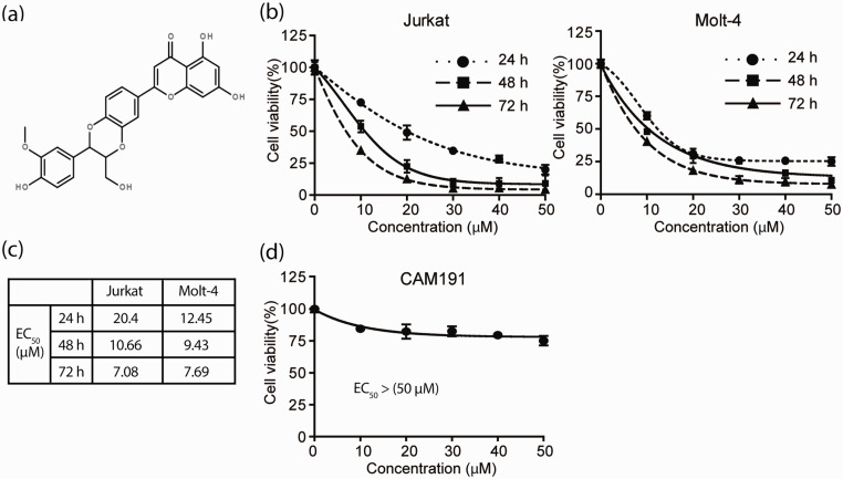
To ascertain whether HD can inhibit proliferation of T-ALL cells in vitro, we first treated Jurkat and Molt-4 with various concentrations of HD (Figure 1(a)), ranging from 10 to 50 µM for up to 72 h. MTT analysis was performed to measure the cell viability. We found that HD can suppress the proliferation of both cell lines in a concentration-dependent manner (Figure 1(b)). The IC50 values ranged from 7 to 20 µM (Figure 1(c)). To determine whether HD specifically inhibits T-ALL cells, we investigated the effect of HD on CAM-191, a normal human lymphocyte cell line. MTT assay confirms that HD shows no significant toxicity to CAM-191 (Figure 1(d)). These results suggested that HD can specifically inhibit T-ALL cell proliferation in vitro.
Figure 1.
HD suppresses the proliferation of T-ALL cells. (a) Chemical structure of HD. (b) T-ALL cells were exposed to various concentrations of HD (0, 10, 20, 30, 40, 50 µM) for 24, 48 and 72 h. MTT analysis was used to determine the cell viability. (c) EC50 values of HD in T-ALL cell lines were calculated by GraphPad Prism. The data represent three independent experiments. (d) Normal human lymphocyte cell CAM-191were treated with different concentrations of HD (0, 10, 20, 30, 40, 50 µM) for 48 h. The cell viability was examined by MTT assay.
HD induces G2/M phase arrest in T-ALL cells
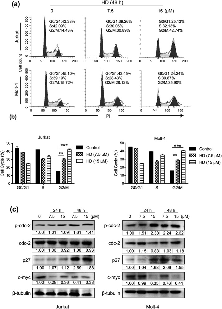
In order to determine whether the decrease of cell viability of HD-treated T-ALL cells is attributed to the induction of cell cycle arrest, flow cytometry was applied to analyze the impact of HD on cell cycle distribution. The results showed that the treatment of either 7.5 or 15 µM HD significantly increased the portion of cells arrested at the G2/M phase, as compared with the control group (Figure 2(a) and (b)). Concurrently, HD treatment elevated the expression of p-cdc2 and p27, while decreased oncogenic c-Myc expression (Figure 2(c)). Together, these results showed that HD could arrest T-ALL cells at the G2/M phase, thereby reducing T-ALL cell viability.
Figure 2.
HD induces T-ALL cell cycle arrest at the G2/M phase. (a) Both cells were treated with HD (0, 7.5, and 15 µM) for 48 h, followed by PI staining and flow cytometry analysis of cell cycle distribution. (b) Cell cycle distribution analysis of (a). HD vs. control, **p< 0.01, ***p< 0.001. (c) The expression levels of cell cycle-related proteins after treatment with HD (0, 7.5, and 15 µM) for 24 and 48 h, with β-tubulin as loading control.
HD induces apoptosis in T-ALL cells
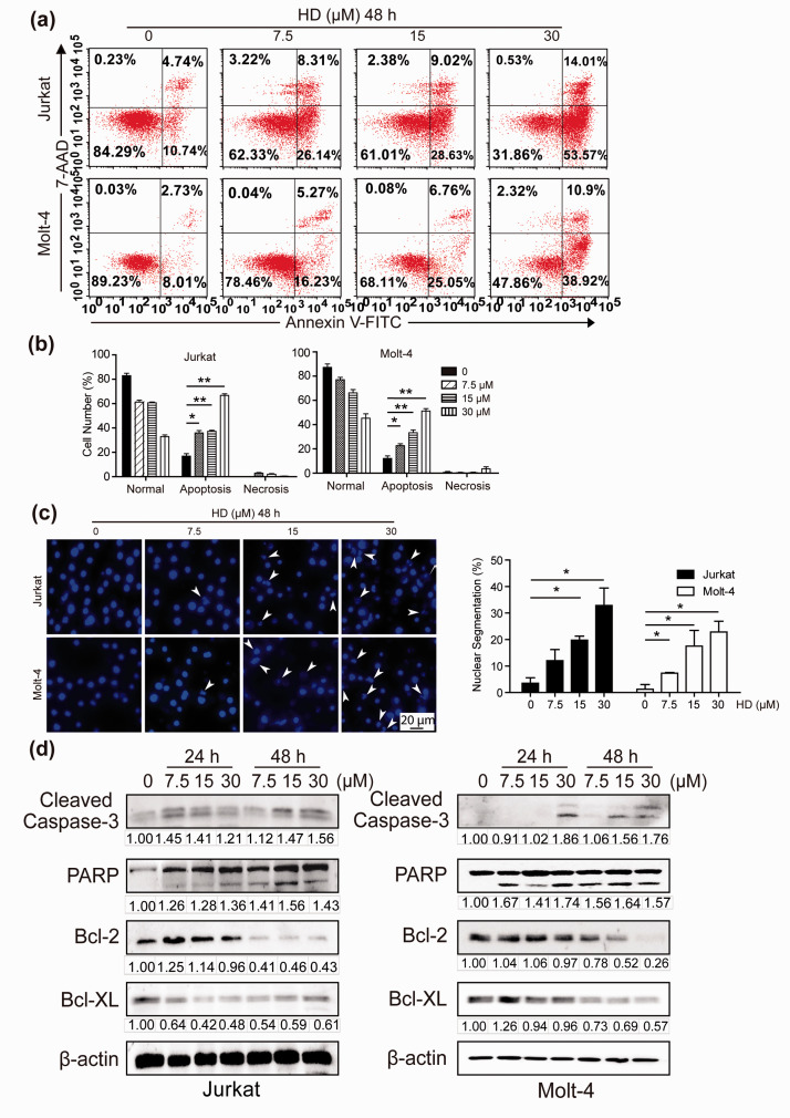
To investigate whether apoptosis is involved in HD’s anti-leukemic activity, we treated both cell lines with 7.5, 15, or 30 µM HD for 48 h. Flow cytometry were carried out to detect the percentage of apoptotic cells. As shown in Figure 3(a) and (b), HD induced concentration-dependent apoptosis in both T-ALL cell lines, with apoptosis rate reaching 66.25% and 52.14% in Jurkat and Molt-4 cells, respectively. For further confirmation, we monitored the change of nuclear morphology. As expected, after treatment with HD for 48 h, cell apoptosis-related phenotypes including chromatin condensation was significantly increased (Figure 3(c)). In addition, HD increased the cleavage and activation of PARP and caspase-3, while Bcl-2 and Bcl-xL were considerably downregulated (Figure 3(d)). Altogether, the results clearly implied that HD induced caspase-dependent apoptosis in T-ALL cells.
Figure 3.
HD induces apoptosis in T-ALL cells. (a) Annexin V/7-AAD double staining and flow cytometry analysis were used to detect the apoptosis ratio of cells treated with HD (0, 7.5, 15, 30 µM) for 48 h. (b) Quantification of the number of apoptotic cells in panel A. HD vs. Control. *p< 0.05, **p< 0.01. (c) After DAPI staining, the nucleus morphology of T-ALL cells treated with HD was imaged and the number of apoptotic cells was quantified. Scale bar, 20 μm. (d) After HD treatment for 24 or 48 h, Western blotting analysis of apoptosis-related proteins was performed. (A color version of this figure is available in the online journal.)
HD induces cytotoxic autophagy in T-ALL cells
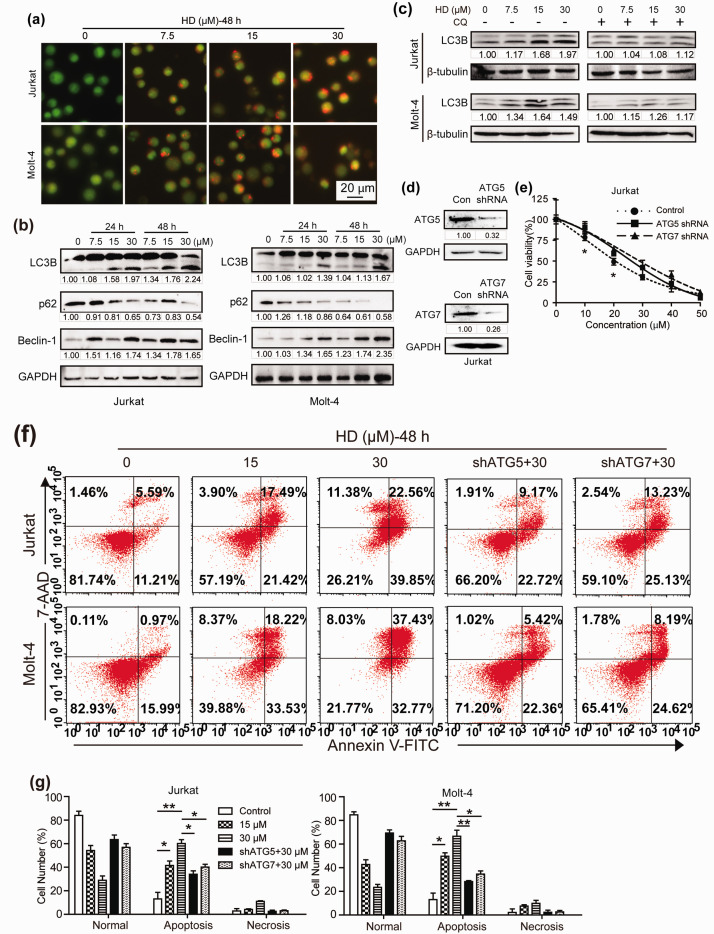
Autophagy and apoptosis are two vital forms of programmed cell death, both of which participate in the regulation of cell death through complex protein intersecting networks. 17 We next studied whether autophagy is related to HD-induced proliferation inhibition. AO staining of HD-treated leukemic cells displayed yellow-orange fluorescence, a specific marker for the occurrence of autophagosomes formation (Figure 4(a)). The occurrence of autophagy was further confirmed by Western blotting analysis, as the expression of LC3-II was clearly elevated after the treatment of HD. Furthermore, HD upregulated Beclin-1 while downregulated p62 (Figure 4(b)). To further confirm autophagy activation by HD, autophagic flux in T-ALL cells was evaluated by detection of LC3-II with or without the addition of chloroquine (CQ 10 µM). The data suggested that HD could enhance autophagic flux in T-ALL cells (Figure 4(c)). As autophagy is crucial in the survival of leukemic cells, 17 in order to clarify the exact function of HD-induced autophagy, we inhibited autophagy in Jurkat cells by knocking down ATG5 and ATG7, both of which are essential molecules for the induction of autophagy (Figure 4(d)). Results showed that inhibition of HD-induced autophagy partially rescued cell viability (Figure 4(e)). To test whether knockdown of ATG5 or ATG7 rescued cell viability via the apoptotic pathways, we assessed the percentage of apoptotic cells in ATG5/7 silenced cells treated with HD. The results showed that knockdown of ATG5 or ATG7 decreased HD-induced cell apoptosis (Figure 4(f) and (g)). These results suggest that HD induced a cytotoxic autophagy, which enhanced the anti-T-ALL efficacy of HD.
Figure 4.
HD induces cytotoxic autophagy in T-ALL cells. (a) Representative images of AO staining in Jurkat and Molt-4 cells treated with various concentration of HD for 48 h were shown. Scale bar, 20 µm. (b) The expression levels of autophagy-specific proteins were detected using Western blotting analysis in cells treated with HD for 24 or 48 h. (c) T-ALL cells were incubated with various concentration of HD with or without CQ (10 µM). The expression of LC3-II was detected by western blotting. (d) Jurkat cells were transiently transfected with ATG5 or ATG7 specific short hairpin RNA (shRNA), and the expression levels of ATG5 and ATG7 were measured using western blotting analysis. (e) After the knockdown of ATG5 or ATG7, Jurkat cells were treated with HD and subjected to MTT analysis. ATG5 shRNA or ATG7 shRNA vs. scramble shRNA as control. (f) ATG5 or ATG7 knockdown cells was subjected to flow cytometry analysis of apoptosis. (g) Quantification of the number of apoptotic cells in panel f. *p< 0.05, **p< 0.01. (A color version of this figure is available in the online journal.)
HD induces ferroptosis in T-ALL cells
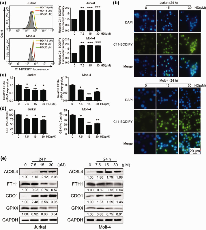
Ferroptosis is recognized to be a programmed oxidative cell death accompanied by lipid peroxidation and iron accumulation, producing reactive oxygen species. 18 Recent studies have shown that autophagy can lead to ferroptosis through ferritin phagocytosis. 19 Thus, whether HD can trigger ferroptosis by autophagy is worth investigating. Upregulation of lipid ROS is a well-characterized ferroptosis marker 20 ; therefore, we examined the effect of HD on lipid ROS levels in Jurkat and Molt-4 cells. The results showed that HD treatment induced rapid accumulation of lipid radicals even at a low concentration, as determined by both flow cytometry and fluorescent imaging using the C11-BODIPY probe (Figure 5(a) and (b)). GPX4 has been proven to participate in the ferroptosis process by scavenging hydroperoxides. 21 Thus, we postulate that HD may induce ferroptosis by inhibiting GPX4. As expected, transcription of GPX4 was decreased by the treatment of HD (Figure 5(c)). Under condition of ferroptosis, GSH depletion plays a crucial role by modulating lipid peroxide level that is normally triggered by decrease of GPX4. 22 Therefore, we investigated GSH level in HD-induced ferroptosis. Unsurprisingly, the GSH level in all HD-treated groups was significantly reduced compared to control (Figure 5(d)). Western blotting analysis further confirmed a concentration-dependent decrease of GPX4 and FTH1, and a concomitant increase of CDO1 and ACSL4, all of which are specific markers of ferroptosis (Figure 5(e)). 23 Collectively, these data confirmed the involvement of ferroptotic cell death in HD-induced proliferation inhibition of T-ALL cells.
Figure 5.
HD induces ferroptosis in T-ALL cells. Lipid ROS production after treatment with HD for 24 h was assessed by both flow cytometry (a) and fluorescent imaging (b) using the C11-BODIPY probe. Scale bar, 20 µm. (c) qRT-PCR was performed to measure the mRNA level of GPX4 after the treatment of HD. (d) The level of GSH was determined in T-ALL cells exposed to 7.5, 15 or 30 µM HD using a GSH assay kit. (e) The expression levels of ferroptosis-related proteins were evaluated by Western blotting. Representative data of three independent experiments is shown. *p< 0.05; **p< 0.01; ***p< 0.001. (A color version of this figure is available in the online journal.)
Inhibition of autophagy reduces HD-induced ferroptosis in T-ALL cells
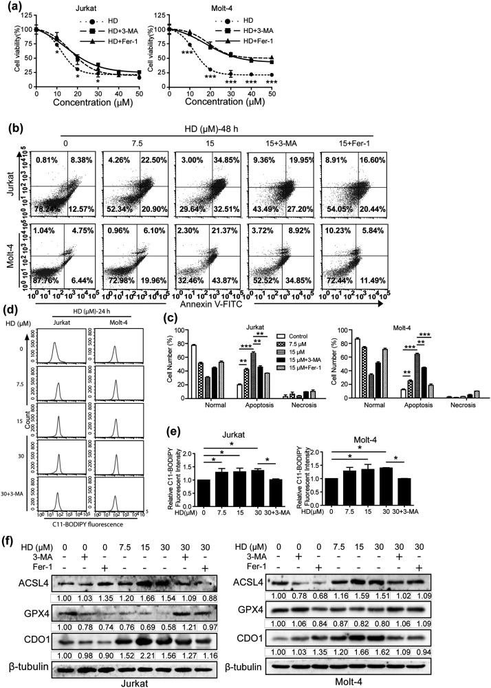
Studies have provided evidence that autophagy contributes to ferroptosis through NCOA4-mediated ferritinophagy, and that autophagy is closely linked to the induction of ferroptosis by regulation of cellular ROS generation.19,24 Thus, to further clarify the crosstalk between autophagy and ferroptosis in HD-treated T-ALL, we first analyzed cell viability after the treatment of HD, in the presence or absence of autophagy inhibitor 3-MA or ferroptosis inhibitor Fer-1. In accordance with the data of Figures 4 and 5, suppression of either autophagy or ferroptosis dramatically abolished HD-induced proliferation inhibition (Figure 6(a)), and suppressed HD-induced T-ALL cells apoptosis (Figure 6(b) and (c)). Collectively, these results further supported the notion that both autophagy and ferroptosis contributed to the suppression of T-ALL in vitro. Next, we investigated whether HD-induced autophagy promotes ferroptosis in T-ALL cells. Data illustrated that the induction of lipid ROS by HD treatment is abolished by the inhibition of autophagy (Figure 6(d) and (e)). Consistently, HD-induced expression of ACSL4 and CDO1 were downregulated in the presence of 3-MA, whereas autophagy inhibition up-regulated GPX4 expression (Figure 6(f)). Altogether, our results suggest that HD-induced ferroptosis is autophagy dependent.
Figure 6.
Inhibition of autophagy reduces HD-induced ferroptosis in T-ALL cells. (a) After treated with HD in the present or absent of autophagy inhibitor 3-MA (2 mM) or ferroptosis inhibitor Fer-1 (10 µM), cell viability was determined by MTT assay. (b) After pre-treatment with either 3-MA (2 mM) or Fer-1 (10 µM) in combination with HD, cell apoptosis ratio was detected by Annexin V/7-AAD double staining and subsequent flow cytometry analysis. (c) Quantification of the number of apoptotic cells in panel b. **p < 0.01, ***p < 0.001. Lipid ROS production after treatment with HD with or without 3-MA (2 mM) for 24 hours was assessed by flow cytometry (d) and quantified in panel e using the C11-BODIPY probe. *p < 0.05. (f) Western blotting analysis of ferroptosis-related proteins in T-ALL cells after pre-treatment with 3-MA (2 mM) or Fer-1 (10 µM) for 2 hours, and then incubated with HD for another 24 h.
Discussion
HD has been proven to possess an anti-cancer capacity in human colon cancer cells and ALL cells by suppression of Wnt/beta-catenin pathway and P-gp, respectively.11,12 In the present study, we first documented that HD inhibited T-ALL cell proliferation in vitro, partially via arresting cells at the G2/M phase and inducing apoptosis. By upregulating the expression of p-cdc2, HD caused delayed progression of T-ALL cells from G2 into mitosis. The down-regulation of c-Myc, an oncogene responsible for promoting cell cycle by activating cyclin and CDK while suppressing p21/p27,25,26 also contributed to the cell cycle arrest and the increase of CDK inhibitor p27. Furthermore, our results showed that HD induced cleavage of PARP and caspase 3, both of which are key executioners of apoptosis specifically cleaved during apoptosis.27,28 At the same time, anti-apoptotic proteins including Bcl-2 and Bcl-xL were significantly downregulated in HD-induced apoptosis. Thus, HD activated the apoptosis pathway to eradicate T-ALL cells that failed to complete the G2/M transition.
Apart from activation of cell cycle arrest and apoptosis, HD also induced cytotoxic autophagy, as evidenced by the up-regulation of autophagy marker Beclin-1 and lipidated LC3-II, as well as the degradation of autophagy substrate p62/SQSTM1. The occurrence of autophagy subsequently contributed to the accumulation of lipid peroxidation and induction of ferroptosis. The term ferroptosis was described for the first time in 2012 to depict a form of cell death activated by iron oxidation. 29 As a caspase-independent form of cell death, ferroptosis is not commonly associated with apoptosis. However, emerging evidence suggests that ferroptosis often shares common pathways with apoptosis. Few studies have reported an interrelationship between ferroptosis and apoptosis: switching apoptosis to ferroptosis 30 and ferroptotic agent-mediated sensitization of apoptosis. 31 Moreover, reports have shown that the product of lipid peroxidation could activate MAPKs pathway through association with ERK, JNK and eventually induce apoptosis. 32 Thus, as a lipid peroxidation inhibitor, 33 Fer-1 may inhibit apoptosis by decreasing lipid ROS in T-ALL cells, as shown in Figure 6(b) and (c). Recent evidence suggests that autophagy is closely related to ferroptosis. By degradation of ferritin in fibroblasts and cancer cells, autophagy increases cellular unstable iron levels through the NCOA4-mediated pathway, thus disrupting cellular iron homeostasis and contributes to ferroptosis. 24 Inhibition of autophagy by knockdown of either ATG5, ATG7, BECN, or LC3B limited erastin-induced lipid peroxidation and ferroptotic cell death.19,24,34 Our study also confirmed that inhibition of autophagy abrogated HD-induced lipid peroxidation and reduced the expression of ferroptosis-related proteins. However, more in-depth investigations are required to elucidate the molecular mechanisms governing the interplay between HD-induced autophagy and ferroptosis.
Flavonolignans are the structural combination of flavonoids and phenylpropanoids (lignans), and have attracted much attention as potent anticancer agents due to their multi-target capacity and minimal adverse effects. The first group of flavonolignans is silymarin complex extracted from milk thistle (Silybum marianum), which has been intensively studied for its hepatoprotection, antioxidant, and anti-cancer properties. 35 Hydnocarpin-type compounds are formally dehydrated analogs of silymarin flavonolignans with flavanone-3-ol (3-hydroxyflavanone) structure. But unlike silybins that can be completely synthesized, 36 hydnocarpin-type flavonolignans are not synthetically available, 8 which largely hindered their scientific study. Our previous finding of converting silibinin to HD under Mitsunobu reaction condition lay the foundation for highly effective and convenient preparation of HD, 16 which shall greatly facilitate the scientific research and clinical application of HD.
In summary, we provide evidence for the first time that, by concurrently targeting multiple pathways mediating cell cycle, apoptosis, and autophagy-dependent ferroptosis, the natural flavonolignan HD possesses anti-leukemic activities in T-ALL cells. Our findings significantly contribute to the understanding of the various biological functions of flavonolignan in T-ALL cells and lay the foundation for future experiments.
Footnotes
AUTHORS’ CONTRIBUTIONS: HJZ and SYL designed the experiments; HHW, GH, LLX, SST and GYK performed the experiments; GZH and LM supplied Hydnocarpin D. HHW and SYL did the statistical analysis; HHW, ZZH and SYL wrote the paper. The final version of the manuscript has the approval of all authors.
DECLARATION OF CONFLICTING INTERESTS: The authors declared no potential conflicts of interest with respect to the research, authorship, and publication of this article.
FUNDING: The author(s) disclosed receipt of the following financial support for the research, authorship, and/or publication of this article: This work was supported by the National Natural Science Foundation of China [grant number 81703549, 81774003]; Zhejiang Provincial Natural Science Fund (grant number LQ19H1600060); Zhejiang Chinese Medical University Research Fund Project (grant number KC201912, 2018ZZ09).
ORCID iDs: Guozheng Huang https://orcid.org/0000-0002-5730-9549
Huajun Zhao https://orcid.org/0000-0001-8643-4261
References
- 1.Pui CH, Robison LL, Look AT. Acute lymphoblastic leukaemia. Lancet 2008; 371:1030–43 [DOI] [PubMed] [Google Scholar]
- 2.Dores GM, Devesa SS, Curtis RE, Linet MS, Morton LM. Acute leukemia incidence and patient survival among children and adults in the United States, 2001-2007. Blood 2012; 119:34–43 [DOI] [PMC free article] [PubMed] [Google Scholar]
- 3.Brown PA, Shah B, Fathi A, Wieduwilt M, Advani A, Aoun P, Barta SK, Boyer MW, Bryan T, Burke PW, Cassaday R, Coccia PF, Coutre SE, Damon LE, DeAngelo DJ, Frankfurt O, Greer JP, Kantarjian HM, Klisovic RB, Kupfer G, Litzow M, Liu A, Mattison R, Park J, Rubnitz J, Saad A, Uy GL, Wang ES, Gregory KM, Ogba N. NCCN guidelines insights: Acute lymphoblastic leukemia, version 12017. J Natl Compr Canc Netw 2017; 15:1091102. [DOI] [PubMed] [Google Scholar]
- 4.Raetz EA, Teachey DTT. Cell acute lymphoblastic leukemia. Hematology Am Soc Hematol Educ Program 2016; 2016:580–8 [DOI] [PMC free article] [PubMed] [Google Scholar]
- 5.Kansagra A, Dahiya S, Litzow M. Continuing challenges and current issues in acute lymphoblastic leukemia. Leuk Lymphoma 2018; 59:526–41 [DOI] [PubMed] [Google Scholar]
- 6.Jahanafrooz Z, Motamed N, Rinner B, Mokhtarzadeh A, Baradaran B. Silibinin to improve cancer therapeutic, as an apoptotic inducer, autophagy modulator, cell cycle inhibitor, and microRNAs regulator. Life Sci 2018; 213:236–47 [DOI] [PubMed] [Google Scholar]
- 7.Sahoo MR, Dhanabal SP, Jadhav AN, Reddy V, Muguli G, Babu UV, Rangesh P. Hydnocarpus: an ethnopharmacological, phytochemical and pharmacological review. J Ethnopharmacol 2014; 154:17–25 [DOI] [PubMed] [Google Scholar]
- 8.Guz NR, Stermitz FR. Synthesis and structures of regioisomeric hydnocarpin-type flavonolignans. J Nat Prod 2000; 63:1140–5 [DOI] [PubMed] [Google Scholar]
- 9.Reddy SV, Tiwari AK, Kumar US, Rao RJ, Rao JM. Free radical scavenging, enzyme inhibitory constituents from antidiabetic ayurvedic medicinal plant hydnocarpus wightiana blume. Phytother Res 2005; 19:277–81 [DOI] [PubMed] [Google Scholar]
- 10.Sharma DK, Hall IH. Hypolipidemic, anti-inflammatory, and antineoplastic activity and cytotoxicity of flavonolignans isolated from hydnocarpus wightiana seeds. J Nat Prod 1991; 54:1298–302 [DOI] [PubMed] [Google Scholar]
- 11.Lee MA, Kim WK, Park HJ, Kang SS, Lee SK. Anti-proliferative activity of hydnocarpin, a natural lignan, is associated with the suppression of wnt/beta-catenin signaling pathway in Colon cancer cells. Bioorg Med Chem Lett 2013; 23:5511–4 [DOI] [PubMed] [Google Scholar]
- 12.Bueno Perez L, Pan L, Sass E, Gupta SV, Lehman A, Kinghorn AD, Lucas DM. Potentiating effect of the flavonolignan (-)-hydnocarpin in combination with vincristine in a sensitive and Pgp-expressing acute lymphoblastic leukemia cell line. Phytother Res 2013; 27:1735–8 [DOI] [PMC free article] [PubMed] [Google Scholar]
- 13.Klionsky DJ, Abdelmohsen K, Abe A, Abedin MJ, Abeliovich H, Acevedo Arozena A, Adachi H, Adams CM, Adams PD, Adeli K, Adhihetty PJ, Adler SG, Agam G, Agarwal R, Aghi MK, Agnello M, Agostinis P, Aguilar PV, Aguirre-Ghiso J, Airoldi EM, Ait-Si-Ali S, Akematsu T, Akporiaye ET, Al-Rubeai M, Albaiceta GM, Albanese C, Albani D, Albert ML, Aldudo J, Algül H, Alirezaei M, Alloza I, Almasan A, Almonte-Beceril M, Alnemri ES, Alonso C, Altan-Bonnet N, Altieri DC, Alvarez S, Alvarez-Erviti L, Alves S, Amadoro G, Amano A, Amantini C, Ambrosio S, Amelio I, Amer AO, Amessou M, Amon A, An Z, Anania FA, Andersen SU, Andley UP, Andreadi CK, Andrieu-Abadie N, Anel A, Ann DK, Anoopkumar-Dukie S, Antonioli M, Aoki H, Apostolova N, Aquila S, Aquilano K, Araki K, Arama E, Aranda A, Araya J, Arcaro A, Arias E, Arimoto H, Ariosa AR, Armstrong JL, Arnould T, Arsov I, Asanuma K, Askanas V, Asselin E, Atarashi R, Atherton SS, Atkin JD, Attardi LD, Auberger P, Auburger G, Aurelian L, Autelli R, Avagliano L, Avantaggiati ML, Avrahami L, Awale S, Azad N, Bachetti T, Backer JM, Bae DH, Bae JS, Bae ON, Bae SH, Baehrecke EH, Baek SH, Baghdiguian S, Bagniewska-Zadworna A, Bai H, Bai J, Bai XY, Bailly Y, Balaji KN, Balduini W, Ballabio A, Balzan R, Banerjee R, Bánhegyi G, Bao H, Barbeau B, Barrachina MD, Barreiro E, Bartel B, Bartolomé A, Bassham DC, Bassi MT, Bast RC, Jr, Basu A, Batista MT, Batoko H, Battino M, Bauckman K, Baumgarner BL, Bayer KU, Beale R, Beaulieu JF, Beck GR, Jr., Becker C, Beckham JD, Bédard PA, Bednarski PJ, Begley TJ, Behl C, Behrends C, Behrens GM, Behrns KE, Bejarano E, Belaid A, Belleudi F, Bénard G, Berchem G, Bergamaschi D, Bergami M, Berkhout B, Berliocchi L, Bernard A, Bernard M, Bernassola F, Bertolotti A, Bess AS, Besteiro S, Bettuzzi S, Bhalla S, Bhattacharyya S, Bhutia SK, Biagosch C, Bianchi MW, Biard-Piechaczyk M, Billes V, Bincoletto C, Bingol B, Bird SW, Bitoun M, Bjedov I, Blackstone C, Blanc L, Blanco GA, Blomhoff HK, Boada-Romero E, Böckler S, Boes M, Boesze-Battaglia K, Boise LH, Bolino A, Boman A, Bonaldo P, Bordi M, Bosch J, Botana LM, Botti J, Bou G, Bouché M, Bouchecareilh M, Boucher MJ, Boulton ME, Bouret SG, Boya P, Boyer-Guittaut M, Bozhkov PV, Brady N, Braga VM, Brancolini C, Braus GH, Bravo-San Pedro JM, Brennan LA, Bresnick EH, Brest P, Bridges D, Bringer MA, Brini M, Brito GC, Brodin B, Brookes PS, Brown EJ, Brown K, Broxmeyer HE, Bruhat A, Brum PC, Brumell JH, Brunetti-Pierri N, Bryson-Richardson RJ, Buch S, Buchan AM, Budak H, Bulavin DV, Bultman SJ, Bultynck G, Bumbasirevic V, Burelle Y, Burke RE, Burmeister M, Bütikofer P, Caberlotto L, Cadwell K, Cahova M, Cai D, Cai J, Cai Q, Calatayud S, Camougrand N, Campanella M, Campbell GR, Campbell M, Campello S, Candau R, Caniggia I, Cantoni L, Cao L, Caplan AB, Caraglia M, Cardinali C, Cardoso SM, Carew JS, Carleton LA, Carlin CR, Carloni S, Carlsson SR, Carmona-Gutierrez D, Carneiro LA, Carnevali O, Carra S, Carrier A, Carroll B, Casas C, Casas J, Cassinelli G, Castets P, Castro-Obregon S, Cavallini G, Ceccherini I, Cecconi F, Cederbaum AI, Ceña V, Cenci S, Cerella C, Cervia D, Cetrullo S, Chaachouay H, Chae HJ, Chagin AS, Chai CY, Chakrabarti G, Chamilos G, Chan EY, Chan MT, Chandra D, Chandra P, Chang CP, Chang RC, Chang TY, Chatham JC, Chatterjee S, Chauhan S, Che Y, Cheetham ME, Cheluvappa R, Chen CJ, Chen G, Chen GC, Chen G, Chen H, Chen JW, Chen JK, Chen M, Chen M, Chen P, Chen Q, Chen Q, Chen SD, Chen S, Chen SS, Chen W, Chen WJ, Chen WQ, Chen W, Chen X, Chen YH, Chen YG, Chen Y, Chen Y, Chen Y, Chen YJ, Chen YQ, Chen Y, Chen Z, Chen Z, Cheng A, Cheng CH, Cheng H, Cheong H, Cherry S, Chesney J, Cheung CH, Chevet E, Chi HC, Chi SG, Chiacchiera F, Chiang HL, Chiarelli R, Chiariello M, Chieppa M, Chin LS, Chiong M, Chiu GN, Cho DH, Cho SG, Cho WC, Cho YY, Cho YS, Choi AM, Choi EJ, Choi EK, Choi J, Choi ME, Choi SI, Chou TF, Chouaib S, Choubey D, Choubey V, Chow KC, Chowdhury K, Chu CT, Chuang TH, Chun T, Chung H, Chung T, Chung YL, Chwae YJ, Cianfanelli V, Ciarcia R, Ciechomska IA, Ciriolo MR, Cirone M, Claerhout S, Clague MJ, Clària J, Clarke PG, Clarke R, Clementi E, Cleyrat C, Cnop M, Coccia EM, Cocco T, Codogno P, Coers J, Cohen EE, Colecchia D, Coletto L, Coll NS, Colucci-Guyon E, Comincini S, Condello M, Cook KL, Coombs GH, Cooper CD, Cooper JM, Coppens I, Corasaniti MT, Corazzari M, Corbalan R, Corcelle-Termeau E, Cordero MD, Corral-Ramos C, Corti O, Cossarizza A, Costelli P, Costes S, Cotman SL, Coto-Montes A, Cottet S, Couve E, Covey LR, Cowart LA, Cox JS, Coxon FP, Coyne CB, Cragg MS, Craven RJ, Crepaldi T, Crespo JL, Criollo A, Crippa V, Cruz MT, Cuervo AM, Cuezva JM, Cui T, Cutillas PR, Czaja MJ, Czyzyk-Krzeska MF, Dagda RK, Dahmen U, Dai C, Dai W, Dai Y, Dalby KN, Dalla Valle L, Dalmasso G, D'Amelio M, Damme M, Darfeuille-Michaud A, Dargemont C, Darley-Usmar VM, Dasarathy S, Dasgupta B, Dash S, Dass CR, Davey HM, Davids LM, Dávila D, Davis RJ, Dawson TM, Dawson VL, Daza P, de Belleroche J, de Figueiredo P, de Figueiredo RC, de la Fuente J, De Martino L, De Matteis A, De Meyer GR, De Milito A, De Santi M, de Souza W, De Tata V, De Zio D, Debnath J, Dechant R, Decuypere JP, Deegan S, Dehay B, Del Bello B, Del Re DP, Delage-Mourroux R, Delbridge LM, Deldicque L, Delorme-Axford E, Deng Y, Dengjel J, Denizot M, Dent P, Der CJ, Deretic V, Derrien B, Deutsch E, Devarenne TP, Devenish RJ, Di Bartolomeo S, Di Daniele N, Di Domenico F, Di Nardo A, Di Paola S, Di Pietro A, Di Renzo L, DiAntonio A, Díaz-Araya G, Díaz-Laviada I, Diaz-Meco MT, Diaz-Nido J, Dickey CA, Dickson RC, Diederich M, Digard P, Dikic I, Dinesh-Kumar SP, Ding C, Ding WX, Ding Z, Dini L, Distler JH, Diwan A, Djavaheri-Mergny M, Dmytruk K, Dobson RC, Doetsch V, Dokladny K, Dokudovskaya S, Donadelli M, Dong XC, Dong X, Dong Z, Donohue TM, Jr, Doran KS, D'Orazi G, Dorn GW, 2nd, Dosenko V, Dridi S, Drucker L, Du J, Du LL, Du L, du Toit A, Dua P, Duan L, Duann P, Dubey VK, Duchen MR, Duchosal MA, Duez H, Dugail I, Dumit VI, Duncan MC, Dunlop EA, Dunn WA, Jr, Dupont N, Dupuis L, Durán RV, Durcan TM, Duvezin-Caubet S, Duvvuri U, Eapen V, Ebrahimi-Fakhari D, Echard A, Eckhart L, Edelstein CL, Edinger AL, Eichinger L, Eisenberg T, Eisenberg-Lerner A, Eissa NT, El-Deiry WS, El-Khoury V, Elazar Z, Eldar-Finkelman H, Elliott CJ, Emanuele E, Emmenegger U, Engedal N, Engelbrecht AM, Engelender S, Enserink JM, Erdmann R, Erenpreisa J, Eri R, Eriksen JL, Erman A, Escalante R, Eskelinen EL, Espert L, Esteban-Martínez L, Evans TJ, Fabri M, Fabrias G, Fabrizi C, Facchiano A, Færgeman NJ, Faggioni A, Fairlie WD, Fan C, Fan D, Fan J, Fang S, Fanto M, Fanzani A, Farkas T, Faure M, Favier FB, Fearnhead H, Federici M, Fei E, Felizardo TC, Feng H, Feng Y, Feng Y, Ferguson TA, Fernández Á F, Fernandez-Barrena MG, Fernandez-Checa JC, Fernández-López A, Fernandez-Zapico ME, Feron O, Ferraro E, Ferreira-Halder CV, Fesus L, Feuer R, Fiesel FC, Filippi-Chiela EC, Filomeni G, Fimia GM, Fingert JH, Finkbeiner S, Finkel T, Fiorito F, Fisher PB, Flajolet M, Flamigni F, Florey O, Florio S, Floto RA, Folini M, Follo C, Fon EA, Fornai F, Fortunato F, Fraldi A, Franco R, Francois A, François A, Frankel LB, Fraser ID, Frey N, Freyssenet DG, Frezza C, Friedman SL, Frigo DE, Fu D, Fuentes JM, Fueyo J, Fujitani Y, Fujiwara Y, Fujiya M, Fukuda M, Fulda S, Fusco C, Gabryel B, Gaestel M, Gailly P, Gajewska M, Galadari S, Galili G, Galindo I, Galindo MF, Galliciotti G, Galluzzi L, Galluzzi L, Galy V, Gammoh N, Gandy S, Ganesan AK, Ganesan S, Ganley IG, Gannagé M, Gao FB, Gao F, Gao JX, García Nannig L, García Véscovi E, Garcia-Macía M, Garcia-Ruiz C, Garg AD, Garg PK, Gargini R, Gassen NC, Gatica D, Gatti E, Gavard J, Gavathiotis E, Ge L, Ge P, Ge S, Gean PW, Gelmetti V, Genazzani AA, Geng J, Genschik P, Gerner L, Gestwicki JE, Gewirtz DA, Ghavami S, Ghigo E, Ghosh D, Giammarioli AM, Giampieri F, Giampietri C, Giatromanolaki A, Gibbings DJ, Gibellini L, Gibson SB, Ginet V, Giordano A, Giorgini F, Giovannetti E, Girardin SE, Gispert S, Giuliano S, Gladson CL, Glavic A, Gleave M, Godefroy N, Gogal RM, Jr, Gokulan K, Goldman GH, Goletti D, Goligorsky MS, Gomes AV, Gomes LC, Gomez H, Gomez-Manzano C, Gómez-Sánchez R, Gonçalves DA, Goncu E, Gong Q, Gongora C, Gonzalez CB, Gonzalez-Alegre P, Gonzalez-Cabo P, González-Polo RA, Goping IS, Gorbea C, Gorbunov NV, Goring DR, Gorman AM, Gorski SM, Goruppi S, Goto-Yamada S, Gotor C, Gottlieb RA, Gozes I, Gozuacik D, Graba Y, Graef M, Granato GE, Grant GD, Grant S, Gravina GL, Green DR, Greenhough A, Greenwood MT, Grimaldi B, Gros F, Grose C, Groulx JF, Gruber F, Grumati P, Grune T, Guan JL, Guan KL, Guerra B, Guillen C, Gulshan K, Gunst J, Guo C, Guo L, Guo M, Guo W, Guo XG, Gust AA, Gustafsson Å B, Gutierrez E, Gutierrez MG, Gwak HS, Haas A, Haber JE, Hadano S, Hagedorn M, Hahn DR, Halayko AJ, Hamacher-Brady A, Hamada K, Hamai A, Hamann A, Hamasaki M, Hamer I, Hamid Q, Hammond EM, Han F, Han W, Handa JT, Hanover JA, Hansen M, Harada M, Harhaji-Trajkovic L, Harper JW, Harrath AH, Harris AL, Harris J, Hasler U, Hasselblatt P, Hasui K, Hawley RG, Hawley TS, He C, He CY, He F, He G, He RR, He XH, He YW, He YY, Heath JK, Hébert MJ, Heinzen RA, Helgason GV, Hensel M, Henske EP, Her C, Herman PK, Hernández A, Hernandez C, Hernández-Tiedra S, Hetz C, Hiesinger PR, Higaki K, Hilfiker S, Hill BG, Hill JA, Hill WD, Hino K, Hofius D, Hofman P, Höglinger GU, Höhfeld J, Holz MK, Hong Y, Hood DA, Hoozemans JJ, Hoppe T, Hsu C, Hsu CY, Hsu LC, Hu D, Hu G, Hu HM, Hu H, Hu MC, Hu YC, Hu ZW, Hua F, Hua Y, Huang C, Huang HL, Huang KH, Huang KY, Huang S, Huang S, Huang WP, Huang YR, Huang Y, Huang Y, Huber TB, Huebbe P, Huh WK, Hulmi JJ, Hur GM, Hurley JH, Husak Z, Hussain SN, Hussain S, Hwang JJ, Hwang S, Hwang TI, Ichihara A, Imai Y, Imbriano C, Inomata M, Into T, Iovane V, Iovanna JL, Iozzo RV, Ip NY, Irazoqui JE, Iribarren P, Isaka Y, Isakovic AJ, Ischiropoulos H, Isenberg JS, Ishaq M, Ishida H, Ishii I, Ishmael JE, Isidoro C, Isobe K, Isono E, Issazadeh-Navikas S, Itahana K, Itakura E, Ivanov AI, Iyer AK, Izquierdo JM, Izumi Y, Izzo V, Jäättelä M, Jaber N, Jackson DJ, Jackson WT, Jacob TG, Jacques TS, Jagannath C, Jain A, Jana NR, Jang BK, Jani A, Janji B, Jannig PR, Jansson PJ, Jean S, Jendrach M, Jeon JH, Jessen N, Jeung EB, Jia K, Jia L, Jiang H, Jiang H, Jiang L, Jiang T, Jiang X, Jiang X, Jiang X, Jiang Y, Jiang Y, Jiménez A, Jin C, Jin H, Jin L, Jin M, Jin S, Jinwal UK, Jo EK, Johansen T, Johnson DE, Johnson GV, Johnson JD, Jonasch E, Jones C, Joosten LA, Jordan J, Joseph AM, Joseph B, Joubert AM, Ju D, Ju J, Juan HF, Juenemann K, Juhász G, Jung HS, Jung JU, Jung YK, Jungbluth H, Justice MJ, Jutten B, Kaakoush NO, Kaarniranta K, Kaasik A, Kabuta T, Kaeffer B, Kågedal K, Kahana A, Kajimura S, Kakhlon O, Kalia M, Kalvakolanu DV, Kamada Y, Kambas K, Kaminskyy VO, Kampinga HH, Kandouz M, Kang C, Kang R, Kang TC, Kanki T, Kanneganti TD, Kanno H, Kanthasamy AG, Kantorow M, Kaparakis-Liaskos M, Kapuy O, Karantza V, Karim MR, Karmakar P, Kaser A, Kaushik S, Kawula T, Kaynar AM, Ke PY, Ke ZJ, Kehrl JH, Keller KE, Kemper JK, Kenworthy AK, Kepp O, Kern A, Kesari S, Kessel D, Ketteler R, Kettelhut Ido C, Khambu B, Khan MM, Khandelwal VK, Khare S, Kiang JG, Kiger AA, Kihara A, Kim AL, Kim CH, Kim DR, Kim DH, Kim EK, Kim HY, Kim HR, Kim JS, Kim JH, Kim JC, Kim JH, Kim KW, Kim MD, Kim MM, Kim PK, Kim SW, Kim SY, Kim YS, Kim Y, Kimchi A, Kimmelman AC, Kimura T, King JS, Kirkegaard K, Kirkin V, Kirshenbaum LA, Kishi S, Kitajima Y, Kitamoto K, Kitaoka Y, Kitazato K, Kley RA, Klimecki WT, Klinkenberg M, Klucken J, Knævelsrud H, Knecht E, Knuppertz L, Ko JL, Kobayashi S, Koch JC, Koechlin-Ramonatxo C, Koenig U, Koh YH, Köhler K, Kohlwein SD, Koike M, Komatsu M, Kominami E, Kong D, Kong HJ, Konstantakou EG, Kopp BT, Korcsmaros T, Korhonen L, Korolchuk VI, Koshkina NV, Kou Y, Koukourakis MI, Koumenis C, Kovács AL, Kovács T, Kovacs WJ, Koya D, Kraft C, Krainc D, Kramer H, Kravic-Stevovic T, Krek W, Kretz-Remy C, Krick R, Krishnamurthy M, Kriston-Vizi J, Kroemer G, Kruer MC, Kruger R, Ktistakis NT, Kuchitsu K, Kuhn C, Kumar AP, Kumar A, Kumar A, Kumar D, Kumar D, Kumar R, Kumar S, Kundu M, Kung HJ, Kuno A, Kuo SH, Kuret J, Kurz T, Kwok T, Kwon TK, Kwon YT, Kyrmizi I, La Spada AR, Lafont F, Lahm T, Lakkaraju A, Lam T, Lamark T, Lancel S, Landowski TH, Lane DJ, Lane JD, Lanzi C, Lapaquette P, Lapierre LR, Laporte J, Laukkarinen J, Laurie GW, Lavandero S, Lavie L, LaVoie MJ, Law BY, Law HK, Law KB, Layfield R, Lazo PA, Le Cam L, Le Roch KG, Le Stunff H, Leardkamolkarn V, Lecuit M, Lee BH, Lee CH, Lee EF, Lee GM, Lee HJ, Lee H, Lee JK, Lee J, Lee JH, Lee JH, Lee M, Lee MS, Lee PJ, Lee SW, Lee SJ, Lee SJ, Lee SY, Lee SH, Lee SS, Lee SJ, Lee S, Lee YR, Lee YJ, Lee YH, Leeuwenburgh C, Lefort S, Legouis R, Lei J, Lei QY, Leib DA, Leibowitz G, Lekli I, Lemaire SD, Lemasters JJ, Lemberg MK, Lemoine A, Leng S, Lenz G, Lenzi P, Lerman LO, Lettieri Barbato D, Leu JI, Leung HY, Levine B, Lewis PA, Lezoualc'h F, Li C, Li F, Li FJ, Li J, Li K, Li L, Li M, Li M, Li Q, Li R, Li S, Li W, Li W, Li X, Li Y, Lian J, Liang C, Liang Q, Liao Y, Liberal J, Liberski PP, Lie P, Lieberman AP, Lim HJ, Lim KL, Lim K, Lima RT, Lin CS, Lin CF, Lin F, Lin F, Lin FC, Lin K, Lin KH, Lin PH, Lin T, Lin WW, Lin YS, Lin Y, Linden R, Lindholm D, Lindqvist LM, Lingor P, Linkermann A, Liotta LA, Lipinski MM, Lira VA, Lisanti MP, Liton PB, Liu B, Liu C, Liu CF, Liu F, Liu HJ, Liu J, Liu JJ, Liu JL, Liu K, Liu L, Liu L, Liu Q, Liu RY, Liu S, Liu S, Liu W, Liu XD, Liu X, Liu XH, Liu X, Liu X, Liu X, Liu Y, Liu Y, Liu Z, Liu Z, Liuzzi JP, Lizard G, Ljujic M, Lodhi IJ, Logue SE, Lokeshwar BL, Long YC, Lonial S, Loos B, López-Otín C, López-Vicario C, Lorente M, Lorenzi PL, Lõrincz P, Los M, Lotze MT, Lovat PE, Lu B, Lu B, Lu J, Lu Q, Lu SM, Lu S, Lu Y, Luciano F, Luckhart S, Lucocq JM, Ludovico P, Lugea A, Lukacs NW, Lum JJ, Lund AH, Luo H, Luo J, Luo S, Luparello C, Lyons T, Ma J, Ma Y, Ma Y, Ma Z, Machado J, Machado-Santelli GM, Macian F, MacIntosh GC, MacKeigan JP, Macleod KF, MacMicking JD, MacMillan-Crow LA, Madeo F, Madesh M, Madrigal-Matute J, Maeda A, Maeda T, Maegawa G, Maellaro E, Maes H, Magariños M, Maiese K, Maiti TK, Maiuri L, Maiuri MC, Maki CG, Malli R, Malorni W, Maloyan A, Mami-Chouaib F, Man N, Mancias JD, Mandelkow EM, Mandell MA, Manfredi AA, Manié SN, Manzoni C, Mao K, Mao Z, Mao ZW, Marambaud P, Marconi AM, Marelja Z, Marfe G, Margeta M, Margittai E, Mari M, Mariani FV, Marin C, Marinelli S, Mariño G, Markovic I, Marquez R, Martelli AM, Martens S, Martin KR, Martin SJ, Martin S, Martin-Acebes MA, Martín-Sanz P, Martinand-Mari C, Martinet W, Martinez J, Martinez-Lopez N, Martinez-Outschoorn U, Martínez-Velázquez M, Martinez-Vicente M, Martins WK, Mashima H, Mastrianni JA, Matarese G, Matarrese P, Mateo R, Matoba S, Matsumoto N, Matsushita T, Matsuura A, Matsuzawa T, Mattson MP, Matus S, Maugeri N, Mauvezin C, Mayer A, Maysinger D, Mazzolini GD, McBrayer MK, McCall K, McCormick C, McInerney GM, McIver SC, McKenna S, McMahon JJ, McNeish IA, Mechta-Grigoriou F, Medema JP, Medina DL, Megyeri K, Mehrpour M, Mehta JL, Mei Y, Meier UC, Meijer AJ, Meléndez A, Melino G, Melino S, de Melo EJ, Mena MA, Meneghini MD, Menendez JA, Menezes R, Meng L, Meng LH, Meng S, Menghini R, Menko AS, Menna-Barreto RF, Menon MB, Meraz-Ríos MA, Merla G, Merlini L, Merlot AM, Meryk A, Meschini S, Meyer JN, Mi MT, Miao CY, Micale L, Michaeli S, Michiels C, Migliaccio AR, Mihailidou AS, Mijaljica D, Mikoshiba K, Milan E, Miller-Fleming L, Mills GB, Mills IG, Minakaki G, Minassian BA, Ming XF, Minibayeva F, Minina EA, Mintern JD, Minucci S, Miranda-Vizuete A, Mitchell CH, Miyamoto S, Miyazawa K, Mizushima N, Mnich K, Mograbi B, Mohseni S, Moita LF, Molinari M, Molinari M, Møller AB, Mollereau B, Mollinedo F, Mongillo M, Monick MM, Montagnaro S, Montell C, Moore DJ, Moore MN, Mora-Rodriguez R, Moreira PI, Morel E, Morelli MB, Moreno S, Morgan MJ, Moris A, Moriyasu Y, Morrison JL, Morrison LA, Morselli E, Moscat J, Moseley PL, Mostowy S, Motori E, Mottet D, Mottram JC, Moussa CE, Mpakou VE, Mukhtar H, Mulcahy Levy JM, Muller S, Muñoz-Moreno R, Muñoz-Pinedo C, Münz C, Murphy ME, Murray JT, Murthy A, Mysorekar IU, Nabi IR, Nabissi M, Nader GA, Nagahara Y, Nagai Y, Nagata K, Nagelkerke A, Nagy P, Naidu SR, Nair S, Nakano H, Nakatogawa H, Nanjundan M, Napolitano G, Naqvi NI, Nardacci R, Narendra DP, Narita M, Nascimbeni AC, Natarajan R, Navegantes LC, Nawrocki ST, Nazarko TY, Nazarko VY, Neill T, Neri LM, Netea MG, Netea-Maier RT, Neves BM, Ney PA, Nezis IP, Nguyen HT, Nguyen HP, Nicot AS, Nilsen H, Nilsson P, Nishimura M, Nishino I, Niso-Santano M, Niu H, Nixon RA, Njar VC, Noda T, Noegel AA, Nolte EM, Norberg E, Norga KK, Noureini SK, Notomi S, Notterpek L, Nowikovsky K, Nukina N, Nürnberger T, O'Donnell VB, O'Donovan T, O'Dwyer PJ, Oehme I, Oeste CL, Ogawa M, Ogretmen B, Ogura Y, Oh YJ, Ohmuraya M, Ohshima T, Ojha R, Okamoto K, Okazaki T, Oliver FJ, Ollinger K, Olsson S, Orban DP, Ordonez P, Orhon I, Orosz L, O'Rourke EJ, Orozco H, Ortega AL, Ortona E, Osellame LD, Oshima J, Oshima S, Osiewacz HD, Otomo T, Otsu K, Ou JH, Outeiro TF, Ouyang DY, Ouyang H, Overholtzer M, Ozbun MA, Ozdinler PH, Ozpolat B, Pacelli C, Paganetti P, Page G, Pages G, Pagnini U, Pajak B, Pak SC, Pakos-Zebrucka K, Pakpour N, Palková Z, Palladino F, Pallauf K, Pallet N, Palmieri M, Paludan SR, Palumbo C, Palumbo S, Pampliega O, Pan H, Pan W, Panaretakis T, Pandey A, Pantazopoulou A, Papackova Z, Papademetrio DL, Papassideri I, Papini A, Parajuli N, Pardo J, Parekh VV, Parenti G, Park JI, Park J, Park OK, Parker R, Parlato R, Parys JB, Parzych KR, Pasquet JM, Pasquier B, Pasumarthi KB, Patschan D, Patterson C, Pattingre S, Pattison S, Pause A, Pavenstädt H, Pavone F, Pedrozo Z, Peña FJ, Peñalva MA, Pende M, Peng J, Penna F, Penninger JM, Pensalfini A, Pepe S, Pereira GJ, Pereira PC, Pérez-de la Cruz V, Pérez-Pérez ME, Pérez-Rodríguez D, Pérez-Sala D, Perier C, Perl A, Perlmutter DH, Perrotta I, Pervaiz S, Pesonen M, Pessin JE, Peters GJ, Petersen M, Petrache I, Petrof BJ, Petrovski G, Phang JM, Piacentini M, Pierdominici M, Pierre P, Pierrefite-Carle V, Pietrocola F, Pimentel-Muiños FX, Pinar M, Pineda B, Pinkas-Kramarski R, Pinti M, Pinton P, Piperdi B, Piret JM, Platanias LC, Platta HW, Plowey ED, Pöggeler S, Poirot M, Polčic P, Poletti A, Poon AH, Popelka H, Popova B, Poprawa I, Poulose SM, Poulton J, Powers SK, Powers T, Pozuelo-Rubio M, Prak K, Prange R, Prescott M, Priault M, Prince S, Proia RL, Proikas-Cezanne T, Prokisch H, Promponas VJ, Przyklenk K, Puertollano R, Pugazhenthi S, Puglielli L, Pujol A, Puyal J, Pyeon D, Qi X, Qian WB, Qin ZH, Qiu Y, Qu Z, Quadrilatero J, Quinn F, Raben N, Rabinowich H, Radogna F, Ragusa MJ, Rahmani M, Raina K, Ramanadham S, Ramesh R, Rami A, Randall-Demllo S, Randow F, Rao H, Rao VA, Rasmussen BB, Rasse TM, Ratovitski EA, Rautou PE, Ray SK, Razani B, Reed BH, Reggiori F, Rehm M, Reichert AS, Rein T, Reiner DJ, Reits E, Ren J, Ren X, Renna M, Reusch JE, Revuelta JL, Reyes L, Rezaie AR, Richards RI, Richardson DR, Richetta C, Riehle MA, Rihn BH, Rikihisa Y, Riley BE, Rimbach G, Rippo MR, Ritis K, Rizzi F, Rizzo E, Roach PJ, Robbins J, Roberge M, Roca G, Roccheri MC, Rocha S, Rodrigues CM, Rodríguez CI, de Cordoba SR, Rodriguez-Muela N, Roelofs J, Rogov VV, Rohn TT, Rohrer B, Romanelli D, Romani L, Romano PS, Roncero MI, Rosa JL, Rosello A, Rosen KV, Rosenstiel P, Rost-Roszkowska M, Roth KA, Roué G, Rouis M, Rouschop KM, Ruan DT, Ruano D, Rubinsztein DC, Rucker EB, 3rd, Rudich A, Rudolf E, Rudolf R, Ruegg MA, Ruiz-Roldan C, Ruparelia AA, Rusmini P, Russ DW, Russo GL, Russo G, Russo R, Rusten TE, Ryabovol V, Ryan KM, Ryter SW, Sabatini DM, Sacher M, Sachse C, Sack MN, Sadoshima J, Saftig P, Sagi-Eisenberg R, Sahni S, Saikumar P, Saito T, Saitoh T, Sakakura K, Sakoh-Nakatogawa M, Sakuraba Y, Salazar-Roa M, Salomoni P, Saluja AK, Salvaterra PM, Salvioli R, Samali A, Sanchez AM, Sánchez-Alcázar JA, Sanchez-Prieto R, Sandri M, Sanjuan MA, Santaguida S, Santambrogio L, Santoni G, Dos Santos CN, Saran S, Sardiello M, Sargent G, Sarkar P, Sarkar S, Sarrias MR, Sarwal MM, Sasakawa C, Sasaki M, Sass M, Sato K, Sato M, Satriano J, Savaraj N, Saveljeva S, Schaefer L, Schaible UE, Scharl M, Schatzl HM, Schekman R, Scheper W, Schiavi A, Schipper HM, Schmeisser H, Schmidt J, Schmitz I, Schneider BE, Schneider EM, Schneider JL, Schon EA, Schönenberger MJ, Schönthal AH, Schorderet DF, Schröder B, Schuck S, Schulze RJ, Schwarten M, Schwarz TL, Sciarretta S, Scotto K, Scovassi AI, Screaton RA, Screen M, Seca H, Sedej S, Segatori L, Segev N, Seglen PO, Seguí-Simarro JM, Segura-Aguilar J, Seki E, Sell C, Seiliez I, Semenkovich CF, Semenza GL, Sen U, Serra AL, Serrano-Puebla A, Sesaki H, Setoguchi T, Settembre C, Shacka JJ, Shajahan-Haq AN, Shapiro IM, Sharma S, She H, Shen CK, Shen CC, Shen HM, Shen S, Shen W, Sheng R, Sheng X, Sheng ZH, Shepherd TG, Shi J, Shi Q, Shi Q, Shi Y, Shibutani S, Shibuya K, Shidoji Y, Shieh JJ, Shih CM, Shimada Y, Shimizu S, Shin DW, Shinohara ML, Shintani M, Shintani T, Shioi T, Shirabe K, Shiri-Sverdlov R, Shirihai O, Shore GC, Shu CW, Shukla D, Sibirny AA, Sica V, Sigurdson CJ, Sigurdsson EM, Sijwali PS, Sikorska B, Silveira WA, Silvente-Poirot S, Silverman GA, Simak J, Simmet T, Simon AK, Simon HU, Simone C, Simons M, Simonsen A, Singh R, Singh SV, Singh SK, Sinha D, Sinha S, Sinicrope FA, Sirko A, Sirohi K, Sishi BJ, Sittler A, Siu PM, Sivridis E, Skwarska A, Slack R, Slaninová I, Slavov N, Smaili SS, Smalley KS, Smith DR, Soenen SJ, Soleimanpour SA, Solhaug A, Somasundaram K, Son JH, Sonawane A, Song C, Song F, Song HK, Song JX, Song W, Soo KY, Sood AK, Soong TW, Soontornniyomkij V, Sorice M, Sotgia F, Soto-Pantoja DR, Sotthibundhu A, Sousa MJ, Spaink HP, Span PN, Spang A, Sparks JD, Speck PG, Spector SA, Spies CD, Springer W, Clair DS, Stacchiotti A, Staels B, Stang MT, Starczynowski DT, Starokadomskyy P, Steegborn C, Steele JW, Stefanis L, Steffan J, Stellrecht CM, Stenmark H, Stepkowski TM, Stern ST, Stevens C, Stockwell BR, Stoka V, Storchova Z, Stork B, Stratoulias V, Stravopodis DJ, Strnad P, Strohecker AM, Ström AL, Stromhaug P, Stulik J, Su YX, Su Z, Subauste CS, Subramaniam S, Sue CM, Suh SW, Sui X, Sukseree S, Sulzer D, Sun FL, Sun J, Sun J, Sun SY, Sun Y, Sun Y, Sun Y, Sundaramoorthy V, Sung J, Suzuki H, Suzuki K, Suzuki N, Suzuki T, Suzuki YJ, Swanson MS, Swanton C, Swärd K, Swarup G, Sweeney ST, Sylvester PW, Szatmari Z, Szegezdi E, Szlosarek PW, Taegtmeyer H, Tafani M, Taillebourg E, Tait SW, Takacs-Vellai K, Takahashi Y, Takáts S, Takemura G, Takigawa N, Talbot NJ, Tamagno E, Tamburini J, Tan CP, Tan L, Tan ML, Tan M, Tan YJ, Tanaka K, Tanaka M, Tang D, Tang D, Tang G, Tanida I, Tanji K, Tannous BA, Tapia JA, Tasset-Cuevas I, Tatar M, Tavassoly I, Tavernarakis N, Taylor A, Taylor GS, Taylor GA, Taylor JP, Taylor MJ, Tchetina EV, Tee AR, Teixeira-Clerc F, Telang S, Tencomnao T, Teng BB, Teng RJ, Terro F, Tettamanti G, Theiss AL, Theron AE, Thomas KJ, Thomé MP, Thomes PG, Thorburn A, Thorner J, Thum T, Thumm M, Thurston TL, Tian L, Till A, Ting JP, Titorenko VI, Toker L, Toldo S, Tooze SA, Topisirovic I, Torgersen ML, Torosantucci L, Torriglia A, Torrisi MR, Tournier C, Towns R, Trajkovic V, Travassos LH, Triola G, Tripathi DN, Trisciuoglio D, Troncoso R, Trougakos IP, Truttmann AC, Tsai KJ, Tschan MP, Tseng YH, Tsukuba T, Tsung A, Tsvetkov AS, Tu S, Tuan HY, Tucci M, Tumbarello DA, Turk B, Turk V, Turner RF, Tveita AA, Tyagi SC, Ubukata M, Uchiyama Y, Udelnow A, Ueno T, Umekawa M, Umemiya-Shirafuji R, Underwood BR, Ungermann C, Ureshino RP, Ushioda R, Uversky VN, Uzcátegui NL, Vaccari T, Vaccaro MI, Váchová L, Vakifahmetoglu-Norberg H, Valdor R, Valente EM, Vallette F, Valverde AM, Van den Berghe G, Van Den Bosch L, van den Brink GR, van der Goot FG, van der Klei IJ, van der Laan LJ, van Doorn WG, van Egmond M, van Golen KL, Van Kaer L, van Lookeren Campagne M, Vandenabeele P, Vandenberghe W, Vanhorebeek I, Varela-Nieto I, Vasconcelos MH, Vasko R, Vavvas DG, Vega-Naredo I, Velasco G, Velentzas AD, Velentzas PD, Vellai T, Vellenga E, Vendelbo MH, Venkatachalam K, Ventura N, Ventura S, Veras PS, Verdier M, Vertessy BG, Viale A, Vidal M, Vieira HL, Vierstra RD, Vigneswaran N, Vij N, Vila M, Villar M, Villar VH, Villarroya J, Vindis C, Viola G, Viscomi MT, Vitale G, Vogl DT, Voitsekhovskaja OV, von Haefen C, von Schwarzenberg K, Voth DE, Vouret-Craviari V, Vuori K, Vyas JM, Waeber C, Walker CL, Walker MJ, Walter J, Wan L, Wan X, Wang B, Wang C, Wang CY, Wang C, Wang C, Wang C, Wang D, Wang F, Wang F, Wang G, Wang HJ, Wang H, Wang HG, Wang H, Wang HD, Wang J, Wang J, Wang M, Wang MQ, Wang PY, Wang P, Wang RC, Wang S, Wang TF, Wang X, Wang XJ, Wang XW, Wang X, Wang X, Wang Y, Wang Y, Wang Y, Wang YJ, Wang Y, Wang Y, Wang YT, Wang Y, Wang ZN, Wappner P, Ward C, Ward DM, Warnes G, Watada H, Watanabe Y, Watase K, Weaver TE, Weekes CD, Wei J, Weide T, Weihl CC, Weindl G, Weis SN, Wen L, Wen X, Wen Y, Westermann B, Weyand CM, White AR, White E, Whitton JL, Whitworth AJ, Wiels J, Wild F, Wildenberg ME, Wileman T, Wilkinson DS, Wilkinson S, Willbold D, Williams C, Williams K, Williamson PR, Winklhofer KF, Witkin SS, Wohlgemuth SE, Wollert T, Wolvetang EJ, Wong E, Wong GW, Wong RW, Wong VK, Woodcock EA, Wright KL, Wu C, Wu D, Wu GS, Wu J, Wu J, Wu M, Wu M, Wu S, Wu WK, Wu Y, Wu Z, Xavier CP, Xavier RJ, Xia GX, Xia T, Xia W, Xia Y, Xiao H, Xiao J, Xiao S, Xiao W, Xie CM, Xie Z, Xie Z, Xilouri M, Xiong Y, Xu C, Xu C, Xu F, Xu H, Xu H, Xu J, Xu J, Xu J, Xu L, Xu X, Xu Y, Xu Y, Xu ZX, Xu Z, Xue Y, Yamada T, Yamamoto A, Yamanaka K, Yamashina S, Yamashiro S, Yan B, Yan B, Yan X, Yan Z, Yanagi Y, Yang DS, Yang JM, Yang L, Yang M, Yang PM, Yang P, Yang Q, Yang W, Yang WY, Yang X, Yang Y, Yang Y, Yang Z, Yang Z, Yao MC, Yao PJ, Yao X, Yao Z, Yao Z, Yasui LS, Ye M, Yedvobnick B, Yeganeh B, Yeh ES, Yeyati PL, Yi F, Yi L, Yin XM, Yip CK, Yoo YM, Yoo YH, Yoon SY, Yoshida K, Yoshimori T, Young KH, Yu H, Yu JJ, Yu JT, Yu J, Yu L, Yu WH, Yu XF, Yu Z, Yuan J, Yuan ZM, Yue BY, Yue J, Yue Z, Zacks DN, Zacksenhaus E, Zaffaroni N, Zaglia T, Zakeri Z, Zecchini V, Zeng J, Zeng M, Zeng Q, Zervos AS, Zhang DD, Zhang F, Zhang G, Zhang GC, Zhang H, Zhang H, Zhang H, Zhang H, Zhang J, Zhang J, Zhang J, Zhang J, Zhang JP, Zhang L, Zhang L, Zhang L, Zhang L, Zhang MY, Zhang X, Zhang XD, Zhang Y, Zhang Y, Zhang Y, Zhang Y, Zhang Y, Zhao M, Zhao WL, Zhao X, Zhao YG, Zhao Y, Zhao Y, Zhao YX, Zhao Z, Zhao ZJ, Zheng D, Zheng XL, Zheng X, Zhivotovsky B, Zhong Q, Zhou GZ, Zhou G, Zhou H, Zhou SF, Zhou XJ, Zhu H, Zhu H, Zhu WG, Zhu W, Zhu XF, Zhu Y, Zhuang SM, Zhuang X, Ziparo E, Zois CE, Zoladek T, Zong WX, Zorzano A, Zughaier SM. Guidelines for the use and interpretation of assays for monitoring autophagy (3rd edition). Autophagy 2016; 12:1–222 [DOI] [PMC free article] [PubMed] [Google Scholar]
- 14.Mizushima N, Komatsu M. Autophagy: renovation of cells and tissues. Cell 2011; 147:728–41 [DOI] [PubMed] [Google Scholar]
- 15.Evangelisti C, Evangelisti C, Chiarini F, Lonetti A, Buontempo F, Neri LM, McCubrey JA, Martelli AM. Autophagy in acute leukemias: a double-edged sword with important therapeutic implications. Biochim Biophys Acta 2015; 1853:14–26 [DOI] [PubMed] [Google Scholar]
- 16.Huang G, Schramm S, Heilmann J, Biedermann D, Křen V, Decker M. Unconventional application of the mitsunobu reaction: selective flavonolignan dehydration yielding hydnocarpins. Beilstein J Org Chem 2016; 12:662–9 [DOI] [PMC free article] [PubMed] [Google Scholar]
- 17.Mukhopadhyay S, Panda PK, Sinha N, Das DN, Bhutia SK. Autophagy and apoptosis: where do they meet? Apoptosis 2014; 19:555–66 [DOI] [PubMed] [Google Scholar]
- 18.Xie Y, Hou W, Song X, Yu Y, Huang J, Sun X, Kang R, Tang D. Ferroptosis: process and function. Cell Death Differ 2016; 23:369–79 [DOI] [PMC free article] [PubMed] [Google Scholar]
- 19.Hou W, Xie Y, Song X, Sun X, Lotze MT, Zeh HJ, 3rd, Kang R, Tang D. Autophagy promotes ferroptosis by degradation of ferritin. Autophagy 2016; 12:1425–8 [DOI] [PMC free article] [PubMed] [Google Scholar]
- 20.Dixon SJ, Lemberg KM, Lamprecht MR, Skouta R, Zaitsev EM, Gleason CE, Patel DN, Bauer AJ, Cantley AM, Yang WS, Morrison B, 3rd, Stockwell BR. Ferroptosis: an iron-dependent form of nonapoptotic cell death. Cell 2012; 149:1060–72 [DOI] [PMC free article] [PubMed] [Google Scholar]
- 21.Yang WS, SriRamaratnam R, Welsch ME, Shimada K, Skouta R, Viswanathan VS, Cheah JH, Clemons PA, Shamji AF, Clish CB, Brown LM, Girotti AW, Cornish VW, Schreiber SL, Stockwell BR. Regulation of ferroptotic cancer cell death by GPX4. Cell 2014; 156:317–31 [DOI] [PMC free article] [PubMed] [Google Scholar]
- 22.Ursini F, Maiorino M. Lipid peroxidation and ferroptosis: the role of GSH and GPx4. Free Radic Biol Med 2020; 152:175–85 [DOI] [PubMed] [Google Scholar]
- 23.Stockwell BR, Friedmann Angeli JP, Bayir H, Bush AI, Conrad M, Dixon SJ, Fulda S, Gascón S, Hatzios SK, Kagan VE, Noel K, Jiang X, Linkermann A, Murphy ME, Overholtzer M, Oyagi A, Pagnussat GC, Park J, Ran Q, Rosenfeld CS, Salnikow K, Tang D, Torti FM, Torti SV, Toyokuni S, Woerpel KA, Zhang DD. Ferroptosis: a regulated cell death nexus linking metabolism, redox biology, and disease. Cell 2017; 171:273–85 [DOI] [PMC free article] [PubMed] [Google Scholar]
- 24.Gao M, Monian P, Pan Q, Zhang W, Xiang J, Jiang X. Ferroptosis is an autophagic cell death process. Cell Res 2016; 26:1021–32 [DOI] [PMC free article] [PubMed] [Google Scholar]
- 25.Doree M, Hunt T. From Cdc2 to Cdk1: when did the cell cycle kinase join its cyclin partner? J Cell Sci 2002; 115:2461–4 [DOI] [PubMed] [Google Scholar]
- 26.Grandori C, Cowley SM, James LP, Eisenman RN. The myc/max/mad network and the transcriptional control of cell behavior. Annu Rev Cell Dev Biol 2000; 16:653–99 [DOI] [PubMed] [Google Scholar]
- 27.Wang ZQ, Stingl L, Morrison C, Jantsch M, Los M, Schulze-Osthoff K, Wagner EF. PARP is important for genomic stability but dispensable in apoptosis. Genes Dev 1997; 11:2347–58 [DOI] [PMC free article] [PubMed] [Google Scholar]
- 28.Cohen GM. Caspases: the executioners of apoptosis. Biochem J 1997; 326:1–16 [DOI] [PMC free article] [PubMed] [Google Scholar]
- 29.Hirschhorn T, Stockwell BR. The development of the concept of ferroptosis. Free Radic Biol Med 2019; 133:130–43 [DOI] [PMC free article] [PubMed] [Google Scholar]
- 30.Zheng DW, Lei Q, Zhu JY, Fan JX, Li CX, Li C, Xu Z, Cheng SX, Zhang XZ. Switching apoptosis to ferroptosis: metal-organic network for High-Efficiency anticancer therapy. Nano Lett 2017; 17:284–91 [DOI] [PubMed] [Google Scholar]
- 31.Hong SH, Lee DH, Lee YS, Jo MJ, Jeong YA, Kwon WT, Choudry HA, Bartlett DL, Lee YJ. Molecular crosstalk between ferroptosis and apoptosis: emerging role of ER stress-induced p53-independent PUMA expression. Oncotarget 2017; 8:115164–78 [DOI] [PMC free article] [PubMed] [Google Scholar]
- 32.Forman HJ, Dickinson DA, Iles KE. HNE–signaling pathways leading to its elimination. Molecular Aspects of Medicine 2003; 24:189–94 [DOI] [PubMed] [Google Scholar]
- 33.Skouta R, Dixon SJ, Wang J, Dunn DE, Orman M, Shimada K, Rosenberg PA, Lo DC, Weinberg JM, Linkermann A, Stockwell BR. Ferrostatins inhibit oxidative lipid damage and cell death in diverse disease models. J Am Chem Soc 2014; 136:45516. [DOI] [PMC free article] [PubMed] [Google Scholar]
- 34.Park E, Chung SW. ROS-mediated autophagy increases intracellular iron levels and ferroptosis by ferritin and transferrin receptor regulation. Cell Death Dis 2019; 10:–822 [DOI] [PMC free article] [PubMed] [Google Scholar]
- 35.Vue B, Chen QH. The potential of flavonolignans in prostate cancer management. Curr Med Chem 2016; 23:3925–50 [DOI] [PubMed] [Google Scholar]
- 36.McDonald BR, Nibbs AE, Scheidt KA. A B, Strategy T. Access the silybins: total synthesis of (-)-isosilybin A. Org Lett 2015; 17:98–101 [DOI] [PMC free article] [PubMed] [Google Scholar]


