Abstract
Dialysis patients have an increased risk of coronavirus disease 2019 (COVID-19)-related mortality. Acute heart failure is a frequent, lethal complication of COVID-19, and it is a risk factor for mortality in hemodialysis patients. Therefore, it is crucial to rapidly distinguish heart failure from COVID-19 pneumonia. Here, we report a case of two episodes of acute dyspnea that were induced by COVID-19 in a peritoneal dialysis (PD) patient. The first episode of acute dyspnea was an exacerbation of heart failure caused by COVID-19 when the patient had a volume overload status due to a peritoneal dialysis catheter malfunction. Heart failure induced by a catheter malfunction was due to omental wrapping, and it was treated with ultrafiltration by hemodialysis and mini-laparotomy. The patient’s acute dyspnea was immediately resolved. The second episode of acute dyspnea was caused by COVID-19 pneumonia, which occurred 1 week after the first episode. This case suggests the importance of identifying heart failure and beginning adequate treatment, in COVID-19 patients with PD.
Keywords: COVID-19, Peritoneal dialysis, Heart failure, Omental wrapping
Introduction
Infection with severe acute respiratory syndrome coronavirus 2 (SARS-CoV-2) was first reported in late 2019. Since then, the virus has rapidly spread, leading to a global pandemic of coronavirus disease 2019 (COVID-19). Chronic kidney disease has been associated with an increased risk of COVID-19-related mortality [1]. COVID-19-related mortality in dialysis patients is 25%, which is markedly higher than that of other populations [2]. In addition to acute respiratory syndrome, sepsis, and acute kidney injury, COVID-19 also induces cardiac involvement such as myocarditis, acute myocardial infarction, and exacerbation of heart failure [3]. For dialysis patients, it is crucial to rapidly distinguish heart failure from COVID-19 pneumonia. Here, we report a case of heart failure induced by COVID-19 and omental wrapping in a peritoneal dialysis (PD) patient.
Case presentation
A 45-year-old man on automated PD (APD) for 6 months for end-stage kidney disease due to diabetic nephropathy was admitted to our hospital to treat PD catheter dysfunction. Echocardiography that had been performed 6 months before revealed a normal left ventricular ejection fraction of 56% and diastolic dysfunction (E/A of 0.63, eʹ of 6 cm/s, and E/eʹ of 11.1). From about 5 months after starting PD, his weight increased from 82.5 kg to 90.0 kg and peripheral edema worsened, although furosemide and tolvaptan doses were increased. To treat volume overload, the glucose concentration of APD was increased from 1.5 to 2.5%, 1 month before admission. At that time, his catheter tip was located at the ideal position. His weight and ultrafiltration volume were not changed despite increasing the APD glucose concentration. In addition, the APD machine alarm sounded with an increased frequency. The day before admission, an abdominal X-ray revealed catheter malposition. We made a diagnosis of volume overload with PD catheter malfunction. His wife was positive for SARS-CoV-2 3 days before the patient’s admission, and thus, he was admitted to the isolation ward as a case of SARS-CoV-2 infection to treat catheter malfunction.
Upon admission, there was no dyspnea. A physical examination revealed the following: body temperature, 37.7 °C; height, 174 cm; weight, 94.0 kg; blood pressure (BP), 161/99 mmHg; heart rate (HR), 71 bpm; and oxygen saturation (SpO2), 98% (room air). Coarse crackle in both lungs and pitting edema in both lower extremities were observed. The blood leukocyte count was 5600/μL and the C-reactive protein (CRP) level was 0.17 mg/dL. The brain natriuretic peptide (BNP) level was increased to 1174 pg/mL. Polymerase chain reaction (PCR) results for SARS-CoV-2 were negative.
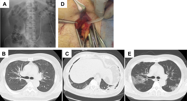
The patient’s clinical course is shown in Table 1. On day 1, the catheterography revealed catheter malposition and dysfunction caused by omental wrapping (Fig. 1A). Fluoroscopic-guided stiff wire manipulation was attempted, but the catheter malposition was not corrected. On the night of day 1, he suddenly presented with a high-grade fever of 38.8 °C, an elevated BP of 210/102 mmHg, diarrhea, and shivering, and his dyspnea progressively worsened with a SpO2 of 82%. Although we strongly suspected that his respiratory failure was caused by COVID-19 pneumonia, a second PCR test for SARS-CoV-2 showed negative results. Cardiac enzyme level (the Troponin I level was 0.029 ng/mL and the CK-MB level was 5 IU/mL) elevation and an ST increase were not observed. Computed tomography (CT) revealed consolidation and bilateral pleural effusion, interlobular septal thickening, and peribronchovascular interstitial thickening, but ground-glass opacities that are typical for COVID-19 pneumonia were not observed (Fig. 1B). Since the patient was isolated due to SARS-CoV-2 infection, it was difficult to perform echocardiography. We speculated on the basis of the acute onset, orthopnoea, elevated blood pressure, previous echocardiography findings of decreased diastolic function, and the CT findings that this dyspnea was due to heart failure with preserved ejection fraction. His respiratory failure was dramatically improved by hemodialysis, and his weight decreased from 94.0 to 86.4 kg.
Table 1.
The patient’s physiological and laboratory data
| Day | − 29 | 1 | 3 | 5 | 7 | 11 | 14 | 35 |
|---|---|---|---|---|---|---|---|---|
| BT, °C | 38.8 | 36.4 | 37.5 | 38.6 | 39 | 36.3 | ||
| SpO2, % | 93 (5 L) | 97 (1 L) | 97 | 99 | 92 (3 L) | 95 | ||
| BW, kg | 90 | 94 | 86.9 | 86.5 | 83 | 83.7 | 82.9 | 82.3 |
| WBC, /μL | 6200 | 5600 | 2000 | 8500 | 4900 | 6600 | 14,200 | 4300 |
| CRP, mg/dL | 0.04 | 0.17 | 0.75 | 0.41 | 0.41 | 2.1 | 1.25 | 0.37 |
| BNP, pg/mL | 461.7 | 1174 | 275 |
BT body temperature, BW body weight, WBC white blood cell, CRP C-reactive protein, BNP brain natriuretic peptide
Fig. 1.
A Catheterography revealed catheter tip dislocation and omental wrapping. Outflow of contrast was limited from the side holes but not from the catheter tip. Contrast defects in the catheter were observed. B Chest CT findings on day 1 revealed bilateral pleural effusion, interlobular septal thickening, and peribronchovascular interstitial thickening. C Chest CT findings on day 4 revealed mild ground-glass opacities. D The catheter tip was wrapped by the omentum. E Chest CT findings on day 8 revealed diffuse ground-glass opacities
Although his respiratory failure was improved, the high-grade fever continued and leukocytopenia developed. Thus, dexamethasone was initiated to treat suspected COVID-19 on day 2. PCR test results for SARS-CoV-2 were positive and ground-glass opacities on CT were observed on day 4 (Fig. 1C). He was diagnosed with COVID-19 pneumonia. On the same day, omental wrapping and catheter malposition were treated by mini-laparotomy (Fig. 1D). Thereafter, his volume status was well controlled. On day 8 after admission, he presented with another high fever, and his respiratory status gradually worsened, with the ground-glass opacities on CT becoming diffuse (Fig. 1E) despite dexamethasone treatment. Intravenous methylprednisolone pulse therapy, which started on day 11, improved his symptoms, and he was discharged on day 20 after admission.
Discussion
In the present case, two episodes of acute dyspnea were induced by COVID-19. The first episode of acute dyspnea was an exacerbation of heart failure caused by SARS-CoV-2 infection because of volume overload status due to a PD catheter malfunction. Therefore, ultrafiltration by hemodialysis rapidly improved his symptoms. The second episode of acute dyspnea caused by COVID-19 pneumonia occurred 1 week after the first episode. In COVID-19, some patients who initially have mild symptoms will subsequently undergo rapid clinical deterioration approximately 1 week after symptom onset [4]. We considered that the time course of this case was important for the management of dyspnea in PD patients with COVID-19.
Heart failure is a common and severe complication in COVID-19 patients. The etiology of COVID-19-induced heart failure is multifactorial, such as cytokine storm, infiltration of the heart by inflammatory cells, hemodynamic changes, increased sympathetic activity and endothelial injury [5]. The presence of congestive heart failure is a risk factor for mortality in hemodialysis patients who were hospitalized for COVID-19 [6]. Since heart failure dramatically worsens and requires urgent care, it is important to confirm heart failure associated with COVID-19 and begin individualized treatment, including diuretics or dialysis [7]. Although several cases of urgent peritoneal dialysis for acute kidney injury and heart failure induced by COVID-19 have been reported, there have been only a few case reports of heart failure caused by COVID-19 in maintenance PD patients to date [8, 9]. Jiang et al. recently reported an observational study in PD patients during the COVID-19 outbreak. Eight PD patients were diagnosed with COVID-19. Among them, four patients developed heart failure and one patient received additional HD to relieve heart failure, as in our case [10]. The lack of elevation in cardiac enzyme levels and the rapid improvement with fluid volume control alone indicate that there was no direct myocardial damage caused by COVID-19.
Since RT-PCR requires a few hours, chest CT is useful to support the clinical suspicion of COVID-19 in an emergency [11]. On the admission day in the present case, we strongly suspected COVID-19 pneumonia, but the PCR results for SARS-CoV-2 were negative twice, and CT findings also indicated heart failure, not COVID-19 pneumonia. However, the high fever and leukocytopenia indicated a viral infection. In addition, our patient presented with diarrhea on day 1. A case series of COVID-19 in hospitalized patients on chronic PD revealed that six of 11 patients had diarrhea [12]. Thus, we started dexamethasone on day 2 because we suspected that COVID-19 induced several symptoms together with heart failure, and the high-grade fever and leukocytopenia were immediately resolved. In the first occurrence of dyspnea, we speculate that cytokine storm or increased sympathetic activity by COVID-19 exacerbated the patient’s volume overload status and finally induced heart failure. The ground-glass opacities and positive PCR SARS-CoV-2 results were first detected when there were no respiratory symptoms on day 4. Usually, when heart failure is exacerbated by infection, symptoms of infection and heart failure are developed at the same time. The first acute episode of dyspnea in our patient was induced by COVID-19 without pneumonia. Nephrologists should consider exacerbation of heart failure when dyspnea occurs in COVID-19 dialysis patients.
To the best of our knowledge, there has been no report of omental wrapping concurrent with a COVID-19 infection. Usually, omental wrapping occurs in the early phase of peritoneal dialysis, and the association between omental wrapping and infections including COVID-19 is unknown [13].
Conclusion
We report a case of a PD patient with heart failure induced by COVID-19 and omental wrapping. Our case includes two occurrences of acute dyspnea due to heart failure and COVID-19 pneumonia. Nephrologists need to differentiate between heart failure and pneumonia in acute dyspnea due to COVID-19 in PD patients.
Footnotes
Publisher's Note
Springer Nature remains neutral with regard to jurisdictional claims in published maps and institutional affiliations.
References
- 1.Gundhi RT, Lynch JB, Del Rio C. Mild or moderate Covid-19. N Engl J Med. 2020;383(18):1757–1766. doi: 10.1056/NEJMcp2009249. [DOI] [PubMed] [Google Scholar]
- 2.Hilbrands LB, Duivenvoorden R, Vart P, Franssen CFM, Hemmelder MH, Jager KJ, et al. Covid-19 related mortality in kidney transplant and dialysis patients: results of the ERACODA collaboration. Nephrol Dial Transplant. 2021;35(11):1973–1983. doi: 10.1093/ndt/gfaa261. [DOI] [PMC free article] [PubMed] [Google Scholar]
- 3.Apetrii M, Enache S, Siriopol D, Burlacu A, Kanbay A, Kanbay M, et al. A brand-new cardiorenal syndrome in the covid-19 setting. Clin Kidney J. 2020;13(3):291–296. doi: 10.1093/ckj/sfaa082. [DOI] [PMC free article] [PubMed] [Google Scholar]
- 4.Huang C, Wang Y, Li X, Ren L, Zhao J, Hu Y, et al. Clinical features of patients infected with 2019 novel coronavirus in Wuhan. Lancet. 2020;395(10223):497–506. doi: 10.1016/S0140-6736(20)30183-5. [DOI] [PMC free article] [PubMed] [Google Scholar]
- 5.Adeghate EA, Eid N, Singh J. Mechanism of COVID-19-induced heart failure: a short review. Heart Fail Rev. 2021;26(2):363–369. doi: 10.1007/s10741-020-10037-x. [DOI] [PMC free article] [PubMed] [Google Scholar]
- 6.Turgutalp K, Ozturk S, Arici M, Eren N, Gorgulu N, Islam M, et al. Determinants of mortality in a large group of hemodialysis patients hospitalized for COVID-19. BMC Nephrol. 2021;22(1):29. doi: 10.1186/s12882-021-02233-0. [DOI] [PMC free article] [PubMed] [Google Scholar]
- 7.Hacquin A, Putot S, Barben J, Chague F, Zeller M, Cottin Y, et al. Bedside chest ultrasound to distinguish heart failure from pneumonia-related dyspnoea in older COVID-19 patients. ESC Heart Fail. 2020;7(6):4424–4428. doi: 10.1002/ehf2.13017. [DOI] [PMC free article] [PubMed] [Google Scholar]
- 8.Nagamato M, Yamada H, Shinozuka K, Shinozuka K, Shimoto M, Yunoki T, et al. Peritoneal dialysis for COVID-19-associated acute kidney injury. Crit Care. 2020;24(1):309. doi: 10.1186/s13054-020-03024-z. [DOI] [PMC free article] [PubMed] [Google Scholar]
- 9.Al-Hwiesh AK, Mohammed AM, Elnokeety M, Al-Hwiesh A-A, Esam S, et al. Successfully treating three patients with acute kidney injury secondary to COVID-19 by peritoneal dialysis: case report and literature review. Perit Dial Int. 2020;40(5):496–498. doi: 10.1177/0896860820953050. [DOI] [PubMed] [Google Scholar]
- 10.Jiang HJ, Tang H, Xiong F, Chen WL, Tian JB, Sun J, et al. COVID-19 in peritoneal dialysis patients. Clin J Am Soc Nephrol. 2021;15(1):1829–1831. doi: 10.2215/CJN.07200520. [DOI] [PMC free article] [PubMed] [Google Scholar]
- 11.Palmisano A, Scotti GM, Lppolito D, Morelli MJ, Vignale D, Gandola D, et al. Chest CT in the emergency department for suspected COVID-19 pneumonia. Radiol Med. 2021;126(3):498–502. doi: 10.1007/s11547-020-01302-y. [DOI] [PMC free article] [PubMed] [Google Scholar]
- 12.Sachdeva M, Uppal NN, Hirsch JS, Ng JH, Malieckal D, Fishbane S, et al. COVID-19 in hospitalized patients on chronic peritoneal dialysis: a case series. Am J Nephrol. 2020;51(8):669–674. doi: 10.1159/000510259. [DOI] [PMC free article] [PubMed] [Google Scholar]
- 13.Moncrief J, Popovich R, Simmons E, He Z. Catheter obstruction with omental wrap stimulated by dialysate exposure. Perit Dial Int. 1993;13(2):S127–S129. doi: 10.1177/089686089301302S34. [DOI] [PubMed] [Google Scholar]


