Correction to: J Exp Clin Cancer Res 39, 234 (2020)
https://doi.org/10.1186/s13046-020-01742-4
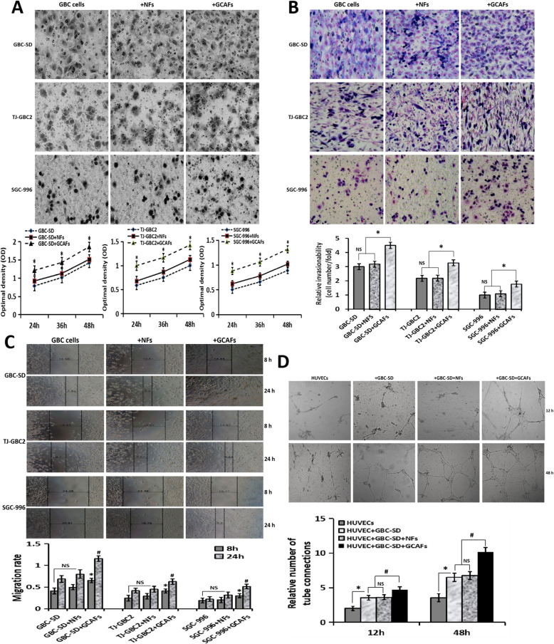
Following publication of the original article [1], the authors identified some minor errors in image-typesetting in Fig. 2, specifically:
Fig. 2a: GBC-SD +NFs panel (top row, middle) has been replaced with the correct image
Fig. 2a: TJ-GBC2 +GCAFs panel (middle row, right) has been replaced with the correct image
Fig. 2a: SGC-996 +NFs panel (bottom row, middle) has been replaced with the correct image
Fig. 2b: SGC-996 +NFs panel (bottom row, middle) has been replaced with the correct image
Fig. 2c: both TJ-GBC2 +NFs 8h and 24h panels (middle row, middle) have been replaced with the correct images
Fig. 2d: +GBC-SD+NFs panel (top row, middle-right) has been replaced with the correct image
Fig. 2.
GCAFs promote the malignant phenotypes of GBC cells/HUVECs. A, Proliferation assay. Compared with NFs, GCAFs significantly promoted the proliferation of GBC cells at 24 h, 36 h and 48 h (vs. GBC cells group and GBC cells+NFs group, all *P < 0.05). B, Transwell invasion assay (Giemsa stain, × 200). The number (relative invasion ability) of cells that invaded through the basement membrane in GBC cell+GCAFs group was significantly more than that of GBC cell group and GBC cell+NFs group (all *P < 0.05). C, Wound healing assay. The cell migration rate of GBC cell+GCAFs group was significantly stronger than that of GBC cell group and GBC cell+NFs group at 8 h and 24 h (all #P < 0.05). D, Tube formation assay. At 12 h: HUVEC group (−); HUVEC+GBC-SD and HUVEC+GBC-SD + NFs (+), (vs. HUVEC, all *P < 0.05); HUVEC+GBC-SD + GCAFs (+), (vs. HUVEC+ GBC-SD or HUVEC+GBC-SD + NFs, all #P < 0.05). At 48 h: obvious tubular formation was observed in all the four groups, the number of tube formed in HUVEC+GBC-SD + GCAFs group was significantly more than that in the other three groups (all #P < 0.01), but no statistical difference was observed between HUVEC+GBC-SD group and HUVEC+GBC-SD + NFs group (P > 0.05)
The corrected figure is given below. The correction does not have any effect on the results or conclusions of the paper. The original article has been corrected.
Footnotes
Mu-Su Pan, Hui Wang and Kamar Hasan Ansari contributed equally to this work.
Contributor Information
Wei Sun, Email: tjshdoc_sw@aliyun.com.
Yue-Zu Fan, Email: fanyuezu@hotmail.com.
Reference
- 1.Pan MS, Wang H, Ansari KH, Li XP, Sun W, Fan YZ. Gallbladder cancer-associated fibroblasts promote vasculogenic mimicry formation and tumor growth in gallbladder cancer via upregulating the expression of NOX4, a poor prognosis factor, through IL-6-JAK-STAT3 signal pathway. J Exp Clin Cancer Res. 2020;39:234. doi: 10.1186/s13046-020-01742-4. [DOI] [PMC free article] [PubMed] [Google Scholar]


