Abstract
Objectives:
The purpose of this prospective, double-blind, randomized clinical trial was to compare the clinical efficiency of nickel-titanium (NiTi) and niobium-titanium-tantalum-zirconium (TiNbTaZr) archwires during initial orthodontic alignment.
Materials and Methods:
All subjects (ages between 12 and 20 years) underwent nonextraction treatment using 0.022-inch brackets. All patients were randomized into two groups for initial alignment with 0.016-inch NiTi archwires (n = 14), or with 0.016-inch TiNbTaZr archwires (n = 14). Digital scans were taken during the course of treatment and were used to compare the improvement in Little's Irregularity Index and the changes in intercanine and intermolar widths.
Results:
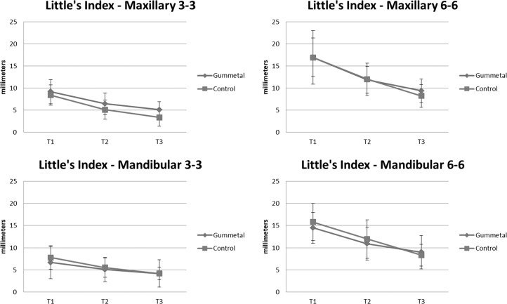
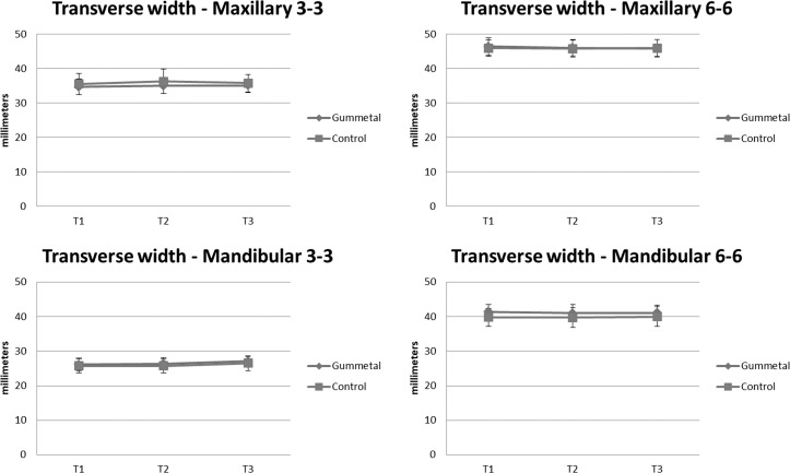
There was approximately a 27% reduction in crowding during the first month with the use of 0.016-inch TiNbTaZr (Gummetal) wire, and an additional 25% decrease in crowding was observed during the next month. There was no significant difference between the two treatment groups in the decrease in irregularity over time (P = .29). There was no significant difference between the two groups in the changes in intercanine and intermolar width (P = .80).
Conclusions:
It can be concluded that Gummetal wires and conventional NiTi wires possess a similar ability to align teeth, and Gummetal wires have additional advantages over conventional NiTi, such as formability and use in patients with nickel allergy.
Keywords: Orthodontic arch wire, Irregularity index, Randomized clinical trial
INTRODUCTION
Since nickel-titanium (NiTi) wires have a high elastic limit and high resilience with a low modulus of elasticity and low rigidity,1,2 they are commonly used as the initial wire in orthodontic treatment. However, there are certainty drawbacks to NiTi wires in certain situations. Some patients may be allergic to the nickel3,4 contained in NiTi wires, although there is some evidence that oral nickel exposure provides for nickel tolerance.5 Nickel sensitivity from orthodontic appliances may be a real, but small, problem.
NiTi wires are also preformed and are not shape formable. In a patient with a different initial arch form, the ideal size of a NiTi wire may not be available, and the use of an inappropriate size of the initial NiTi wire may cause canine and/or molar expansion or contraction. In patients for which no archwire expansion is desired, or for which it is not desired to level the curve of Spee, a shape-formable archwire is more appropriate. Additionally, NiTi does not hold bends well, meaning that detailing bends need to wait until stainless-steel or TMA archwires are placed later in treatment. While detailing bends are typically not placed in the initial archwire, these bends can be useful from the start when ideal bracket placement is not possible as a result of partial eruption, rotations, crowding, or other factors.
When the situations described above occur, appropriate alternative archwires are needed. One such type of wire with the potential to address all of the scenarios is a niobium-based titanium archwire, with the chemical formula niobium-titanium-tantalum-zirconium (TiNbTaZr), the trade name Gummetal (Rocky Mountain Morita Corporation, Tokyo, Japan).6 The wire is nickel-free, shape formable, and produces light-continuous forces. There have been several published lab studies6–10 testing the properties of niobium-based archwires and several animal studies8,11,12 testing the safety and allergenicity of titanium-niobium alloys. However, no randomized clinical trials comparing Gummetal to NiTi in human patients have been published.
In 2009, in “A Systematic Review of Clinical Trials of Aligning Archwires,”2 Riley and Bearn identified seven in vivo clinical trials investigating aligning archwires. Of the seven studies, four were chosen for quality assessment, but a meta-analysis was not possible due to a lack of homogeneity in study design. Most of the studies failed to show a significant difference between the wires being tested. Only one of the four trials showed a statistically significant difference, but the clinical significance was questionable. So that a meta-analysis can be possible in the future, one of the recommendations from the systematic review was that future clinical trials use a valid and reproducible measurement system such as Little's Irregularity Index to measure alignment.
The purpose of this study was to use digital models to compare the effectiveness of Gummetal wires to NiTi wires as the initial archwire in aligning teeth.
MATERIALS AND METHODS
The study protocol was reviewed and approved by the institutional review board of The Ohio State University.
This experiment was a randomized, double-blind, prospective clinical trial involving patients undergoing orthodontic treatment. Subjects were recruited from The Ohio State University Orthodontic Clinic, and informed consent was obtained. Participation in this study was completely optional. Inclusion criteria were (1) age between 12 and 20 years; (2) confirmation that all permanent teeth were fully erupted from first molar to first molar; (3) no history of trauma involving periodontal structures or bisphosphonate use; (4) no known allergy to nickel or to any other metal; (5) no root abnormalities visible on the patient's panoramic radiograph, such as developmentally short roots, resorption, or dilacerations; (6) no periodontal disease, as determined by radiographs, and documentation of no pocket depths greater than 3 mm; and (7) Little's Irregularity Index greater than 2 mm, and treated without any extractions.
With an alpha risk of .05, a sample size of 28 subjects (14 in each group) was required to detect a difference of 1 mm in tooth movement with a power of 0.80. A difference of 1.0 mm in tooth movement was chosen because any differences of less than 1.0 mm could not be considered clinically meaningful. Subjects were assigned into either the control group (0.016-inch round NiTi wires; GAC Sentalloy, DENTSPLY GAC, Islandia, NY; n = 14) or the experimental (0.016-inch round TiNbTaZr wires: Gummetal, Rocky Mountain Morita Corporation; n = 14) treatment group using simple randomization according to a computer-generated randomization list.
A 0.022-inch slot straight wire orthodontic appliance (Forestadent®, Pforzheim, Germany) was used on all patients. No additional active or passive adjunctive appliances, such as a quad helix, rapid palatal expander, transpalatal arch, or lower lingual holding arch, were used on these patients. Both maxillary and mandibular arches were studied. Before giving the wires to the operators, the Gummetal wires were contoured to the original archform based on pretreatment study models by the first author. For the NiTi group, because the archforms cannot be modified, the operators were given a stock of Sentalloy wires according to canine width.
During the experimental period, the archwire was re-ligated, without modifications to the wire, to the brackets or to the teeth (no interproximal reduction) using elastomeric modules (DENTSPLY GAC). Once brackets were bonded by the orthodontic resident, the same archwire (0.016-inch NiTi in half the patients, and 0.016-inch Gummetal in the other half) was re-ligated for two subsequent appointments at 4–6-week intervals.
A digital scan was obtained on each patient before bonding the brackets. Then, every 4–6 weeks for an additional two appointments, another digital scan was obtained to measure tooth movement. The scanner used in this study was manufactured by Trios, and the software used to make the measurements was 3Shape OrthoAnalyzer (TRIOS; 3Shape, Copenhagen, Denmark). After the digital scans were performed on the patients, Little's Irregularity Index was calculated for each patient. Although this index can be influenced greatly by a single tooth and may not detect vertical discrepancies, it is traditionally applied to this type of study.13,14 Transverse widths of the digital models were measured by a dentist rater who was blinded as to which wire was used in each patient. Canine measurements were made from cusp tip to cusp tip, and molar measurements were made from central fossa to central fossa. Research assistants (undergraduate students) were trained to use the OrthoAnalyzer software and were blinded to the type of archwire used in each patient. The rater performed each measurement twice in order to calculate intrarater reliability. The repeated measurements occurred at least 2 weeks apart.
Statistical Analysis
Intraclass correlation coefficients were calculated to determine intrarater reliability for Little's Irregularity Index and transverse measurements for each jaw. Initial between-group measurements in age were assessed by the randomization test, while those for occlusal class and race/ethnicity were evaluated using the Fisher exact test. Differences in gender were evaluated by the chi-square test.
Both the Little's Irregularity Index and transverse width-dependent variables were analyzed using a repeated-measure, mixed-model analysis of variance (ANOVA). The independent variables were jaw, tooth, assessment period, and wire type. Patient sex was a random variable.
RESULTS
Sample demographics and characteristics are presented in Table 1. There were no significant differences between the two groups in age, gender, race/ethnicity, and occlusal class (P > .05).
Table 1.
Demographic Distribution of Subjects
| Variable |
Treatment Groups |
P-Value |
||
| All | Gummetal | NiTi | ||
| (N = 28) |
(N = 14) |
(N = 14) |
||
| Age, y | .32 | |||
| Mean | 15.43 | 16.50 | ||
| Standard deviation | 2.31 | 3.27 | ||
| Minimum | 13 | 12 | ||
| Maximum | 19 | 20 | ||
| Gender, No. (%) | .7 | |||
| Female | 17 (61) | 9 (640) | 8 (57) | |
| Male | 11 (39) | 5 (36) | 6 (43) | |
| Race/ethnicity, No. (%) | 1.00 | |||
| Caucasian | 21 (75) | 11 (79) | 10 (36) | |
| African American | 2 (7) | 1 (7) | 1 (7) | |
| Asian | 3 (11) | 1 (7) | 2 (14) | |
| Hispanic | 2 (7) | 1 (7) | 1 (7) | |
| Occlusal class, No. (%) | .17 | |||
| Class I | 16 (57) | 6 (43) | 10 (71) | |
| Class II | 9 (32) | 5 (36) | 4 (29) | |
| Class III | 3 (11) | 3 (21) | 0 (0) | |
The range for the intrarater reliability for Little's Irregularity Index and transverse measurements resulted in 0.56–0.86 and 0.93–0.99, respectively.
A summary of the changes in Little's Irregularity Index and in transverse width is presented in Figures 1 and 2. With both types of wires, Little's Irregularity Index decreased over time in each jaw, both in the anterior region and for the full arch. Although the maxilla presented with greater initial irregularity than the mandible, the difference in the reduction in irregularity over time between the two jaws was not significant. With both wires, there was minimal change in the transverse dimensions over time.
Figure 1.
Graph of changes in Little's Irregularity Index.
Figure 2.
Graph of changes in transverse width.
For irregularity, ANOVA showed a significant effect for jaw, tooth, assessment period, tooth × period interaction, and jaw × wire type interaction. For the transverse measurements, the ANOVA revealed a significant effect for jaw, tooth, jaw × tooth interaction, jaw × wire type interaction, and tooth × wire type interaction.
DISCUSSION
This study found that both the control and the experimental wire reduced Little's Irregularity Index in patients and that there was no significant difference between the two types of wires for alignment. There was also no statistically significant difference in the transverse expansion caused by the two types of wires, despite the fact that the Gummetal wires were pre-formed to the pretreatment archform. In fact, changes in transverse width were very minimal for both treatment groups, and there was no statistically significant canine or molar expansion in either treatment group. This may be the case because the flexible wires delivered little force over a limited amount of time and were not capable of changing the arch form under these circumstances.
When looking at the irregularity of the full arch for both treatment groups, irregularity decreased by 27% during the first 4–6 months of treatment and then decreased by another 25% during the next 4–6 months of treatment; these differences were statistically significant. When looking at the irregularity of the anterior region only for both treatment groups, irregularity decreased by 31% during the first 4–6 months of treatment, and this amount was statistically significant. The decrease in anterior irregularity during the second 4–6 months of treatment was 24% but was not statistically significant, indicating a plateauing or slowing of alignment as the irregularity decreased.
These results are in line with those of previous studies; however, other studies sometimes used different measurement systems and performed measurements at different intervals, making precise comparisons difficult. Celikoglu et al.15 measured the change in irregularity in the mandibular anterior segment over 8 weeks when using 0.014-inch NiTi with both conventional and self-ligating brackets. They found that irregularity decreased by 3.84 mm in the self-ligating group and by 2.93 mm in the conventional ligation group. These numbers are in line with the current results of a decrease of 2.48 mm in the Gummetal group and 3.6 mm in the control group in terms of mandibular anterior irregularity.
Abdelrahman et al.16 measured initial irregularity of the mandibular anterior segment as well as irregularity after 8 weeks. They found that 0.014-inch superelastic NiTi reduced irregularity by 4.76 mm, 0.014-inch thermal NiTi reduced irregularity by 4.86 mm, and 0.014-inch Nitinol reduced irregularity by 4.75 mm during this time period. These subjects experienced a greater decrease in irregularity than did the current study's subjects; in this study, a decrease of 2.48 mm was found in the Gummetal group and a decrease of 3.6 mm in the NiTi group. The difference is likely due to dissimilarities in initial irregularity.
The study of Ong et al.17 did not keep the initial wire in place for a set amount of time; instead, they kept the initial wire in place until the clinician felt that rectangular wires could be ligated. They then measured the reduction in irregularity that had occurred up to this point in the mandibular anterior region. They found that during the observation time, 0.014-inch Nitinol reduced irregularity by 4.4 mm; 0.014-inch Sentalloy reduced irregularity by 4.8 mm; and 0.014-inch Damon CuNiTi reduced irregularity by 3.7 mm. These numbers are higher than the current study's numbers of 2.48 mm in the Gummetal group and 3.6 mm in the NiTi group. The difference is likely due to a longer observation period in the study of Ong et al. Ong et al. then placed rectangular wires of various cross sections into the brackets. They found that irregularity decreased by an additional 0.2 mm with 0.017 × 0.017-inch heat-activated NiTi, an additional 1.1 mm with 0.016 × 0.022-inch Bioforce wires, and an additional 0.1 mm with 0.014 × 0.025-inch Damon CuNiTi wires, showing that the majority of the reduction in irregularity occurs in the initial portion of treatment with light round wires.17
Sebastian18 did not report irregularity; this study measured mean total tooth movement at 4, 8, and 12 weeks in the mandibular anterior region. The study found that 0.016-inch NiTi led to a total of 1.552 mm tooth movement after 4 weeks, 2.327 mm after 8 weeks, and 3.103 after 12 weeks. These results were in contrast to 0.016-inch coaxial NiTi, which led to a total of 4.934 mm tooth movement after 4 weeks, 7.4 mm after 8 weeks, and 9.867 mm after 12 weeks.18 Their findings of 2.327 mm total tooth movement after 8 weeks and 3.103 mm after 12 weeks for the conventional NiTi group are comparable to the current findings of 2.48 mm in the Gummetal group and 3.6 mm in the control group.
Other studies have also measured changes in transverse width during treatment. Celikoglu et al.15 compared the effects of self-ligating vs conventional brackets on transverse expansion of the mandibular arch when using 0.014-inch NiTi wires. After 8 weeks of treatment with 0.014-inch NiTi, they found 0.87 mm expansion in mandibular intercanine width in the self-ligating group and 0.59 mm expansion in the conventional bracket group. During this same time period, Celikoglu et al. found 0.29 mm expansion in mandibular intermolar width in the self-ligating group and 0.35 mm expansion in the conventional bracket group. The results of Celikoglu et al. of less than 1 mm intercanine and intermolar expansion are similar to the results that were found in the current study.
The original a priori power analysis indicated a power of 0.95 to detect a difference of ±1 mm in tooth movement. However, a post hoc power analysis revealed that the power for the Little index was 0.503 for a difference of ±1 mm and 0.230 for a ±1-mm difference for the transverse measurements, both of which are inadequate. However, if the differences were increased to ±2 mm for the Little index and to ±2.5 mm for the transverse measurements, the post hoc power would increase to 0.976 and to 0.861 for the Little index and transverse width measures, respectively. Consequently, readers should be aware that a realistic lower threshold for detecting tooth movement as a result of wire type in this study is in the range of 2.0 to 2.5 mm.
Although the two types of wires in the current study showed no difference in performance, other factors were statistically significant. Little's Irregularity Index improved over time with both types of wires. There were differences in the initial irregularity between the mandible and the maxilla, with the maxilla presenting with greater initial irregularity; however, the difference in their change in irregularity over time was not significant. There were also differences in comparing the full arch to the anterior segment, indicating that the patients' crowding was not confined to the anterior segments. In terms of transverse widths, there were differences in the maxilla vs the mandible, and there were differences between the molar width and the canine width.
It should be noted that although the performance of the two wires was shown to be comparable, after removing the wires from the patients' mouths at the end of the study, the Gummetal wires appeared to undergo more permanent distortion than did the NiTi wires. Because Gummetal wires are shape formable when bent at a sharp enough angle, they also have the ability to experience deformation when ligated into brackets on teeth that are severely displaced. In cases of severe crowding, it is possible that Gummetal would underperform NiTi because of Gummetal's shape-formable properties. It may be suggested that omega loops could be bent and adjusted to the malocclusion with Gummetal to prevent flaring and expansion. However, at the same time, permanent distortion may result in less flexibility of the wire, and, thus, it may take more time in the case of leveling cases with severe crowding. A future study focusing exclusively on severe crowding could test this theory, and clinicians will need to learn the limits of these wires during clinical use.
This study produced similar results to previous laboratory studies. In vitro studies showed that Gummetal has low stiffness, low Young's modulus, and generates light forces,7,19 meaning that its clinical performance should be comparable to NiTi. Suzuki et al.8 used niobium-titanium archwires and NiTi archwires to produce buccal tooth movement in rats, and they found no difference in terms of tooth movement between the two types of wires. The current study confirms these results in humans.
Previous laboratory studies7 have shown Gummetal to have a torsional stiffness sufficient to produce third-order corrections. Therefore, it is suggested that Gummetal wires could be used to generate torque in patients. A future clinical study comparing the performance of rectangular Gummetal to either stainless steel or TMA could confirm this finding.
In an in vivo experiment, it is not possible to account for every possible difference among subjects because no two subjects present with the exact same malocclusion. Potential confounding factors include differences in bone density and occlusal forces among subjects. The goal of this study was to compare how these two types of wires behave in a clinical setting. To determine precise differences between the two types of wires, a laboratory study would be appropriate. Additionally, a larger sample size in an in vivo study might reveal smaller differences in performance between the two types of wires that this study was not able to detect, but any such difference would likely be of minimal clinical relevance. In addition, a study focusing exclusively on patients with severe crowding may reveal a difference in performance between the two wires. There are no published studies examining the use of Gummetal wires in extraction cases. A study of the same archwires over a longer duration of treatment would be unlikely to demonstrate differences between the treatment groups because there was a greater reduction in irregularity during the first month of treatment than during the second month; re-ligating the same wires for additional months would lead to diminishing returns.
CONCLUSIONS
Based on the present findings, it was concluded that Gummetal could be used as an alternative initial archwire to NiTi wires.
Since Gummetal wire is nickel-free, there is a possibility that this wire could be used in patients who have a nickel allergy. This wire also could be used in cases in which prevention of unwanted flaring or expansion at the initial stage of leveling is desired.
However, further study is necessary to investigate the usefulness of Gummetal wire with different wire sizes and in various situations in order to conclude that Gummetal has advantage(s) over other wires.
ACKNOWLEDGMENT
We thank the Delta Dental Foundation for providing financial support for this research through the Dental Master's Thesis Award Program.
REFERENCES
- 1.Kapila S, Sachdeva R. Mechanical properties and clinical applications of orthodontic wires. Am J Orthod Dentofacial Orthop. 1989;96:100–109. doi: 10.1016/0889-5406(89)90251-5. [DOI] [PubMed] [Google Scholar]
- 2.Riley M, Bearn D. A systematic review of clinical trials of aligning archwires. J Orthod. 2009;36:42–51. doi: 10.1179/14653120722914. [DOI] [PubMed] [Google Scholar]
- 3.Veien NK, Borchorst E, Hattel T, Laurberg G. Stomatitis or systemically-induced contact dermatitis from metal wire in orthodontic materials. Contact Dermatitis. 1994;30:210–213. doi: 10.1111/j.1600-0536.1994.tb00645.x. [DOI] [PubMed] [Google Scholar]
- 4.Eliades T, Athanasiou AE. In vivo aging of orthodontic alloys: implications for corrosion potential, nickel release, and biocompatibility. Angle Orthod. 2002;72:222–237. doi: 10.1043/0003-3219(2002)072<0222:IVAOOA>2.0.CO;2. [DOI] [PubMed] [Google Scholar]
- 5.Noble J, Ahing SI, Karaiskos NE, Wiltshire WA. Nickel allergy and orthodontics, a review and report of two cases. Br Dent J. 2008;204:297–300. doi: 10.1038/bdj.2008.198. [DOI] [PubMed] [Google Scholar]
- 6.Dalstra M, Denes G, Melsen B. Titanium-niobium, a new finishing wire alloy. Clin Orthod Res. 2000;3:6–14. doi: 10.1034/j.1600-0544.2000.030103.x. [DOI] [PubMed] [Google Scholar]
- 7.Murakami T, Iijima M, Muguruma T, Yano F, Kawashima I, Mizoguchi I. High-cycle fatigue behavior of beta-titanium orthodontic wires. Dent Mater J. 2015;34:189–195. doi: 10.4012/dmj.2014-012. [DOI] [PubMed] [Google Scholar]
- 8.Suzuki A, Kanetaka H, Shimizu Y, et al. Orthodontic buccal tooth movement by nickel-free titanium-based shape memory and superelastic alloy wire. Angle Orthod. 2006;76:1041–1046. doi: 10.2319/083105-306. [DOI] [PubMed] [Google Scholar]
- 9.Sidambe AT. Biocompatibility of Advanced Manufactured Titanium Implants-A Review. Materials (Basel) 2014;7:8168–8188. doi: 10.3390/ma7128168. [DOI] [PMC free article] [PubMed] [Google Scholar]
- 10.Niinomi M. Fatigue performance and cyto-toxicity of low rigidity titanium alloy, Ti-29Nb-13Ta4.6Zr. Biomaterials. 2003;24:2673–2683. doi: 10.1016/s0142-9612(03)00069-3. [DOI] [PubMed] [Google Scholar]
- 11.Tanaka H, Mori Y, Noro A, et al. Apatite formation and biocompatibility of a low Young's modulus Ti-Nb-Sn alloy treated with anodic oxidation and hot water. PLoS One. 2016;11:e0150081. doi: 10.1371/journal.pone.0150081. [DOI] [PMC free article] [PubMed] [Google Scholar]
- 12.Bai Y, Deng Y, Zheng Y, et al. Characterization, corrosion behavior, cellular response and in vivo bone tissue compatibility of titanium-niobium alloy with low Young's modulus. Mater Sci Eng C Mater Biol Appl. 2016;59:565–576. doi: 10.1016/j.msec.2015.10.062. [DOI] [PubMed] [Google Scholar]
- 13.Dowling AH, Burns A, Macauley D, Garvey TM, Fleming GJ. Can the intra-examiner variability of Little's Irregularity Index be improved using 3D digital models of study casts? J Dent. 2013;41:1271–1280. doi: 10.1016/j.jdent.2013.08.020. [DOI] [PubMed] [Google Scholar]
- 14.O'Rourke N, Albeedh H, Sharma P, Johal A. Effectiveness of bonded and vacuum-formed retainers: a prospective randomized controlled clinical trial. Am J Orthod Dentofacial Orthop. 2016;150:406–415. doi: 10.1016/j.ajodo.2016.03.020. [DOI] [PubMed] [Google Scholar]
- 15.Celikoglu M, Bayram M, Nur M, Kilkis D. Mandibular changes during initial alignment with SmartClip self-ligating and conventional brackets: a single-center prospective randomized controlled clinical trial. Korean J Orthod. 2015;45:78–94. doi: 10.4041/kjod.2015.45.2.89. [DOI] [PMC free article] [PubMed] [Google Scholar]
- 16.Abdelrahman RSh, Al-Nimri KS, Al Maaitah EF. A clinical comparison of three aligning archwires in terms of alignment efficiency: a prospective clinical trial. Angle Orthod. 2015;85:434–439. doi: 10.2319/041414-274.1. [DOI] [PMC free article] [PubMed] [Google Scholar]
- 17.Ong E, Ho C, Miles P. Alignment efficiency and discomfort of three orthodontic archwire sequences: a randomized clinical trial. J Orthod. 2011;38:32–39. doi: 10.1179/14653121141218. [DOI] [PubMed] [Google Scholar]
- 18.Sebastian B. Alignment efficiency of superelastic coaxial nickel-titanium vs superelastic single-stranded nickel-titanium in relieving mandibular anterior crowding: a randomized controlled prospective study. Angle Orthod. 2012;82:703–708. doi: 10.2319/072111-460.1. [DOI] [PMC free article] [PubMed] [Google Scholar]
- 19.Gordin DM, Ion R, Vasilescu C, Drob SI, Cimpean A, Gloriant T. Potentiality of the “Gum Metal” titanium-based alloy for biomedical applications. Mater Sci Eng C Mater Biol Appl. 2014;44:362–370. doi: 10.1016/j.msec.2014.08.003. [DOI] [PubMed] [Google Scholar]


