PLOS ONE logoLink to PLOS ONE
. 2021 Jul 19;16(7):e0254186. doi: 10.1371/journal.pone.0254186

Unaffected fellow eye neovascularization in patients with type 3 neovascularization: Incidence and risk factors

Jae Hyuck Kwak 1,2, Woo Kyung Park 1,2, Rae Young Kim 1,2, Mirinae Kim 1,2, Young-Gun Park 1,2, Young-Hoon Park 1,2,*
Editor: Yuhua Zhang3
PMCID: PMC8289018  PMID: 34280215

Abstract

Purpose

To evaluate the incidence and risk factors of neovascularization in unaffected fellow eyes of patients diagnosed with type 3 neovascularization in Korea.

Methods

This retrospective study included 93 unaffected fellow eyes of 93 patients diagnosed with type 3 neovascularization. For initial type 3 neovascularization diagnosis, optical coherence tomography and angiography were conducted. These baseline data were compared between patients with and without neovascularization in their fellow eyes during the follow-up period.

Results

The mean follow-up period was 66.1±31.1 months. Neovascularization developed in 49 (52.8%) fellow eyes after a mean period of 29.5±19.6 months. In the fellow eye neovascularization group, the incidence of soft drusen and reticular pseudodrusen was significantly higher than that in the non-neovascularization group (83.7% vs. 36.5%, p<0.001; 67.3% vs. 40.9%, p = 0.017, respectively), but the choroidal vascularity index (CVI) showed a significantly lower value (60.7±2.0% vs. 61.7±2.5%; p = 0.047). The presence of reticular pseudodrusen was related with the duration from baseline to development of fellow eye neovascularization (p = 0.038).

Conclusion

Neovascularization developed in 52.8% of unaffected fellow eyes. The presence of soft drusen, reticular pseudodrusen, and lower CVI values can be considered risk factors of neovascularization in unaffected fellow eyes of patients with type 3 neovascularization. The lower CVI values suggest that choroidal ischemic change may affect the development of choroidal neovascularization in these patients.

Introduction

Retinal angiomatous proliferation (RAP) is a type of neovascular age-related macular degeneration (nAMD). Yannuzzi et al [1] first proposed the term RAP in 2001 and described that in this disease, the neovascular membranes originate from the proliferation of intraretinal capillaries, that is related with a telangiectatic response. The main distinction between RAP and other types of nAMD is that RAP is characterized by the intraretinal location of the neovessels. Therefore, Freund et al proposed an alternative term “type 3 neovascularization” for this type of nAMD to emphasize the location of the neovascular complex independent of its origin [2]. The frequency of type 3 neovascularization has been reported to be 15–20% among nAMD cases in Caucasians [24], and 4.5–11.1% in Asian patients [57].

Numerous studies have reported that type 3 neovascularization has a poor prognosis and requires appropriate treatment to prevent disease progression [810]. In addition, many studies have suggested that type 3 neovascularization tends to bilateral involvement [1113]. Gross et al [12] reported that fellow eye type 3 neovascularization developed within 3 years in 100% of patients with unilateral type 3 neovascularization. In a recent study, Chang et al [13] reported an incidence of 38.3% of unaffected fellow eye neovascularization in patients with type 3 neovascularization during mean 27.8-month follow-up. These inconsistencies in the reported results could be due to variation in reporting baseline characteristics and follow-up period among studies. Nevertheless, all these studies indicate a high risk of unaffected fellow eye involvement in patients with type 3 neovascularization. Therefore, identifying the risk factors for fellow eye neovascularization is crucial for preventing bilateral poor visual outcomes in these patients.

Although epidemiologic studies of the risk factors for type 3 neovascularization are lacking, recent studies have shown that reticular pseudodrusen [13,14], larger drusen density, and thinner subfoveal choroidal thickness [1517] were related with type 3 neovascularization.

Controversies remain regarding the origin and pathophysiology of type 3 neovascularization. Yannuzzi et al [4] suggested that type 3 neovascularization may consist of two neovascular change sites, one is in the retina and the other located in the choroid. Compared to the choroid, the retina is relatively easy to analyze through various reliable image tools, so several in vivo studies have confirmed the intraretinal origin of the initial angiogenic process [1820]. However, unlike the retina, evaluating the choroid is difficult due to the lack of reliable analytic tools. Recently, the subfoveal choroidal thickness (SFCT) [2123] and choroidal vascularity index (CVI) [24] have been employed as useful optic coherence tomography (OCT)-based parameters to quantify structural and vascular alterations of the choroid.

The purpose of this study was to evaluate the incidence of unaffected fellow eye neovascular change in Korean type 3 neovascularization patients and identify various risk factors, including choroidal quantitative parameters that may affect the development of fellow eye neovascularization.

Methods

This study followed the tenets of the Declaration of Helsinki and was approved by the Institutional Review Board of Seoul St. Mary’s Hospital, The Catholic University of Korea. Informed consent was waived due to the retrospective nature of this study.

We retrospectively reviewed the medical records of all treatment-naive Korean patients who were diagnosed with unilateral type 3 neovascularization at the Department of Ophthalmology and Visual Science of Seoul St. Mary’s Hospital between January 2008 and March 2018 and were followed up for more than 2 years. On the first visit, complete ophthalmic examination was performed, including measurement of best-corrected visual acuity, slit-lamp biomicroscopy, fundus photography, spectral-domain OCT (SD-OCT), fluorescein angiography (FA), and indocyanine green angiography (ICGA) (Spectralis HRA+OCT; Heidelberg Engineering, Heidelberg, Germany). All patients in this study were diagnosed and determined stage by two retinal specialists (W.K.P. and Y.G.P.) based on the findings of funduscopy, SD-OCT, FA, and ICGA. Yannuzzi’s classification method [25] was applied in this study to determine stage. Not all fellow eyes showed any other form of neovascular maculopathy or macular edema at the initial examination. Patients with high myopia and other eye diseases, such as advanced degenerative macular disorders, epiretinal membrane, macular hole, retinal vascular obstruction, central serous chorioretinopathy, and uveitis in the fellow eye were excluded from our study.

The presence of drusen was determined using fundus photos and SD-OCT. Large soft drusen (>125 μm) and reticular pseudodrusen were confirmed by SD-OCT. The SFCT was measured as the vertical distance from the hyper reflective line corresponding to Bruch’s membrane beneath the retinal pigment epithelium under the fovea to the inner scleral border using the software-based calipers of the OCT viewer. To measure the CVI, we adapted the OCT B-scan image based technique proposed by Agrawal et al. [26] In brief, image binarization and processing were performed using Image J software (version 1.52; provided in the public domain by the National Institutes of Health, Bethesda, MD, USA; http://imagej.nih.gov/ij/). In this technique, Niblack autolocal threshold was applied, and other compensation modes were not used. All images and measurements were evaluated by two retinal specialists (J.H.K. and Y.H.P.), who were masked to fellow eye information and angiography. In case of disagreement, a third retinal specialist (M.K.) finally determined the results.

The patients were followed up every 1 to 3 months according to their therapeutic plan. Best-corrected visual acuity measurement, fundus photography, and SD-OCT were conducted as a routine process. When neovascularization was suspected due to newly detected subretinal fluid, macular edema, retinal hemorrhage, and pigment epithelial detachment, FA and ICGA were performed to confirm the presence of neovascularization.

We divided the patients into two groups based on the development of neovascularization in the fellow eye during the follow-up period. The patients’ baseline characteristics, including age, sex, follow-up duration, history of diabetes mellitus or hypertension, presence of large soft drusen and reticular pseudodrusen in the fellow eyes, and the SFCT and CVI in the fellow eyes at the initial ophthalmic examination, were compared between the two groups.

For Statistical analysis, we use IBM SPSS software (IBM Corp., Armonk, NY, USA). The independent t-test, chi-square test, and Fisher’s exact test were employed to compare the baseline data between the two groups. The duration from baseline to development of fellow eye neovascularization, with or without risk factors, was compared using independent t-test. The cumulative proportions of fellow eye neovascularization-free patients, with and without large soft drusen and reticular pseudodrusen, was compared using Kaplan-Meier analysis and Log rank test. Continuous data are expressed as mean ± standard deviation. The results were considered statistically significant at p-value <0.05.

Results

A total of 93 patients (18 men and 75 women) with a mean age of 77.2±6.0 years were included in this study. The mean follow-up duration was 66.1±31.1 months. Large soft drusen and reticular pseudodrusen were detected in more than 50% of fellow eyes (61.3% and 54.8%, respectively). The mean SFCT was 162.4±66.7 μm and the mean CVI was 61.2± 2.3%. The patients’ baseline characteristics and clinical features are presented in Table 1.

Table 1. Baseline characteristics and clinical features of patients presenting with unilateral type 3 neovascularization.

Baseline characteristics and clinical features n = 93
Age, years 77.2±6.0
Sex, male (%) 18 (19.4%)
Follow-up duration, months 66.1±31.1
Diabetes mellitus, no.(%) 16 (17.2%)
Hypertension, no.(%) 32 (34.4%)
Large soft drusen of the fellow eye, no.(%) 57 (61.3%)
Reticular pseudodrusen of the fellow eye, no. (%) 51 (54.8%)
Subfoveal choroidal thickness of the fellow eye, μm 162.4±66.7
Choroidal vascularity index of the fellow eye, % 61.2±2.3
Type 3 neovascularization stage, no. (%)
1 21(22.6%)
2 33(35.5%)
3 39(41.9%)

All continuous data are presented as means ± standard deviations.

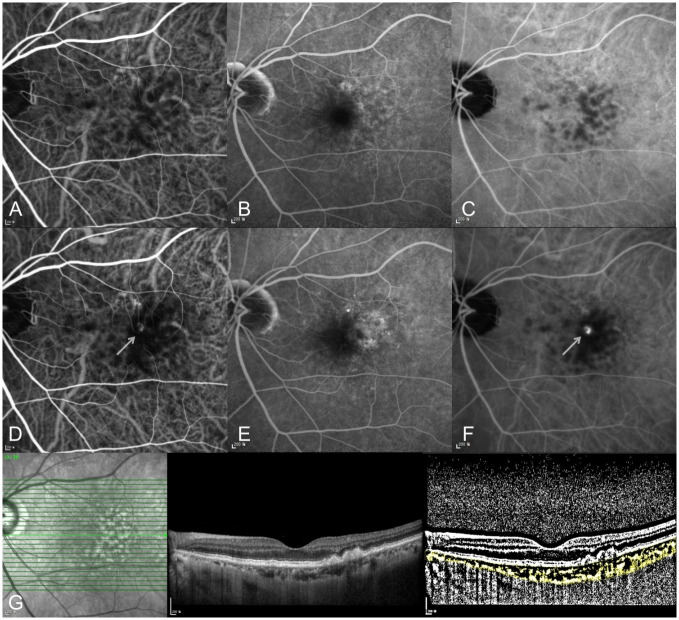
Forty-nine patients (52.8%) developed neovascularization in the unaffected fellow eye during the follow-up period. The mean duration from baseline to development of fellow eye neovascularization was 29.6±19.6 (range 5–95) months. The cumulative incidence of neovascularization in the fellow eyes was 10.8% at 1 year and 23.7% at 2 years. Among the 49 fellow eyes that developed neovascularization, 42 (85.7%) presented with type 3 neovascularization and 7 had other types of nAMD. A representative case of type 3 neovascularization development in the fellow eye is shown in Fig 1.

Fig 1. A 72-year-old woman with right eye type 3 neovascularization.

Fig 1

(A-C) At initial diagnosis, no neovascular lesion was detected in her left eye at early-to-late phase on indocyanine green and fluorescein angiography. (A) early phase indocyanine green angiography, (B) late phase and fluorescein angiography, (C) late phase indocyanine green angiography. (D-F) Ten months later, type 3 neovascularization progressed in her left eye. The arrow indicates the neovascular lesion on indocyanine green angiography. (D) early phase indocyanine green angiography, (E) late phase and fluorescein angiography, (F) late phase indocyanine green angiography. (G) OCT B-scan and binarized image to measure the choroidal vascularity index (CVI) in the left eye at initial diagnosis of type 3 neovascularization in the right eye.

The comparison data of baseline characteristics and clinical features between patients, with and those without fellow eye neovascularization to find the risk factors related with development of neovascularization in fellow eyes, is summarized in Table 2. The incidence of soft drusen and reticular pseudodrusen was significantly higher in the neovascularization group than in the non-neovascularization group (p<0.001 and p = 0.017, respectively). However, the CVI showed a significantly lower value in the neovascularization group (p = 0.047). Other risk factors, including age (p = 0.606), diabetes mellitus (p = 0.091), hypertension (p = 0.350), SFCT (p = 0.176), and baseline type 3 neovascularization stage (p = 0.980), showed no significant differences between the two groups.

Table 2. Factors associated with neovascularization in the fellow eye of patients presenting with unilateral type 3 neovascularization.

Risk factors Fellow eye NV(+) n = 49 Fellow eye NV(-) n = 44 p-value
Age, years 76.9 ± 5.5 77.6 ± 6.5 0.606
Sex, male (%) 9 (18.4%) 9 (20.5%) 0.799*
Follow-up duration, months 68.4 ± 31.4 63.5 ± 31.0 0.458
Diabetes mellitus, no.(%) 12 (24.5%) 4 (9.1%) 0.091
Hypertension, no.(%) 19 (38.8%) 13 (30.0%) 0.350*
Large soft drusen of the fellow eye, no. (%) 41 (83.7%) 16 (36.46%) <0.001*
 Reticular pseudodrusen of the fellow eye, no. (%) 33 (67.3%) 18 (40.9%) 0.017*
Subfoveal choroidal thickness of the fellow eye, μm 156.9 ± 53.4 167.9 ± 78.3 0.176
Choroidal vascular index of the fellow eye, % 60.7 ± 2.0 61.7 ± 2.5 0.047
Type 3 neovascularization stage, no. (%) 0.980*
 1 11 (22.4%) 10 (22.7%)
 2 17 (34.7%) 16 (36.4%)
 3 21 (42.9%) 18 (40.9%)

All continuous data are presented as means ± standard deviations.

p-value by independent t-test,

* p-value by chi-square test,

p-value by Fisher’s exact test,

NV, neovascularization.

Among the factors that showed significant differences between the groups, only reticular pseudodrusen was associated with the duration from baseline to development of fellow eye neovascularization (p = 0.038). In patients with reticular pseudodrusen in the neovascularization group, the mean duration was 25.6±15.5 months.

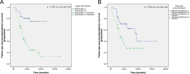
The cumulative proportions of fellow eye neovascularization-free patients, with and without large soft drusen, and reticular pseudodrusen are shown in Fig 2. The mean neovascularization-free period was 53.2±8.0 and 97.2±7.1 months in fellow eyes, with and without large soft drusen, respectively (p<0.001). In addition, this period was 52.7±6.6 months in the presence of reticular pseudodrusen and 100.1±13.2 months in the absence of reticular pseudodrusen (p = 0.004).

Fig 2. Kaplan-Meier curve analysis of fellow eye neovascularization-free patients.

Fig 2

Comparison of Kaplan-Meier curves indicating the cumulative proportion of fellow eye neovascularization-free patients among those with and these without (A) large soft drusen and (B) reticular pseudodrusen.

Discussion

In this study, we investigated the incidence and risk factors of neovascularization in unaffected fellow eyes in Korean unilateral type 3 neovascularization patients. During follow-up period, 49 of 93 patients (52.8%) developed neovascular changes in the unaffected fellow eyes, and the mean interval to binocular involvement was 29.6 months. The presence of large soft drusen and reticular pseudodrusen, as well as lower CVI values, were significantly correlated with the incidence of fellow eye neovascularization.

Neovascularization developed in more than half of unaffected fellow eyes in our study, and this result shows that type 3 neovascularization tends to involve bilaterally. This presents a similar trend as the results reported in previous studies [1113], but there is a difference in the rate of fellow eye involvement. Since few similar studies have been conducted in Asians, the answer to the inconsistency in incidence of unaffected fellow eye neovascularization is thought to be found in subsequent studies.

Regarding the risk factors for fellow eye neovascularization in type 3 neovascularization, previous studies have reported that a larger drusen density, thinner subfoveal choroidal thickness [1517], and reticular pseudodrusen [13,14] were associated with the incidence of RAP. More recently, Masaaki et al. [27] reported that hypoautofluorescence on near-infrared autofluorescence imaging may be associated with early-stage type 3 neovascularization. Furthermore, a large soft drusen (≥125 μm in diameter) is a well-known risk factor for nAMD [28]. Our results tend to support the results of these previous studies. The mean time to bilateral involvement in patients with these types of drusen was also consistent with the trends reported in previous studies. However, to the best of our knowledge, few studies have analyzed the CVI in patients with type 3 neovascularization. Moreover, type 3 neovascularization predominantly involves both eyes; thus, the CVI analysis in the unaffected fellow eye conducted in this study would be valuable.

Since some histopathological studies reported that the choroid may play a significant role in the pathogenesis of nAMD [2932], investigation of structural and vascular alterations in the choroid has been a major issue to understand nAMD. However, due to the lack of reliable imaging tools capable of sufficient evaluation in clinical conditions, OCT-based parameters, including SFCT and CVI, have been considered as a valuable alternative due to their noninvasive nature and ease of quantification. According to Agrawal et al. [33] the CVI was reduced in the affected eye of patients with nAMD despite the unchanged choroidal thickness because the CVI may be more sensitive in the analysis of choroidal vascular components. In the current study, we found that a lower CVI may be associated with the development of neovascularization in the unaffected fellow eye of type 3 neovascularization patients. Reduced choroidal vascularity could lead to choroidal ischemic change, and this condition might affect the formation of choroidal neovascularization. According to a recently published study, melanin deficiency may occurs in retinal pigment epithelium cells as a result of intensive macular stress, and there is a possibility that type 3 neovascularization may occur due to several cytokines including vascular endothelial growth factors secreted during this period [27]. Here, intensive macular stress can be induced from ischemic choroidal condition, so our study is thought to show a trend that is not different from the results of previous studies. Thus, these results show the possibility that low CVI values in the unaffected fellow eyes of patients with type 3 neovascularization may be associated with progressed choroidal ischemic change and possible subclinical disease. Based on the results of the current study, we expect that the early evaluation of the CVI in the unaffected fellow eyes of patients with type 3 neovascularization may help in establishing a proper follow-up plan.

The mean SFCT of the fellow eye in unilateral RAP was 162.4 μm in this study, and the difference between the two groups was not statistically significant. Most of the nAMD types that developed in the fellow eye were type 3 in our study (85.7%), but the mean SFCT of the fellow eyes was not significantly lower in the group with neovascularization. The reason for this result is thought to be that the SFCT can be influenced by multiple factors, including axial length, age, and intraocular pressure [34]. Therefore, it would be difficult to evaluate the SFCT as a single risk factor for predicting development of fellow eye neovascular change in unilateral type 3 neovascularization.

To the best of our knowledge, this is the first study investigating the influence of the CVI in the development of fellow eye neovascularization in patients with unilateral type 3 neovascularization. However, our study had several limitations. First, this study was conducted as a single center study. A single center study with limited number of study collective may have limited power to verify incidence of the neovascularization of the fellow eyes related type 3 neovascularization in Korean population. Second, the measurement of the SFCT and CVI was performed manually because there is no automated software. In addition, in the current CVI theory, dark and white pixels in the binarized image were assumed to present the vascular and stromal structures, which were not histologically confirmed. However, there is no definite evidence to rebut this theory and previous empirical studies strongly support this theory [33]. Third, optical coherence tomography angiography (OCTA) was not included as routine ophthalmic examination in this study. In cases without retinal fluid, early type 3 neovascularization lesion may be present on OCTA. Further studies including OCTA findings are needed and will provide more information to determine risk factors. Finally, as a result of this study, the mean difference between the CVI values in the two groups was 1%, which was not large, and the p-value also showed that the significance level was not very high (p = 0.047). However, based on the results of this study, we believe that the results of this study can be reinforced by conducting further studies with well-calculated sample sizes.

In conclusion, the incidence of neovascularization in the unaffected fellow eyes of patients with unilateral type 3 neovascularization was 52.8% during the follow-up period (≥ 2 years) and the mean interval to bilateral involvement was 29.6 months. Determined factors related with the development of neovascularization in the fellow eyes were large soft drusen and reticular pseudodrusen. Moreover, this study demonstrated that choroidal ischemic change of the fellow eye, confirmed by a lower CVI value, might affect choroidal neovascularization. At present, the CVI is measured manually, but if an automated CVI analysis tool becomes available, we expect that lower CVI values in the fellow eye could be considered as a useful indicator of risk for neovascularization, which can be easily referenced in clinical practice.

Supporting information

S1 File

(XLSX)

Data Availability

All relevant data are within the manuscript and its Supporting information files.

Funding Statement

This study was supported by the Basic Science Research Program through the National Research Foundation of Korea (NRF-2020R1F1A1074898). URL of funder website: https://www.nrf.re.kr. Initials of the authors who received each award: YHP. The funders had no role in study design, data collection and analysis, decision to publish, or preparation of the manuscript.

References

  • 1.Yannuzzi LA, Negrão S, Iida T, Carvalho C, Rodriguez-Coleman H, Slakter J, et al. RETINAL ANGIOMATOUS PROLIFERATION IN AGE-RELATED MACULAR DEGENERATION. Retina (Philadelphia, Pa). 2001;21(5):416–34. doi: 10.1097/00006982-200110000-00003 [DOI] [PubMed] [Google Scholar]
  • 2.Freund KB, Ho IV, Barbazetto IA, Koizumi H, Laud K, Ferrara D, et al. Type 3 neovascularization: the expanded spectrum of retinal angiomatous proliferation. Retina (Philadelphia, Pa). 2008;28(2):201–11. doi: 10.1097/IAE.0b013e3181669504 [DOI] [PubMed] [Google Scholar]
  • 3.Cohen SY, Creuzot-Garcher C, Darmon J, Desmettre T, Korobelnik JF, Levrat F, et al. Types of choroidal neovascularisation in newly diagnosed exudative age-related macular degeneration. British Journal of Ophthalmology. 2007;91(9):1173–6. doi: 10.1136/bjo.2007.115501 [DOI] [PMC free article] [PubMed] [Google Scholar]
  • 4.Yannuzzi LA, Freund KB, Takahashi BS. Review of Retinal Angiomatous Proliferation or Type 3 Neovascularization. Retina (Philadelphia, Pa). 2008;28(3):375–84. [DOI] [PubMed] [Google Scholar]
  • 5.Liu Y, Wen F, Huang S, Luo G, Yan H, Sun Z, et al. Subtype lesions of neovascular age-related macular degeneration in Chinese patients. Graefe’s Archive for Clinical and Experimental Ophthalmology. 2007;245(10):1441–5. doi: 10.1007/s00417-007-0575-8 [DOI] [PubMed] [Google Scholar]
  • 6.Maruko I, Iida T, Saito M, Nagayama D, Saito K. Clinical characteristics of exudative age-related macular degeneration in Japanese patients. American journal of ophthalmology. 2007;144(1):15–22. doi: 10.1016/j.ajo.2007.03.047 [DOI] [PubMed] [Google Scholar]
  • 7.Song SJ, Youm DJ, Chang Y, Yu HG. Age-related macular degeneration in a screened South Korean population: prevalence, risk factors, and subtypes. Ophthalmic Epidemiol. 2009;16(5):304–10. [PubMed] [Google Scholar]
  • 8.Bottoni F, Massacesi A, Cigada M, Viola F, Musicco I, Staurenghi G. Treatment of Retinal Angiomatous Proliferation in Age-Related Macular Degeneration: A Series of 104 Cases of Retinal Angiomatous Proliferation. Archives of Ophthalmology. 2005;123(12):1644–50. doi: 10.1001/archopht.123.12.1644 [DOI] [PubMed] [Google Scholar]
  • 9.Scott AW, Bressler SB. Retinal angiomatous proliferation or retinal anastomosis to the lesion. Eye. 2010;24(3):491–6. doi: 10.1038/eye.2009.311 [DOI] [PubMed] [Google Scholar]
  • 10.Kuhn D, Meunier I, Soubrane G, Coscas G. Imaging of Chorioretinal Anastomoses in Vascularized Retinal Pigment Epithelium Detachments. Archives of Ophthalmology. 1995;113(11):1392–8. doi: 10.1001/archopht.1995.01100110052025 [DOI] [PubMed] [Google Scholar]
  • 11.Campa C, Harding SP, Pearce IA, Beare NAV, Briggs MC, Heimann H. Incidence of neovascularization in the fellow eye of patients with unilateral retinal angiomatous proliferation. Eye. 2010;24(10):1585–9. doi: 10.1038/eye.2010.88 [DOI] [PubMed] [Google Scholar]
  • 12.Gross NE, Aizman A, Brucker A, Klancnik JMJ, Yannuzzi LA. NATURE AND RISK OF NEOVASCULARIZATION IN THE FELLOW EYE OF PATIENTS WITH UNILATERAL RETINAL ANGIOMATOUS PROLIFERATION. Retina (Philadelphia, Pa). 2005;25(6):713–8. doi: 10.1097/00006982-200509000-00005 [DOI] [PubMed] [Google Scholar]
  • 13.Chang YS, Kim JH, Yoo SJ, Lew YJ, Kim J. Fellow-eye neovascularization in unilateral retinal angiomatous proliferation in a Korean population. Acta ophthalmologica. 2016;94(1):e49–e53. doi: 10.1111/aos.12748 [DOI] [PubMed] [Google Scholar]
  • 14.Sawa M, Ueno C, Gomi F, Nishida K. INCIDENCE AND CHARACTERISTICS OF NEOVASCULARIZATION IN FELLOW EYES OF JAPANESE PATIENTS WITH UNILATERAL RETINAL ANGIOMATOUS PROLIFERATION. Retina (Philadelphia, Pa). 2014;34(4):761–7. doi: 10.1097/01.iae.0000434566.57189.37 [DOI] [PubMed] [Google Scholar]
  • 15.Kim JH, Kim JR, Kang SW, Kim SJ, Ha HS. Thinner choroid and greater drusen extent in retinal angiomatous proliferation than in typical exudative age-related macular degeneration. American journal of ophthalmology. 2013;155(4):743–9, 9.e1–2. doi: 10.1016/j.ajo.2012.11.001 [DOI] [PubMed] [Google Scholar]
  • 16.Marques JP, Laíns I, Costa M, Pires I, da Luz Cachulo M, Figueira J, et al. RETINAL ANGIOMATOUS PROLIFERATION: A Quantitative Analysis of the Fundoscopic Features of the Fellow Eye. Retina (Philadelphia, Pa). 2015;35(10):1985–91. doi: 10.1097/IAE.0000000000000619 [DOI] [PubMed] [Google Scholar]
  • 17.Yamazaki T, Koizumi H, Yamagishi T, Kinoshita S. Subfoveal choroidal thickness in retinal angiomatous proliferation. Retina (Philadelphia, Pa). 2014;34(7):1316–22. doi: 10.1097/IAE.0000000000000086 [DOI] [PubMed] [Google Scholar]
  • 18.Querques G, Souied EH, Freund KB. How Has High-Resolution Multimodal Imaging Refined Our Understanding of the Vasogenic Process in Type 3 Neovascularization? Retina (Philadelphia, Pa). 2015;35(4):603–13. doi: 10.1097/IAE.0000000000000487 [DOI] [PubMed] [Google Scholar]
  • 19.Tan AC, Dansingani KK, Yannuzzi LA, Sarraf D, Freund KB. TYPE 3 NEOVASCULARIZATION IMAGED WITH CROSS-SECTIONAL AND EN FACE OPTICAL COHERENCE TOMOGRAPHY ANGIOGRAPHY. Retina (Philadelphia, Pa). 2017;37(2):234–46. doi: 10.1097/IAE.0000000000001343 [DOI] [PubMed] [Google Scholar]
  • 20.Sacconi R, Sarraf D, Garrity S, Freund KB, Yannuzzi LA, Gal-Or O, et al. Nascent Type 3 Neovascularization in Age-Related Macular Degeneration. Ophthalmol Retina. 2018;2(11):1097–106. doi: 10.1016/j.oret.2018.04.016 [DOI] [PubMed] [Google Scholar]
  • 21.Jonas JB, Forster TM, Steinmetz P, Schlichtenbrede FC, Harder BC. CHOROIDAL THICKNESS IN AGE-RELATED MACULAR DEGENERATION. Retina (Philadelphia, Pa). 2014;34(6):1149–55. doi: 10.1097/IAE.0000000000000035 [DOI] [PubMed] [Google Scholar]
  • 22.Imamura Y, Fujiwara T, Margolis R, Spaide RF. Enhanced depth imaging optical coherence tomography of the choroid in central serous chorioretinopathy. Retina (Philadelphia, Pa). 2009;29(10):1469–73. doi: 10.1097/IAE.0b013e3181be0a83 [DOI] [PubMed] [Google Scholar]
  • 23.Koizumi H, Yamagishi T, Yamazaki T, Kawasaki R, Kinoshita S. Subfoveal choroidal thickness in typical age-related macular degeneration and polypoidal choroidal vasculopathy. Graefe’s Archive for Clinical and Experimental Ophthalmology. 2011;249(8):1123–8. doi: 10.1007/s00417-011-1620-1 [DOI] [PubMed] [Google Scholar]
  • 24.Agrawal R, Ding J, Sen P, Rousselot A, Chan A, Nivison-Smith L, et al. Exploring choroidal angioarchitecture in health and disease using choroidal vascularity index. Progress in Retinal and Eye Research. 2020;77:100829. doi: 10.1016/j.preteyeres.2020.100829 [DOI] [PubMed] [Google Scholar]
  • 25.Yannuzzi LA, Negrão S, Iida T, Carvalho C, Rodriguez-Coleman H, Slakter J, et al. Retinal angiomatous proliferation in age-related macular degeneration. Retina (Philadelphia, Pa). 2001;21(5):416–34. doi: 10.1097/00006982-200110000-00003 [DOI] [PubMed] [Google Scholar]
  • 26.Agrawal R, Chhablani J, Tan KA, Shah S, Sarvaiya C, Banker A. CHOROIDAL VASCULARITY INDEX IN CENTRAL SEROUS CHORIORETINOPATHY. Retina (Philadelphia, Pa). 2016;36(9):1646–51. doi: 10.1097/IAE.0000000000001040 [DOI] [PubMed] [Google Scholar]
  • 27.Saito M, Itagaki K, Sekiryu T. Fundus autofluorescence of retinal angiomatous proliferation. PLOS ONE. 2020;15(12):e0243458. doi: 10.1371/journal.pone.0243458 [DOI] [PMC free article] [PubMed] [Google Scholar]
  • 28.Mitchell P, Liew G, Gopinath B, Wong TY. Age-related macular degeneration. Lancet. 2018;392(10153):1147–59. doi: 10.1016/S0140-6736(18)31550-2 [DOI] [PubMed] [Google Scholar]
  • 29.Ramrattan RS, van der Schaft TL, Mooy CM, de Bruijn WC, Mulder PG, de Jong PT. Morphometric analysis of Bruch’s membrane, the choriocapillaris, and the choroid in aging. Invest Ophthalmol Vis Sci. 1994;35(6):2857–64. [PubMed] [Google Scholar]
  • 30.Spraul CW, Lang GE, Grossniklaus HE. Morphometric analysis of the choroid, Bruch’s membrane, and retinal pigment epithelium in eyes with age-related macular degeneration. Investigative Ophthalmology & Visual Science. 1996;37(13):2724–35. [PubMed] [Google Scholar]
  • 31.McLeod DS, Taomoto M, Otsuji T, Green WR, Sunness JS, Lutty GA. Quantifying Changes in RPE and Choroidal Vasculature in Eyes with Age-Related Macular Degeneration. Investigative Ophthalmology & Visual Science. 2002;43(6):1986–93. [PubMed] [Google Scholar]
  • 32.Mullins RF, Johnson MN, Faidley EA, Skeie JM, Huang J. Choriocapillaris Vascular Dropout Related to Density of Drusen in Human Eyes with Early Age-Related Macular Degeneration. Investigative Ophthalmology & Visual Science. 2011;52(3):1606–12. doi: 10.1167/iovs.10-6476 [DOI] [PMC free article] [PubMed] [Google Scholar]
  • 33.Agrawal R, Ding J, Sen P, Rousselot A, Chan A, Nivison-Smith L, et al. Exploring choroidal angioarchitecture in health and disease using choroidal vascularity index. Prog Retin Eye Res. 2020;77:100829. doi: 10.1016/j.preteyeres.2020.100829 [DOI] [PubMed] [Google Scholar]
  • 34.Agrawal R, Gupta P, Tan K-A, Cheung CMG, Wong T-Y, Cheng C-Y. Choroidal vascularity index as a measure of vascular status of the choroid: Measurements in healthy eyes from a population-based study. Sci Rep. 2016;6:21090. doi: 10.1038/srep21090 [DOI] [PMC free article] [PubMed] [Google Scholar]

Decision Letter 0

Yuhua Zhang

30 Mar 2021

PONE-D-21-05303

Unaffected fellow eye neovascularization in patients with retinal angiomatous proliferation: incidence and risk factors

PLOS ONE

Dear Dr. Park,

Thank you for submitting your manuscript to PLOS ONE. After careful consideration, we feel that it has merit but does not fully meet PLOS ONE’s publication criteria as it currently stands. Therefore, we invite you to submit a revised version of the manuscript that addresses the points raised during the review process.

The academic editor suggests using 'Type 3 MNV' to replace the term 'retinal angiomatous proliferation or RAP.' Please not the recent publication on consensus nomenclature for reporting neovascular AMD.

Spaide RF, Jaffe GJ, Sarraf D, Freund KB, Sadda SR, Staurenghi G, Waheed NK, Chakravarthy U, Rosenfeld PJ, Holz FG, Souied EH, Cohen SY, Querques G, Ohno-Matsui K, Boyer D, Gaudric A, Blodi B, Baumal CR, Li X, Coscas GJ, Brucker A, Singerman L, Luthert P, Schmitz-Valckenberg S, Schmidt-Erfurth U, Grossniklaus HE, Wilson DJ, Guymer R, Yannuzzi LA, Chew EY, Csaky K, Mones JM, Pauleikhoff D, Tadayoni R, Fujimoto J (2020) Consensus Nomenclature for Reporting Neovascular Age-Related Macular Degeneration Data: Consensus on Neovascular Age-Related Macular Degeneration Nomenclature Study Group. Ophthalmology 127 (5):616-636. doi:10.1016/j.ophtha.2019.11.004

Please submit your revised manuscript by May 31,2021. If you will need more time than this to complete your revisions, please reply to this message or contact the journal office at plosone@plos.org. When you're ready to submit your revision, log on to https://www.editorialmanager.com/pone/ and select the 'Submissions Needing Revision' folder to locate your manuscript file.

Please include the following items when submitting your revised manuscript:

  • A rebuttal letter that responds to each point raised by the academic editor and reviewer(s). You should upload this letter as a separate file labeled 'Response to Reviewers'.

  • A marked-up copy of your manuscript that highlights changes made to the original version. You should upload this as a separate file labeled 'Revised Manuscript with Track Changes'.

  • An unmarked version of your revised paper without tracked changes. You should upload this as a separate file labeled 'Manuscript'.

If you would like to make changes to your financial disclosure, please include your updated statement in your cover letter. Guidelines for resubmitting your figure files are available below the reviewer comments at the end of this letter.

If applicable, we recommend that you deposit your laboratory protocols in protocols.io to enhance the reproducibility of your results. Protocols.io assigns your protocol its own identifier (DOI) so that it can be cited independently in the future. For instructions see: http://journals.plos.org/plosone/s/submission-guidelines#loc-laboratory-protocols. Additionally, PLOS ONE offers an option for publishing peer-reviewed Lab Protocol articles, which describe protocols hosted on protocols.io. Read more information on sharing protocols at https://plos.org/protocols?utm_medium=editorial-email&utm_source=authorletters&utm_campaign=protocols.

We look forward to receiving your revised manuscript.

Kind regards,

Yuhua Zhang

Academic Editor

PLOS ONE

Journal Requirements:

When submitting your revision, we need you to address these additional requirements.

1. Please ensure that your manuscript meets PLOS ONE's style requirements, including those for file naming. The PLOS ONE style templates can be found at

https://journals.plos.org/plosone/s/file?id=wjVg/PLOSOne_formatting_sample_main_body.pdf and

https://journals.plos.org/plosone/s/file?id=ba62/PLOSOne_formatting_sample_title_authors_affiliations.pdf

2. If you are reporting a retrospective study of medical records or archived samples, please ensure that you have discussed whether all data were fully anonymized before you accessed them. If patients provided informed written consent to have data from their medical records used in research, please include this information.

3. We note that you have indicated that data from this study are available upon request. PLOS only allows data to be available upon request if there are legal or ethical restrictions on sharing data publicly. For information on unacceptable data access restrictions, please see http://journals.plos.org/plosone/s/data-availability#loc-unacceptable-data-access-restrictions.

In your revised cover letter, please address the following prompts:

a) If there are ethical or legal restrictions on sharing a de-identified data set, please explain them in detail (e.g., data contain potentially identifying or sensitive patient information) and who has imposed them (e.g., an ethics committee). Please also provide contact information for a data access committee, ethics committee, or other institutional body to which data requests may be sent.

b) If there are no restrictions, please upload the minimal anonymized data set necessary to replicate your study findings as either Supporting Information files or to a stable, public repository and provide us with the relevant URLs, DOIs, or accession numbers. Please see http://www.bmj.com/content/340/bmj.c181.long for guidelines on how to de-identify and prepare clinical data for publication. For a list of acceptable repositories, please see http://journals.plos.org/plosone/s/data-availability#loc-recommended-repositories.

We will update your Data Availability statement on your behalf to reflect the information you provide.

[Note: HTML markup is below. Please do not edit.]

Reviewers' comments:

Reviewer's Responses to Questions

Comments to the Author

1. Is the manuscript technically sound, and do the data support the conclusions?

The manuscript must describe a technically sound piece of scientific research with data that supports the conclusions. Experiments must have been conducted rigorously, with appropriate controls, replication, and sample sizes. The conclusions must be drawn appropriately based on the data presented.

Reviewer #1: Partly

Reviewer #2: No

**********

2. Has the statistical analysis been performed appropriately and rigorously?

Reviewer #1: Yes

Reviewer #2: Yes

**********

3. Have the authors made all data underlying the findings in their manuscript fully available?

The PLOS Data policy requires authors to make all data underlying the findings described in their manuscript fully available without restriction, with rare exception (please refer to the Data Availability Statement in the manuscript PDF file). The data should be provided as part of the manuscript or its supporting information, or deposited to a public repository. For example, in addition to summary statistics, the data points behind means, medians and variance measures should be available. If there are restrictions on publicly sharing data—e.g. participant privacy or use of data from a third party—those must be specified.

Reviewer #1: No

Reviewer #2: No

**********

4. Is the manuscript presented in an intelligible fashion and written in standard English?

PLOS ONE does not copyedit accepted manuscripts, so the language in submitted articles must be clear, correct, and unambiguous. Any typographical or grammatical errors should be corrected at revision, so please note any specific errors here.

Reviewer #1: Yes

Reviewer #2: Yes

**********

5. Review Comments to the Author

Please use the space provided to explain your answers to the questions above. You may also include additional comments for the author, including concerns about dual publication, research ethics, or publication ethics. (Please upload your review as an attachment if it exceeds 20,000 characters)

Reviewer #1: I think that the findings could be of interest to the readers and this is one of the largest series reporting type 3 MNVs.

However, I think that there are some issues that need to be solved:

1° - I have some concerns about the CVI analysis.

- Which type of analysis was performed? Based on one b-scan? Which one? Or a 3D CVI analysis? Please specify.

- Which kind of threshold was applied? An auto-local threshold or not? Was there a compensation mode used in the algorithm?

- I have some concerns about the results. Only a value of 2 for the SD in the results of CVI is a little strange. The % expressed by CVI, is useful very variable between subjects, with higher levels of SD (as reported by the results of several papers). Do you have a technical explanation of that?

2° - The main result of the paper is based on the difference in CVI. However, in %, the mean difference between the two groups was only 1% (60.7% vs 61.7%). This is a very little difference and I am not sure that it is clinically relevant and repeatable. How was calculated the sample size? Which margin of error? Which power? Which alpha? Because it is very important to conduct an analysis based on the sample size. In fact, if the sample is too large for the expected results, some difference could be statistically significant only because the sample is too large. This is a very important point because the sample here is large, the difference is small (60.7% vs 61.7%), and the p-value was at the limit of significance (0.047).

3° - I think that is better to refer to type 3 MNV as “Type 3 MNV” (Freund KB, Ho IV, Barbazetto IA, Koizumi H, Laud K, Ferrara D, Matsumoto Y, Sorenson JA, Yannuzzi L. Type 3 neovascularization: the expanded spectrum of retinal angiomatous proliferation. Retina. 2008 Feb;28(2):201-11.) instead of RAP. Indeed, the term “RAP” was coined based on a retinal origin of type 3 MNV. On the other hand, Type 3 MNV is independent of the origin of the lesion (retinal or choroidal origin).

4°- Several in vivo studies, suggested the retinal origin of the type 3 MNV (i.e. Sacconi R, Sarraf D, Garrity S, Freund KB, Yannuzzi LA, Gal-Or O, Souied E, Sieiro A, Corbelli E, Carnevali A, Querques L, Bandello F, Querques G. Nascent Type 3 Neovascularization in Age-Related Macular Degeneration. Ophthalmol Retina. 2018 Nov;2(11):1097-1106.). I think that the authors should cite it.

Reviewer #2: To Authors,

The concept of this paper is slightly interesting.

The study may be the first study for investigating the influence of the CVI in the development of fellow eye neovascularization in patients with unilateral RAP. However, the significant difference to be just “p=0.047”, which should be too low for making people to have common sense.

At line 193-199, the authors mentioned,

“In the current study, we revealed that a lower CVI was associated with the development of neovascularization in the unaffected fellow eye of RAP patients. Reduced choroidal vascularity could lead to choroidal ischemic change, and this condition might affect the formation of choroidal neovascularization. Moreover, this result may suggest that lower CVI values in the unaffected fellow eyes of patients with RAP indicate progressed choroidal ischemic change and possible subclinical disease. Based on the results of the current study, we recommend early evaluation of the CVI in the unaffected fellow eyes of patients with RAP and establishing a proper follow-up plan.”

However, this study showed the significant difference to be just “p=0.047”, which should be too low for making people to have common sense. The authors should rewrite the paragraph.

In the Discussion section, the authors did make no mention of the study of PLoS ONE 15(12): e0243458. https://doi.org/10.1371/journal.pone.0243458, which is the latest publication on the topic about onset of RAP lesions. This paper must be presented and discussed in the Discussion section of the paper by the authors

**********

6. PLOS authors have the option to publish the peer review history of their article (what does this mean?). If published, this will include your full peer review and any attached files.

If you choose “no”, your identity will remain anonymous but your review may still be made public.

Do you want your identity to be public for this peer review? For information about this choice, including consent withdrawal, please see our Privacy Policy.

Reviewer #1: No

Reviewer #2: No

[NOTE: If reviewer comments were submitted as an attachment file, they will be attached to this email and accessible via the submission site. Please log into your account, locate the manuscript record, and check for the action link "View Attachments". If this link does not appear, there are no attachment files.]

While revising your submission, please upload your figure files to the Preflight Analysis and Conversion Engine (PACE) digital diagnostic tool, https://pacev2.apexcovantage.com/. PACE helps ensure that figures meet PLOS requirements. To use PACE, you must first register as a user. Registration is free. Then, login and navigate to the UPLOAD tab, where you will find detailed instructions on how to use the tool. If you encounter any issues or have any questions when using PACE, please email PLOS at figures@plos.org. Please note that Supporting Information files do not need this step.

PLoS One. 2021 Jul 19;16(7):e0254186. doi: 10.1371/journal.pone.0254186.r002

Author response to Decision Letter 0


11 May 2021

Reviewer #1: I think that the findings could be of interest to the readers and this is one of the largest series reporting type 3 MNVs.

However, I think that there are some issues that need to be solved:

1° - I have some concerns about the CVI analysis.

- Which type of analysis was performed? Based on one b-scan? Which one? Or a 3D CVI analysis? Please specify.

Response) An OCT B-scan image based and Agrawal’s method was adapted. We add this in the methods section, page 4, line 99.

- Which kind of threshold was applied? An auto-local threshold or not? Was there a compensation mode used in the algorithm?

Response) Niblack auto-local threshold was applied, and other compensation modes were not used. We add this in the methods section, page 5, line 101-102

- I have some concerns about the results. Only a value of 2 for the SD in the results of CVI is a little strange. The % expressed by CVI, is useful very variable between subjects, with higher levels of SD (as reported by the results of several papers). Do you have a technical explanation of that?

Response) Thank you for pointing this out. In this study, we measured CVI according to the method of Agrawal et al, as mentioned in the methods section, and there were no technical differences. And referring to CVI in existing literatures (Rupesh Agrawal, Jianbin Ding, Parveen Sen, Andres Rousselot, Amy Chan, Lisa Nivison-Smith, Xin Wei, Sarakshi Mahajan, Ramasamy Kim, Chitaranjan Mishra, Manisha Agarwal, Min Hee Suh, Saurabh Luthra, Marion R. Munk, Carol Y. Cheung, Vishali Gupta, Exploring choroidal angioarchitecture in health and disease using choroidal vascularity index, Progress in Retinal and Eye Research, Volume 77, 2020,100829), it has been reported that the SD value of CVI varies from less than 1 to more than 10 in various diseases.

2° - The main result of the paper is based on the difference in CVI. However, in %, the mean difference between the two groups was only 1% (60.7% vs 61.7%). This is a very little difference and I am not sure that it is clinically relevant and repeatable. How was calculated the sample size? Which margin of error? Which power? Which alpha? Because it is very important to conduct an analysis based on the sample size. In fact, if the sample is too large for the expected results, some difference could be statistically significant only because the sample is too large. This is a very important point because the sample here is large, the difference is small (60.7% vs 61.7%), and the p-value was at the limit of significance (0.047).

Response) Thank you for pointing this out. Since this study was conducted in a retrospectively medical chart review method, the sample size could not be calculated at the research design stage. In fact, there is a lot of debate about whether it makes sense for post-hoc power analysis, and it is said that If a sample is selected, outcomes are no longer random and power analysis becomes meaningless for this particular study sample. (Zhang Y, Hedo R, Rivera A, et. al Post hoc power analysis: is it an informative and meaningful analysis? General Psychiatry 2019;32:e100069. doi: 10.1136/gpsych-2019-100069). Therefore, we have not added any information about post-hoc power analysis to the manuscript. However, we added to the discussion section that one of the limitations of this study was that the mean difference between the CVI results is small and the p-value was close to the upper limit of the statistical significance. (Revised manuscript with tract changes, Discussion, page 10, line 231-235)

3° - I think that is better to refer to type 3 MNV as “Type 3 MNV” (Freund KB, Ho IV, Barbazetto IA, Koizumi H, Laud K, Ferrara D, Matsumoto Y, Sorenson JA, Yannuzzi L. Type 3 neovascularization: the expanded spectrum of retinal angiomatous proliferation. Retina. 2008 Feb;28(2):201-11.) instead of RAP. Indeed, the term “RAP” was coined based on a retinal origin of type 3 MNV. On the other hand, Type 3 MNV is independent of the origin of the lesion (retinal or choroidal origin).

Response) We very much appreciate this comment and totally agree with you. We have revised entire manuscript and added related information to the introduction section, page 3, line 49-52 in revised manuscript with tract changes.

4°- Several in vivo studies, suggested the retinal origin of the type 3 MNV (i.e. Sacconi R, Sarraf D, Garrity S, Freund KB, Yannuzzi LA, Gal-Or O, Souied E, Sieiro A, Corbelli E, Carnevali A, Querques L, Bandello F, Querques G. Nascent Type 3 Neovascularization in Age-Related Macular Degeneration. Ophthalmol Retina. 2018 Nov;2(11):1097-1106.).

I think that the authors should cite it

Response) Thank you for the comments. We add this in the introduction section, page 3, line 69-71 in revised manuscript with tract changes.

Reviewer #2: To Authors,

The concept of this paper is slightly interesting.

The study may be the first study for investigating the influence of the CVI in the development of fellow eye neovascularization in patients with unilateral RAP. However, the significant difference to be just “p=0.047”, which should be too low for making people to have common sense.

Response) We very much appreciate this comment and totally agree with you. We added to the discussion section that one of the limitations of this study was that the p-value was close to the upper limit of the statistical significance. (Revised manuscript with tract changes, Discussion, page 10, line 231-235)

At line 193-199, the authors mentioned,

“In the current study, we revealed that a lower CVI was associated with the development of neovascularization in the unaffected fellow eye of RAP patients. Reduced choroidal vascularity could lead to choroidal ischemic change, and this condition might affect the formation of choroidal neovascularization. Moreover, this result may suggest that lower CVI values in the unaffected fellow eyes of patients with RAP indicate progressed choroidal ischemic change and possible subclinical disease. Based on the results of the current study, we recommend early evaluation of the CVI in the unaffected fellow eyes of patients with RAP and establishing a proper follow-up plan.”

However, this study showed the significant difference to be just “p=0.047”, which should be too low for making people to have common sense. The authors should rewrite the paragraph.

Response) Thank you for pointing this out. As per your comments, we have revised the paragraph. (Revised manuscript with tract changes, Discussion, page 9, line 204-212)

Attachment

Submitted filename: Response to Reviewers.docx

Decision Letter 1

Yuhua Zhang

14 Jun 2021

PONE-D-21-05303R1

Unaffected fellow eye neovascularization in patients with type 3 neovascularization: incidence and risk factors

PLOS ONE

Dear Dr. Park,

Thank you for submitting your manuscript to PLOS ONE. After careful consideration, we feel that it has merit but does not fully meet PLOS ONE’s publication criteria as it currently stands. Therefore, we invite you to submit a revised version of the manuscript that addresses the points raised during the review process.

I hope you can address the concerns raised by Reviewer 2, though you do not have to agree with the comments. 

Please submit your revised manuscript by 07/10/2021. If you will need more time than this to complete your revisions, please reply to this message or contact the journal office at plosone@plos.org. When you're ready to submit your revision, log on to https://www.editorialmanager.com/pone/ and select the 'Submissions Needing Revision' folder to locate your manuscript file.

Please include the following items when submitting your revised manuscript:

  • A rebuttal letter that responds to each point raised by the academic editor and reviewer(s). You should upload this letter as a separate file labeled 'Response to Reviewers'.

  • A marked-up copy of your manuscript that highlights changes made to the original version. You should upload this as a separate file labeled 'Revised Manuscript with Track Changes'.

  • An unmarked version of your revised paper without tracked changes. You should upload this as a separate file labeled 'Manuscript'.

If you would like to make changes to your financial disclosure, please include your updated statement in your cover letter. Guidelines for resubmitting your figure files are available below the reviewer comments at the end of this letter.

If applicable, we recommend that you deposit your laboratory protocols in protocols.io to enhance the reproducibility of your results. Protocols.io assigns your protocol its own identifier (DOI) so that it can be cited independently in the future. For instructions see: http://journals.plos.org/plosone/s/submission-guidelines#loc-laboratory-protocols. Additionally, PLOS ONE offers an option for publishing peer-reviewed Lab Protocol articles, which describe protocols hosted on protocols.io. Read more information on sharing protocols at https://plos.org/protocols?utm_medium=editorial-email&utm_source=authorletters&utm_campaign=protocols.

We look forward to receiving your revised manuscript.

Kind regards,

Yuhua Zhang

Academic Editor

PLOS ONE

Additional Editor Comments (if provided):

I hope you can address the comments from reviewer 2.

[Note: HTML markup is below. Please do not edit.]

Reviewers' comments:

Reviewer's Responses to Questions

Comments to the Author

1. If the authors have adequately addressed your comments raised in a previous round of review and you feel that this manuscript is now acceptable for publication, you may indicate that here to bypass the “Comments to the Author” section, enter your conflict of interest statement in the “Confidential to Editor” section, and submit your "Accept" recommendation.

Reviewer #1: All comments have been addressed

Reviewer #2: (No Response)

**********

2. Is the manuscript technically sound, and do the data support the conclusions?

The manuscript must describe a technically sound piece of scientific research with data that supports the conclusions. Experiments must have been conducted rigorously, with appropriate controls, replication, and sample sizes. The conclusions must be drawn appropriately based on the data presented.

Reviewer #1: Yes

Reviewer #2: Partly

**********

3. Has the statistical analysis been performed appropriately and rigorously?

Reviewer #1: Yes

Reviewer #2: Yes

**********

4. Have the authors made all data underlying the findings in their manuscript fully available?

The PLOS Data policy requires authors to make all data underlying the findings described in their manuscript fully available without restriction, with rare exception (please refer to the Data Availability Statement in the manuscript PDF file). The data should be provided as part of the manuscript or its supporting information, or deposited to a public repository. For example, in addition to summary statistics, the data points behind means, medians and variance measures should be available. If there are restrictions on publicly sharing data—e.g. participant privacy or use of data from a third party—those must be specified.

Reviewer #1: Yes

Reviewer #2: No

**********

5. Is the manuscript presented in an intelligible fashion and written in standard English?

PLOS ONE does not copyedit accepted manuscripts, so the language in submitted articles must be clear, correct, and unambiguous. Any typographical or grammatical errors should be corrected at revision, so please note any specific errors here.

Reviewer #1: Yes

Reviewer #2: Yes

**********

6. Review Comments to the Author

Please use the space provided to explain your answers to the questions above. You may also include additional comments for the author, including concerns about dual publication, research ethics, or publication ethics. (Please upload your review as an attachment if it exceeds 20,000 characters)

Reviewer #1: The authors should be congratulated because they address all the reviewers' comments. I have no more issues.

Reviewer #2: I could not find your comments about the following question.

In the Discussion section, the authors did make no mention of the study of PLoS ONE 15(12): e0243458. https://doi.org/10.1371/journal.pone.0243458, which is the latest publication on the topic about onset of RAP lesions (type 3 MNV). This paper must be presented and discussed in the Discussion section of the paper by the authors.

**********

7. PLOS authors have the option to publish the peer review history of their article (what does this mean?). If published, this will include your full peer review and any attached files.

If you choose “no”, your identity will remain anonymous but your review may still be made public.

Do you want your identity to be public for this peer review? For information about this choice, including consent withdrawal, please see our Privacy Policy.

Reviewer #1: No

Reviewer #2: No

[NOTE: If reviewer comments were submitted as an attachment file, they will be attached to this email and accessible via the submission site. Please log into your account, locate the manuscript record, and check for the action link "View Attachments". If this link does not appear, there are no attachment files.]

While revising your submission, please upload your figure files to the Preflight Analysis and Conversion Engine (PACE) digital diagnostic tool, https://pacev2.apexcovantage.com/. PACE helps ensure that figures meet PLOS requirements. To use PACE, you must first register as a user. Registration is free. Then, login and navigate to the UPLOAD tab, where you will find detailed instructions on how to use the tool. If you encounter any issues or have any questions when using PACE, please email PLOS at figures@plos.org. Please note that Supporting Information files do not need this step.

PLoS One. 2021 Jul 19;16(7):e0254186. doi: 10.1371/journal.pone.0254186.r004

Author response to Decision Letter 1


16 Jun 2021

Reviewer #1: The authors should be congratulated because they address all the reviewers' comments. I have no more issues.

Response) We very much appreciate your comment.

Reviewer #2: I could not find your comments about the following question.

In the Discussion section, the authors did make no mention of the study of PLoS ONE 15(12): e0243458. https://doi.org/10.1371/journal.pone.0243458, which is the latest publication on the topic about onset of RAP lesions (type 3 MNV). This paper must be presented and discussed in the Discussion section of the paper by the authors.

Response) Thank you for pointing this out. The study mentioned above suggested that melanin deficiency in RPE cells is a result of intensive macular stress and that neovascularization may occur in association with several cytokines secreted during this period. Here, intensive macular stress can be induced from ischemic choroidal condition, so our study is thought to show a trend that is not different from the results of the above study. We add this in the discussion section, page 9, line 191-193 and 208-212 in revised manuscript with tract changes.

Attachment

Submitted filename: Response to Reviewers2.docx

Decision Letter 2

Yuhua Zhang

22 Jun 2021

Unaffected fellow eye neovascularization in patients with type 3 neovascularization: incidence and risk factors

PONE-D-21-05303R2

Dear Dr. Park,

We’re pleased to inform you that your manuscript has been judged scientifically suitable for publication and will be formally accepted for publication once it meets all outstanding technical requirements.

Within one week, you’ll receive an e-mail detailing the required amendments. When these have been addressed, you’ll receive a formal acceptance letter and your manuscript will be scheduled for publication.

An invoice for payment will follow shortly after the formal acceptance. To ensure an efficient process, please log into Editorial Manager at http://www.editorialmanager.com/pone/, click the 'Update My Information' link at the top of the page, and double check that your user information is up-to-date. If you have any billing related questions, please contact our Author Billing department directly at authorbilling@plos.org.

If your institution or institutions have a press office, please notify them about your upcoming paper to help maximize its impact. If they’ll be preparing press materials, please inform our press team as soon as possible -- no later than 48 hours after receiving the formal acceptance. Your manuscript will remain under strict press embargo until 2 pm Eastern Time on the date of publication. For more information, please contact onepress@plos.org.

Kind regards,

Yuhua Zhang

Academic Editor

PLOS ONE

Additional Editor Comments (optional):

Reviewers' comments:

Acceptance letter

Yuhua Zhang

7 Jul 2021

PONE-D-21-05303R2

Unaffected fellow eye neovascularization in patients with type 3 neovascularization: incidence and risk factors

Dear Dr. Park:

I'm pleased to inform you that your manuscript has been deemed suitable for publication in PLOS ONE. Congratulations! Your manuscript is now with our production department.

If your institution or institutions have a press office, please let them know about your upcoming paper now to help maximize its impact. If they'll be preparing press materials, please inform our press team within the next 48 hours. Your manuscript will remain under strict press embargo until 2 pm Eastern Time on the date of publication. For more information please contact onepress@plos.org.

If we can help with anything else, please email us at plosone@plos.org.

Thank you for submitting your work to PLOS ONE and supporting open access.

Kind regards,

PLOS ONE Editorial Office Staff

on behalf of

Dr. Yuhua Zhang

Academic Editor

PLOS ONE

Associated Data

    This section collects any data citations, data availability statements, or supplementary materials included in this article.

    Supplementary Materials

    S1 File

    (XLSX)

    Attachment

    Submitted filename: Response to Reviewers.docx

    Attachment

    Submitted filename: Response to Reviewers2.docx

    Data Availability Statement

    All relevant data are within the manuscript and its Supporting information files.


    Articles from PLoS ONE are provided here courtesy of PLOS

    RESOURCES