Abstract
In humans, GART [phosphoribosylglycinamide formyltransferase (EC 2.1.2.2) / phosphoribosylglycinamide synthetase (EC 6.3.4.13) / phosphoribosylaminoimidazole synthetase (EC 6.3.3.1)] is a trifunctional protein which catalyzes the second, third, and fifth reactions of the ten step de novo purine synthesis (DNPS) pathway. The second step of DNPS is conversion of phosphoribosylamine (5-PRA) to glycineamide ribonucleotide (GAR). 5-PRA is extremely unstable under physiological conditions and is unlikely to accumulate in the absence of GART activity. Recently, a HeLa cell line null mutant for GART was constructed via CRISPR-Cas9 mutagenesis. This cell line, crGART, is an important cellular model of DNPS inactivation that does not accumulate DNPS pathway intermediates. In the current study, we characterized the crGART versus HeLa transcriptomes in purine-supplemented and purine-depleted growth conditions. We observed multiple transcriptome changes and discuss pathways and ontologies particularly relevant to Alzheimer disease and Down syndrome. We selected the Cluster of Differentiation (CD36) gene for initial analysis based on its elevated expression in crGART versus HeLa as well as its high basal expression, high log2 value, and minimal P-value.
Introduction
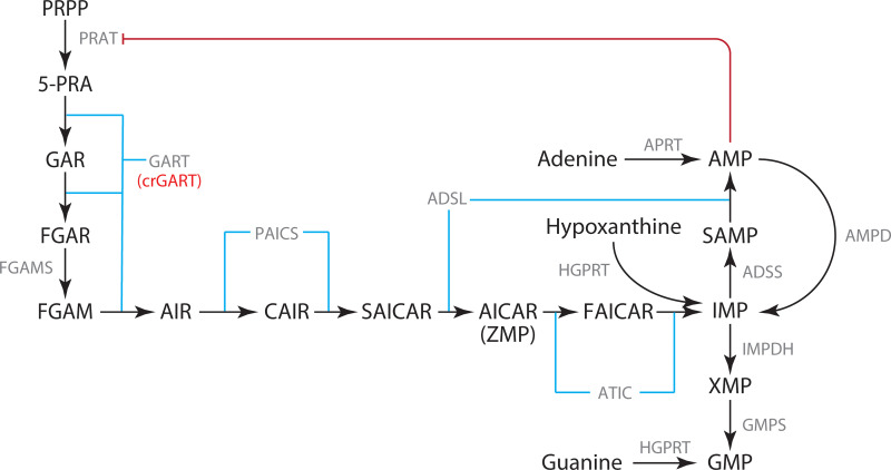
De novo purine biosynthesis (DNPS) is one of the oldest and most fundamental biochemical pathways [1]. In mammals, starting with phosphoribosyl pyrophosphate (PRPP), the six enzyme, ten step pathway produces inosine monophosphate (IMP), which is subsequently converted to guanosine monophosphate (GMP) or adenosine monophosphate (AMP) via two more enzymatic reactions (Fig 1). Purines are critical as building blocks and carriers of genetic information in the form of RNA and DNA, intra and intercellular signaling molecules, energy currency, and substrates and co-enzymes. While salvage pathways can produce purine nucleoside monophosphates from free purine bases (such as adenine, guanine and hypoxanthine) and PRPP, ultimately all purines are produced by DNPS. DNPS is upregulated at the G1/S phase and is critical during cellular division [2], most likely to supply purines for DNA replication and elevated RNA transcription. Given its importance in cellular division, and that supply/transport of free purine bases across placental membranes is inefficient [3, 4], DNPS is critical in mammalian development, including embryonic development. In mammals, the trifunctional GART enzyme catalyzes steps 2, 3, and 5 of DNPS (Fig 1). The human GART gene is located on Hsa21 and is therefore present in three copies in Down syndrome (DS, Trisomy 21), the most common genetic cause of intellectual disability in humans. Triplication of the GART gene has been hypothesized to be related to the pathology associated with DS [5]. GART expression is also altered in certain cancers [6, 7] and is likely important for neurological metabolism and development [8, 9].
Fig 1. De novo purine synthesis (DNPS) pathway.
DNPS mediates the conversion of PRPP to IMP. IMP is subsequently converted to AMP or GMP. The HeLa GART KO, crGART, is indicated.
The GART gene also encodes a monofunctional GARS protein via alternative transcription [5]. This alternative transcript includes a polyadenylation signal located in the intron separating the last GARS domain exon from the first AIRS domain exon. In human, mouse and Drosophila, this transcript has an in-frame TAA stop codon which is part of the 5′ donor splice site. This gene organization is not found in bacteria, lower eukaryotes, or plants. The function of GARS or the reason for this conservation of the gene structure confined to animals is not known. Characterization of the crGART transcriptome should allow identification of pathways in which GART plays a regulatory role and a better understanding of the GART gene and present an opportunity to assess the role of monofunctional GARS.
Functional mutations in DNPS genes are extremely rare in humans and have so far been limited to the adenylosuccinate lyase (ADSL), 5-aminoimidazole-4-carboxamide ribonucleotide formyltransferase / inosine monophosphate cyclohydrolase (ATIC), and phosphoribosylaminoimidazole carboxylase / phosphoribosylaminoimidazole succinocarboxamide synthetase (PAICS) genes. To date, fewer than 100 patients have been identified with ADSL deficiency. Until quite recently, only one patient had been identified with AICA-ribosiduria (ATIC deficiency), but now, three more individuals (two are siblings) have been identified [10]. Finally, two individuals (siblings) have been identified with PAICS deficiency [11]. ADSL deficiency is a spectrum disorder with three generalized classes: neonatal fatal, severe, or mild to moderate. Features of ADSL deficiency include seizures, psychomotor retardation, respiratory failure, and craniofacial abnormalities [12]. AICA-ribosiduria is characterized by mental retardation, blindness, epilepsy, and craniofacial and body dysmorphic features [6, 9]. The siblings identified with PAICS deficiency died within three days of birth and exhibited craniofacial abnormalities and body dysmorphic features [11]. The majority of these mutations are amino acid point mutations ultimately resulting in decreased enzymatic activity. Thus far, no functional mutations in GART have been reported. Taken together, these data suggest that defects in DNPS are usually embryonic fatal.
DNPS nulls for ADSL, ATIC, and GART were recently generated in HeLa cells via CRISPR-Cas9 induced mutagenesis [13]. The ADSL and ATIC mutants (crADSL and crATIC) accumulate DNPS pathway intermediates when cultured in purine free media (the DNPS pathway is up-regulated in the absence of exogenous purines). These intermediates, SAICAR and ZMP respectively, are regulators of transcription, and we have presented evidence for transcriptional regulation via DNPS deficiency and intermediate accumulation ([14] and [15]). We were unable to detect intermediate accumulation in the GART mutant (crGART). This is expected since the initial substrate for GART is phosphoribosylamine (5-PRA), which is extremely unstable under physiological conditions and hydrolyses to ribose-5 phosphate; half-life 5 seconds [16]. We hypothesize that changes in transcription due to GART knockout in this cell line are due to the deficit in DNPS, and not intermediate accumulation. To evaluate the crGART transcriptome, we employed RNA-seq to compare the crGART and HeLa transcriptomes in adenine-supplemented and adenine-depleted conditions. Our results indicate that GART null cells have a large number of differentially expressed genes (DEG) and may have an important role in embryogenesis, neural development, and perhaps special relevance to Alzheimer’s disease (AD) and Down syndrome (DS). We focus on one DEG, Cluster of Differentiation 36 (CD36). CD36 shows a high log2 value for expression in crGART versus HeLa as well as a very high base mean value for transcript expression and a minimal P-value. In addition, CD36 is well-characterized. CD36 encodes a scavenger receptor that functions as a master regulator of long chain fatty acid (LCFA) metabolism. CD36 has been implicated in numerous health conditions, such as obesity, dyslipidemia, atherosclerosis, and AD. Given GART triplication and purine dysregulation in DS and the role of CD36 in conditions reminiscent of DS, we hypothesize that CD36 may have important roles in DS and associated conditions.
Materials and methods
Methods were performed as previously described ([14] and [15]), except as noted.
Cell culture
Briefly, cells were maintained on 60 mm TPP (Techno Plastic Products, AG, Switzerland) plates in Dulbecco’s Modified Eagle Medium (DMEM, Gibco) supplemented with 10% fetal calf serum (FCS), 30 μM adenine, and normocin (Invivogen).
At two and one days prior to starvation induction, complete medium was replaced. Ten to twelve hours prior to adenine starvation, medium was changed to DMEM 10% FCS with 100 μM adenine. This concentration completely inhibits DNPS [17] and intermediate accumulation [14, 15, 18]. At ~50–70% confluence, medium was changed to DMEM supplemented with 10% fetal calf macroserum (FCM), normocin, with or without 100 μM adenine for 10 hour incubation. FCM is FCS dialyzed against saline using a 3.5 kDa molecular weight cutoff to deplete purines present in FCS.
HPLC analysis to detect metabolite accumulation
HPLC-EC analysis was performed as previously described ([14, 18] and [15]). No DNPS intermediates were detected in adenine-supplemented or -starved cells.
RNA-seq
RNA-seq was performed as previously described ([14] and [15]).
Data processing
RNA-seq sequence (FASTQ format by the Genomics and Microarray Core Facility at the University of Colorado, Denver) was aligned to the Ensembl98 [19] Homo_sapiens.GRCh38.98 transcriptome using salmon version 1.0.0 [20], then differential gene expression analysis was performed using DESeq2 version 1.26.0 [21] with independent hypothesis weighting (IHW) [22] and approximate posterior estimation for GLM coefficients (apeglm) [23], R version 3.5.3 [24]. Differentially expressed genes are listed in S1 Table.
ClueGO analysis
The Cytoscape (version 3.7.2) app ClueGO (version 2.5.5) [25] was employed for ontology and pathway analysis of lists of differentially expressed genes (DEGs). A list of 300 DEGs with the top 150 most positive log2 and 150 most negative log2 values was used to query gene ontology (GO) databases (UniProt-GOA_08.01.2020 for Biological process, Cellular component, and Molecular function) and Reactome databases (Pathway_08.01.2020 and Reactions_08.01.2020). See S1–S6 Figs in S1 File and S2 Table.
qPCR validation of DEGs
The RNA-seq analysis was validated by qPCR of six DEGs covering a wide range of RNA-seq log2 fold differences (similar to previously described [12]). Samples were collected and processed approximately one year after collection and processing of samples for RNA-seq. Detection and amplification was performed with an Applied Biosystems QuantStudio7 Flex System and SsoAdvanced Sybr Green (BioRad cat #1725270). Candidate gene primers were obtained from IDT PrimeTime service: ALPP (Hs.PT.56a.38602874.g), DPYSL3 (Hs.PT.58.39796068), GATA3 (Hs.PT.58.19431110), IQGAP2 (Hs.PT.58.28018594), OASL (Hs.PT.58.50426392), TWIST1 (Hs.PT.58.18940950) and ACTB (Hs.PT.39a.22214847). Ct values and RNA-seq log2 values were normalized to ACTB (β-actin).
PANTHER analysis
PANTHER Overrepresentation Test analysis via the Gene Ontology Consortium portal (https://www.geneontology.com) was performed to confirm and augment the ClueGO results. Unlike ClueGO, PANTHER is capable of using large gene lists. Three gene lists were used to query PANTHER. The complete list of significant DEGs, a list of DEGs unique to the crGART versus WT comparison (i.e., not present in previous analyses of crADSL and crATIC), and a list of DEGs common to the three mutants compared to WT were used to query Gene Ontology biological process, cellular component, and molecular function. See Results and S3–S5 Tables. The “Test Type” option was set for “Fisher’s Exact” and “Correction” was set to “Calculate False Discovery Rate”. The GO ontology database release date was 2020-02-21.
GART transfection
crGART cells were transfected with pCMV-GART-K1 [26] using Lipofectamine 2000 as per manufacturer’s protocol (Invitrogen). Transfectants were cultured in high glucose DMEM, 10% fetal calf serum (FCS) supplemented with normocin, Geneticin (ThermoFisher Scientific), and 30 μM adenine (3 x 10⁻⁵ M). Stable transfectants were selected for Geneticin resistance and then checked for purine (adenine) growth requirement.
Protein isolation
Confluent cell cultures (60 mm plates) were rinsed 2X with 1X PBS, then 500 μl cell lysis buffer (1% Triton X-100, 20 mM Tris pH 7.5, 50 mM NaCl, 5mM MgCl2) was added. The plates were scraped and lysate was collected and transferred to 1.5 ml Eppendorf tubes. Lysates were centrifuged for 30 minutes at 16,000 x g. The supernatant was collected and protein concentration was measured by Bradford assay (Sigma).
Western blot
Total HeLa, crATIC, crGART, and crGART/K1-GART (transfected cells) protein (30 μg/lane) was separated by SDS-PAGE (8–16% gradient tris-glycine gel) for 40 minutes at ~225 V. Gels were wet-blotted to nitrocellulose (Amersham Protran 0.45 μM NC, buffer: 25 mM Tris, 200 mM glycine, 20% methanol) at 200 mA for 1 h. Blots were blocked 30 minutes in block solution (1% BSA, 0.1% Tween-20, 1X TBS, 0.01% sodium azide, pH 7.4) then incubated for 1.5 h at RT with either CD36 (Abcam ab133625, 1:500 dilution) or PPARγ (Cell Signaling 2430S, 1:250 dilution) and β-actin (Cell Signaling 8457, 1:1000 dilution) rabbit primary antibodies in primary Ab solution (5% BSA, 0.1% Tween-20, 0.01% sodium azide, 1X TBS, pH 7.4). Blots were washed 3X for 5 minutes in 1X TBS. Blots were then incubated in goat anti-rabbit IgG-AP conjugate (Bio-Rad, 170–6518) diluted 1:10,000 in block solution for 1 hour. Three more washes (5 minutes each in 1X TBS) were performed, then blots were treated with CDP-Star® Substrate with Nitro-Block-II™ Enhancer (Applied Biosystems, T2218) for five minutes and visualized using a Bio-Rad Chemidoc Imaging System. See S1 Raw images for unaltered immunoblot images with annotation.
Metabolon metabolomic analysis
Cells were grown and starvation induced in 100 mm TPP cell culture dishes as described above. Cells were harvested according to Metabolon’s prescribed protocol. Briefly, cells were washed in 1X PBS and treated with Detachin (Genlantis) to release cells from the plate. Cells were transferred to 15 ml conical tubes and 10 ml of experimental condition media was added. Cells were pelleted at 1000 x g for 5 minutes, the pellet was aspirated, resuspended in 1 X PBS and pelleted again, aspirated, and stored at -80°C until shipment to Metabolon on dry ice. Samples were analyzed by Metabolon for metabolomic analysis using ultra high-performance liquid chromatography/tandem accurate mass spectrometry (UHPLC/MS/MS). Peak detection and identification, determination of relative concentrations, and statistical analysis were provided by Metabolon (http://www.metabolon.com).
Results
Adenine is required for proliferative growth of crGART
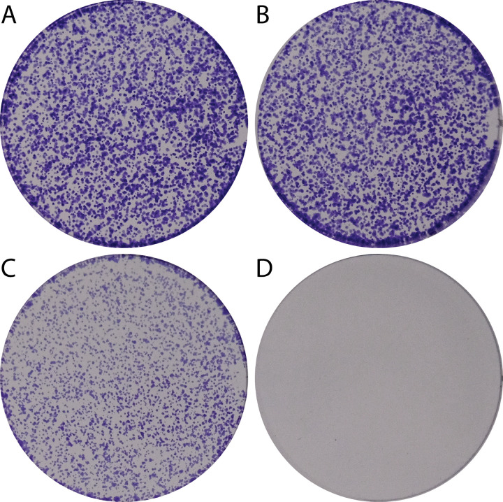
Given that GART catalyzes three of the ten steps in the DNPS pathway, we hypothesized that crGART cells would exhibit proliferative arrest in adenine-deprived conditions. HeLa and crGART cells were cultured in complete-serum media overnight and then subsequently cultured in media supplemented with dialyzed-serum (FCM) with or without supplemental adenine. HeLa cells showed proliferative growth in both media conditions while crGART showed proliferative growth in only adenine-supplemented conditions (Fig 2).
Fig 2. crGART requires adenine for growth.
HeLa (A,B) and crGART (C,D) cells were plated and cultured in 30 μM adenine-supplemented (A,C) or adenine-depleted (B,D) media. Plates were stained with crystal violet.
DNPS intermediates were not detected in adenine starved crGART cells
Next, we assessed whether the crGART cells accumulate any detectable DNPS intermediates. We demonstrated previously that crADSL [14] and crATIC [15] cells accumulate substrate when cultured in adenine free media. crGART cells were cultured as previously described and metabolites extracted. HPLC-EC analysis did not indicate accumulation of any DNPS intermediates. This is consistent with previous work demonstrating that 5-PRA is highly unstable and breaks down in approximately five seconds under cellular conditions to ribose-5 phosphate [12, 14, 27]. This suggests that crGART is likely useful as a model of general DNPS deficiency.
Transcriptome analysis of crGART versus HeLa identified differentially expressed genes
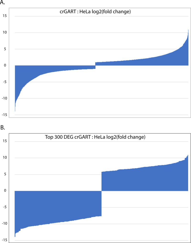
To investigate the effect of GART KO on the transcriptome, we compared the crGART and HeLa transcriptomes after culture in adenine-supplemented and adenine-depleted conditions. 4218 genes were significantly differentially expressed by cell type. This represents a log(2)counts crGART: HeLa fold change range of -13.94 to 10.93 (Fig 3A). For ClueGO analysis, we used the “top 300” DEG list (see Materials and Methods 2.5), which represents a log(2)counts fold change range of -13.74 to -7.67 and 5.83 to 10.93 (Fig 3B). Positive values indicate enrichment in crGART and negative log2 values indicate enrichment in HeLa. Principal component analysis shows clustering by cell type and supplementation (Fig 4).
Fig 3. Plot of log(2) values crGART: HeLa DEG fold change.
A. All significant DEGs B. Significant DEGs in the “top 300” list (see Methods).
Fig 4. Principal component analysis of crGART and HeLa replicates.
PCA shows a robust difference by cell type and a much smaller difference by adenine supplementation.
CD36
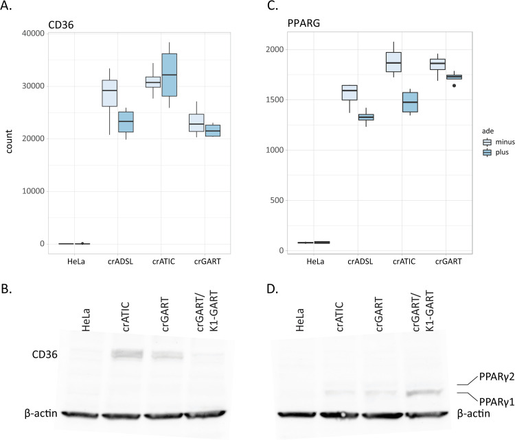
One of the largest magnitude DEGs we obtained was for the CD36 gene. Transcript levels for the Cluster of Differentiation 36 (CD36) gene are highly elevated in crGART versus HeLa (Fig 5A, S1 Table) and in crADSL [14] and crATIC [15], which suggests that DNPS regulates expression of the gene. In addition, CD36 exhibits an extremely large base mean value, indicating high levels of transcript, as well as zero P-value (S1 Table). The elevated expression of CD36 correlates with elevated expression of the Peroxisome Proliferator Activated Receptor γ (PPARG) gene in the mutant cell lines (Fig 5C). CD36 and PPARγ are major components of a pathway that regulates fatty acid storage, triglyceride synthesis, glucose uptake, and fatty acid metabolism [28].
Fig 5. CD36 and PPARγ.
A. Box plot showing CD36 RNA-seq transcript levels in HeLa and DNPS mutants B. CD36 immunoblot C. Box plot showing PPARG RNA-seq transcript levels D. PPARγ immunoblot. The predicted MW of CD36 is 75 kDa, and the predicted MW of PPARγ1 and PPARγ2 are 53 and 57 kDa. The MW of β-actin is 45 kDa.
To evaluate CD36 and PPARγ protein expression, we performed immunoblot analysis, which confirmed elevated expression in crATIC and crGART, and relative absence of the two proteins in HeLa (representative blots: Fig 5B and 5D). Our results also show that elevated CD36 expression is partially rescued in crGART cells stably transfected with a CHO-K1 GART expression construct.
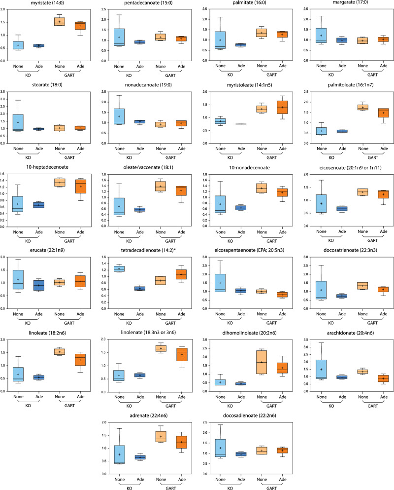
In a separate set of experiments, we conducted a metabolomic analysis of crGART and crGART stably transfected with a GART expression construct via the Metabolon Global Metabolic Panel analysis. In this analysis, 22 long-chain fatty acids were identified, and of these, about 15 are markedly elevated in the transfected cells (Fig 6, S6 Table). This is especially interesting given that CD36 is an important regulator of long-chain fatty acid metabolism [29] and that CD36 expression is reduced in the transfected cells.
Fig 6. LCFA Box plots.
Box plots supplied by Metabolon, Inc. show LCFA levels in crGART (KO) cells and transfected crGART (GART) cells. Ade indicates that cells were grown in purine supplemented media, None indicates purine starvation.
ClueGO analysis: Functional enrichment analysis employing Gene Ontology and Reactome databases
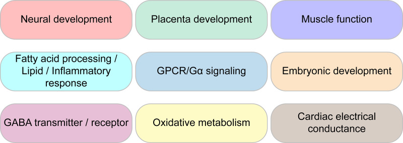
In order to assess what processes or pathways are affected by ablation of the GART gene, we performed GO and Reactome functional analysis of DEGs. We queried the Gene Ontology database (Biological process, Cellular component, and Molecular function) as well as the Reactome knowledgebase (Pathways and Reactions) for this analysis. We obtained a very large number of terms but will focus only on terms of special interest to our laboratory (Fig 7). All data is available in supplementary material.
Fig 7. Prominent GO ontologies from ClueGO analysis.
For the GO terms associated with Biological process, 70 GO groups were identified with 548 GO terms. Related GO groups were organized into neural function, development, muscle function, fatty acids, cardiac function, G-protein coupled activity, oxides, and lipids categories. Within neural function, associated terms include neuron migration, dendrites, axon, hippocampus, neuroblast proliferation, synapse, GABA, and catecholamines. DNPS is known to be important for neural function and diseases associated with DNPS exhibit strong neurological phenotypes. Interestingly, another category identified was development. Development includes disparate terms, ranging from organ morphogenesis related terms such as renal, pancreas, and prostate development, to mesenchyme morphogenesis, stem cell differentiation, alkaline phosphatase activity, blood vessel morphogenesis, epithelial cell differentiation, bone development, endoderm, etc. Placental development included labyrinthine development and female pregnancy. For muscle function terms, we obtained smooth muscle function, myotubes and sarcomere organization, vasoconstriction, actin filament movement, and striated and smooth muscle contraction. Cardiac function was also enriched, including sinoatrial and atrioventricular node function terms, as well as atria and ventricle development. Oxidation related terms such as nitric oxide and superoxide metabolism were found. Lipid terms included fatty acids, phospholipase activity, phosphatidyl metabolic process, PI3K, and inflammatory response centered around IL-1 and TNFα. Given the importance of purines in development and neurobiology, the observed enrichment in related terms was expected. However, enrichment in cardiac terms involving electrical conduction was unexpected and may suggest a novel line of inquiry. The enrichment for placental development terms was also unexpected and suggests a purine requirement early in development and that DNPS may play an important role in blastocyst formation.
For the GO terms associated with Cellular component, 12 groups were identified with 26 terms. Terms include voltage gated potassium channels, I-band and intercalated disc, gap junction, GABA synapse and various other synaptic membrane terms.
For the GO terms associated with Molecular function, 25 groups were identified with 54 terms. Term categories included neuronal and ion transmembrane function, development, phospholipid activity, cytokine, etc. In the neuronal category, neuropeptide, acetylcholine receptor, potassium ion gate channels, and neurotransmitter transmembrane transporter terms were prominent. For phospholipids, notable terms included lipase activity, phospholipase (A2, 1, and C) activity, phosphatidylcholine acylhydrolase activity, phosphatidylinositol 3 kinase, and nitric oxide synthase. Enrichment for Syndecan protein binding and G-protein coupled peptide receptor terms is consistent with the G-protein coupled activity category in the Biological process results above.
Reactome Pathways knowledgebase showed 14 groupings of 29 terms. Categories include diseases associated with O-glycosylation of proteins, peptide hormone metabolism, rhodopsin-like receptors, and GPCR and G-alpha signaling. Neuronal and development terms were identified, as well as TFAP-2 which is heavily involved in neural-crest development. Consistent with the GABA terms we obtained in Biological process, we identified GABA receptor and GABA B receptor terms. This suggests that neural deficits associated with DNPS or GART deficiency may be associated with alteration of GABAergic signaling.
Reactome Reactions knowledgebase showed 10 groupings of 24 terms. Term categories included proteoglycans and GPI anchors, GPCR, GEF, and Gi activated ligands. Keratin filaments was also noted.
ClueGO networks, pie charts, and ontology histograms are shown in S1–S5 Figs in S1 File. Since the Biological Process analysis identified a very large number of ontologies, we also performed an analysis with a more stringent cutoff (P < 0.001, see S6 Fig in S1 File).
Corroboration of Gene Ontology analysis via PANTHER analysis
PANTHER analysis with the complete list of significant DEGs was consistent with the ClueGO results and revealed new ontology terms such as DNA and RNA binding, polymerase, and transcription regulation (S3 Table).
Comparison of crATIC, crADSL and crGART DEGs
To get a better sense of which DEGs are due specifically to ablation of the GART gene, we identified the significant DEGs unique to the crGART vs HeLa comparison. There were 1282 genes that changed significantly in the crGART vs HeLa comparison but did not change in the crADSL vs HeLa [14] and crATIC vs HeLa comparisons [15]. The list of genes was used to query PANTHER via the Gene Ontology web portal (www.geneontology.org). We obtained Biological process ontologies related to RNA splicing and mitochondrion organization (S4 Table).
We also identified DEGs common to all three mutants compared to HeLa. PANTHER functional enrichment analysis of the list of 7745 genes returned numerous Biological process ontologies, including regulation of protein localization to plasma membrane, regulation of osteoblast differentiation, positive regulation of I-kappaB kinase/NF-kappaB signaling, heart morphogenesis, establishment of vesicle localization, Ras protein signal transduction, embryonic organ morphogenesis, and many others (S5 Table). This result illustrates the importance of DNPS in multiple aspects of cell development and function.
Validation of RNA-seq by qPCR of candidate genes
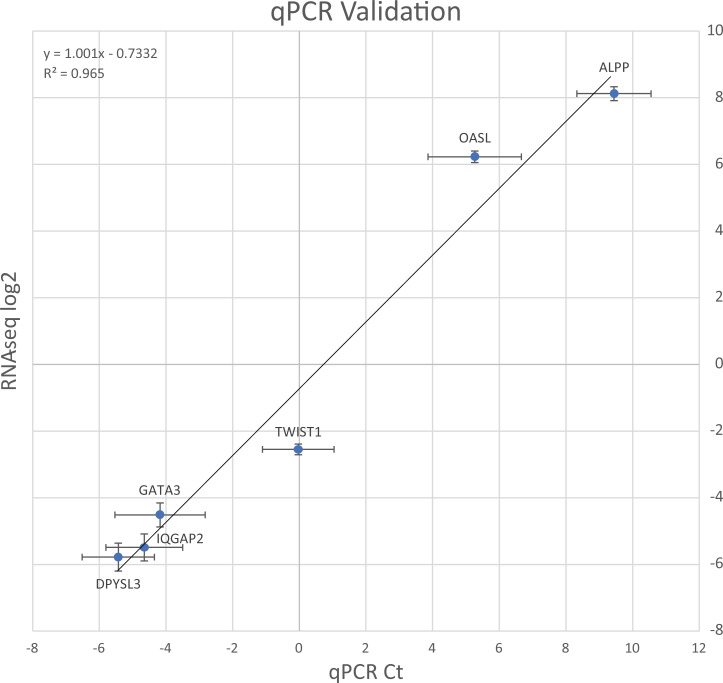
To confirm our RNA-seq results, we performed qPCR to measure transcript levels for ALPP, DPYSL3, GATA3, IQGAP2, OASL and TWIST1, and normalized to ACTB. These genes span a wide range of transcript levels. The transcript level results (Ct values) are consistent with the log(2)fold values for these genes from our RNA-seq analysis (Fig 8).
Fig 8. Validation of RNA-seq by qPCR.
RNA-seq log2 values are plotted on the Y axis and qPCR Ct values are plotted on the X axis. Error bars indicate standard error.
Discussion
In this study, we evaluated the purine dietary requirement for crGART and performed HPLC-EC to detect accumulation of metabolites during purine starvation. We compared the crGART and HeLa transcriptomes via RNA-seq analysis in adenine-supplemented and adenine-depleted conditions.
The purpose of adenine supplementation is twofold: 1) to provide purines necessary for cell survival (which are absent in FCM) and 2) to eliminate DNPS [30, 31], preventing intermediate accumulation. Adenine phosphoribosyltransferase (APRT) catalyzes the conversion of adenine and phosphoribosyl pyrophosphate (PRPP) to AMP and pyrophosphate (PPi). APRT is present in all mammalian tissues and is uniquely responsible for metabolic adenine salvage from dietary sources [32]. Once AMP has been produced, it can be converted to IMP by AMP deaminase (AMPD), and then converted to GMP by IMP dehydrogenase (IMPDH, converts IMP into XMP) and GMP synthase (converts XMP into GMP, see Fig 1) [33]. Salvage could also be accomplished via supplementation with hypoxanthine (Fig 1) but not guanine. GMP can be produced via HGPRT catalysis of guanine, but IMP (and AMP) cannot (Fig 1). We chose adenine supplementation rather than hypoxanthine supplementation for consistency with studies employing the crADSL cell line, in which salvage via hypoxanthine is disrupted. Interestingly, the APRT and HPRT1 genes (HPRT1 encodes the HGPRT protein) both show mildly reduced expression in crGART (log2 values -0.14 and -0.48, respectively). The level of APRT expression is adequate for salvage since we can rescue the crGART phenotype with adenine supplementation.
The GART gene encodes a trifunctional protein and also a monofunctional GARS protein via alternative transcription. The conversion of 5-PRA to GAR is the first reaction and is catalyzed by the GARS domain of the trifunctional protein [16]. As discussed previously, 5-PRA is extremely unstable under physiological conditions, and is unlikely to accumulate. Our results show that crGART requires purine (adenine) supplementation for proliferative growth, but apparently does not accumulate pathway intermediates during purine starvation. In the crADSL and crATIC DNPS-KO models, metabolic substrates (SAICAR and ZMP respectively) readily accumulate during purine starvation and there is strong evidence that both substrates alter cellular processes [34–36]. Since the crGART line does not accumulate detectable DNPS metabolic intermediates during purine starvation, it is likely a useful model of strict DNPS deficiency.
The biological significance of the monofunctional GARS protein has not yet been elucidated. One hypothesis is that GARS may interact with PRAT to facilitate transfer of 5-PRA, the unstable product of PRAT, to GARS/GART thus increasing the rate of DNPS [12, 32]. This would imply that lack of GARS would inhibit DNPS. The pCMV-GART-K1 expression construct that we used to transfect crGART encodes only the trifunctional GART gene, not the monofunctional GARS gene. The transfected cell line is able to synthesize levels of purines adequate for growth, but purine levels may be less adequate, or inadequate, for other DNPS functions. This would be consistent with the observation that the transfected crGART cell line does not exhibit fully restored inhibition of CD36 expression. Since the GART gene is trisomic in DS, GARS overexpression may play a role in elevated purine levels in DS. Other hypotheses exist, for example, the monofunctional GARS protein may have a role in release of metabolites from the de novo purine pathway so they can be used in other metabolic pathways. The crGART cell line should be invaluable for investigating the role of the monofunctional GARS protein.
Typically, in “omics” studies, an extremely large amount of data is produced and it is difficult to completely evaluate all of it. We employed ClueGO and PANTHER analysis, which are both limited in that they rely on the gene ontology (GO) and similar databases. GO is curated both manually and computationally and is, at any given time, a work in progress. This means that an analysis at a given time may be very different from an analysis performed six months later. ClueGO has the additional limitation that it cannot handle very long lists of genes, which is why we employed a list of 300 genes with the largest magnitude changes. There are limitations to RNA-seq as well. The number of sample replicates, sequencing depth, computation approaches, technical issues, etc. will all affect the outcome of the analysis. Fortunately, the sequence and DEGs are both available, and can be re-evaluated in the future as often as desired. This helps to minimize limitations, at least to some extent. Unfortunately, it was not feasible for us to evaluate all the DEGs we identified, but we present the data and ontology analyses so interested investigators can follow up on DEGs of interest. For example, in a list of the “top 25” genes (excluding gene models and pseudogenes) with the highest log2 values (indicating greatly elevated expression in crGART versus HeLa, see S1 Table), the CD36 gene stands out because of its very high baseMean value (indicating high transcript levels) and P-value. But there are other notable candidates for further analysis. For example, four of these genes are transcription factors: CEBPE, CREB3L3, LEF1, and LHX1.
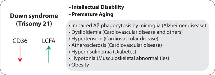
We noted that the CD36 gene is highly expressed in all three DNPS-KO transcriptomes that we have characterized (crADSL, crATIC, and crGART), with average log2(fold change) value of about 8.5 (Fig 5A). We have shown that CD36 protein expression is elevated in crATIC and crGART versus HeLa (Fig 5B) and that protein expression is partially rescued (reduced) in crGART cells stably transfected with a CHO-K1 GART expression construct. Finally, we show that numerous LCFA are elevated in transfected crGART, which correlates with and may be related to reduced CD36 expression in these cells. CD36 is involved in fatty acid uptake and metabolism [29], inflammation cascade [37], angiogenesis, apoptosis, thrombosis, atherosclerosis, Alzheimer’s disease, and insulin resistance [38]. Interestingly, many of these phenotypes are seen in DS and associated conditions. We hypothesize that CD36 is regulated by DNPS and is dysregulated in DS. This hypothesis is supported by an RNA-seq analysis of age and sex matched fibroblast cell lines derived from people with DS and normosomic controls showing that CD36 expression is reduced two-fold in the DS lines (see supplemental table from [39]). As such, reduced CD36 expression/elevated LCFA levels may play an important role in DS phenotypes and associated conditions (Fig 9).
Fig 9. Suggested role of CD36 in Down syndrome.
We hypothesize that reduced CD36 expression and increased LCFA levels may be important in intellectual disability and premature aging, as well as co-morbidities characteristic of DS.
The PPARG gene is also highly expressed in all three DNPS-KO transcriptomes (Fig 5C) and protein expression is elevated in crATIC and crGART (Fig 5D). PPARG encodes PPARγ, an inducible transcription factor that is considered a master regulator in lipid metabolism, with roles in fat, carbohydrate, and general energy metabolism, as well as insulin sensitivity, cell proliferation and differentiation, inflammation, and cancer [40]. A CD36-PPARγ axis regulates fatty acid storage, triglyceride synthesis, glucose uptake, and fatty acid metabolism [28].
In addition to CD36 and PPARG, our RNA-seq analysis led to identification of numerous DEGs and gene ontologies from ClueGO functional enrichment analysis. Many of these were consistent with our previous analyses of the crADSL and crATIC mutants and are likely to be due to DNPS deficiency. These results support the hypothesis that DNPS is essential in development and that alterations in DNPS and intermediate metabolite accumulation both regulate the transcriptome. This is consistent with previous work demonstrating that DNPS is upregulated in the G1/S phase cell cycle interface [3, 4], is critical in embryogenesis which is marked by rapid cellular division, and that purines are essential in development [8] especially in neural development [41].
We also obtained DEGs and gene ontologies potentially relevant to DS. This is perhaps unsurprising given that 1) the GART gene is located on Hsa21 and is triplicated in DS, 2) GART expression is dysregulated in DS and 3) purine levels are also dysregulated in DS [42]. In addition, although the current work involves a GART null model, it is likely that at least some pathways affected by absence of GART are also affected by increase in GART, although the relationship is unlikely to be symmetrical or straightforward. Ontologies include terms relevant to placental development, neural development and cognition, cardiac development, and Alzheimer disease.
People with DS are at elevated risk for developmental as well as aging related disorders. In addition to intellectual disability, common disorders include hypotonia [43], congenital heart malformation, disease of pulmonary circulation, cardiac arrest, hypotension, infantile spasm, epilepsy, OSA (sleeping disorders), dementia, hypothyroidism, and obesity [44]. Our transcriptomic analysis revealed multiple terms potentially relating GART and DNPS to these disorders. Ontological groupings show enrichment in terms associated with organismal development, cardiac electrophysiology, neural function/transmission and GABA, placenta, alkaline phosphatase activity, phospholipase activity, Amyloid β, and muscle function.
DS is most commonly associated with intellectual disability [44]. Previous work has demonstrated that defects in purine metabolism and/or purine concentration in the developing brain are related to intellectual disabilities [5, 8]. DS brains show decreased neuronal density by mass and volume in various brain regions (e.g. cortex, hippocampus, cerebellum), which occurs during development, possibly during gestation [45]. Purinergic signaling regulates axon guidance and growth as well as establishment of correct synaptic contacts [8] and is crucial for CNS development [46]. The GART enzyme exhibits altered spatiotemporal expression in the developing nervous system in DS. Typically, levels of GART are highly expressed in the developing cerebellum and decrease precipitously post-partum. However, in DS, GART levels persist and decrease later in development [5].
Our ClueGO results include terms related to alkaline phosphatase function, specifically differential expression of ALPP, ALPI, ALPG, and ALPL. ALPP, ALPI, and ALPG show elevated expression in crGART. ALPL, which encodes tissue non-specific alkaline phosphatase (TNAP), is elevated in HeLa. TNAP is a membrane bound extracellular enzyme present in mineralizing bones, renal tissue, and the central nervous system (CNS) [47]. In studies of murine CNS development TNAP was found to be associated with the neural tube [48] and its expression is highest in early embryonic development associating with neural precursor and progenitor cells [49]. TNAP expression has also been observed during synaptic formation and maturation [50], promoting axonal growth [51]. TNAP also has a role in the hydrolysis of extracellular nucleotides [8, 52]. Extracellular ATP was found to induce migration of neural progenitor cells [53], indicating that TNAP may play a role in regulating extracellular ATP pools for migratory events. Another TNAP function is found in the metabolism of Vitamin B6, which is a cofactor for enzymes involved in neurotransmitter (e.g. GABA) synthesis [54]. Not much is known about the direct role of TNAP in proliferation and differentiation. However, given the function and spatiotemporal expression of TNAP, it is likely that TNAP is directly involved in purinergic signaling or regulation of the extracellular purine pool. Hence, altered TNAP levels may potentially be deleterious during CNS development.
Gamma-amino butyric acid (GABA) is the main inhibitory neurotransmitter in healthy adult brains and has been of particular interest in DS [45, 55]. Analysis of fetal brain tissue has shown a smaller hippocampus and decreased GABA neurotransmitters in DS which suggests impaired neurogenesis or migration of GABAergic interneurons [56]. DS patients exhibit an increased incidence of epileptic seizures [57], children exhibit sleep disturbances [58] and hyperactivity [59]. These conditions may be partially due to abnormal GABA signaling. Studies employing DS murine models have shown that altered GABA signaling results in synaptic excitatory/inhibitory signal imbalance, impaired synaptic plasticity [45, 55, 60], and learning and memory deficits [61, 62]. The enrichment for GABA-related terms within our analysis suggests that GART and DNPS may play a role in these processes.
Alzheimer’s disease (AD) is a form of dementia characterized by accumulation of neural amyloid β plaques, Tau neurofibrillary tangles, and chronic neural inflammation. Recent work supports the hypothesis that inflammation plays a critical role in AD, and therapeutic intervention designed to reduce neural inflammation shows promise. Inflammation is typically mediated by cytokine and chemokine secretion as well as fatty acid metabolism [63]. Chemokines and cytokines (such as IL-1β and TNFɑ) are secreted in response to injury and act as proinflammatory signals, resulting in clearance cell recruitment to the damaged tissue [63]. Fatty acid derivatives, specifically lipoxins (derived from ω-6 via phospholipaseA2 and lipoxygenase catalysis of arachidonic acid) [64] and ω-3 metabolites such as maresins, resolvins, and protectins [65] are all potent anti-inflammation mediators. ω-6 fatty acid is typically stored in the phospholipid bilayer as arachidonic acid, which is then cleaved by phospholipase activity and then metabolized via secondary enzymes to eicosanoids and lipoxins [66]. Coincidentally, extracellular ATP signaling plays a crucial role in inflammation through the purinergic P2 and P1 receptors [67]. DNPS and GART levels may play important roles in these processes.
We obtained 1282 DEGs unique to the crGART versus HeLa comparison (i.e., DEGs not found in the crADSL vs HeLa [14] and crATIC vs HeLa comparisons [15]). These DEGs mapped to Biological process ontologies related to RNA splicing and mitochondrion organization (S4 Table). It is unclear why these genes are significantly changed only in the crGART cell line. One possibility is that the absence of GART may have an effect unrelated, or peripherally related, to DNPS that affects the transcription of these genes.
In conclusion, our results indicate that DNPS deficiency affects the cellular transcriptome, significantly altering the expression of over 4000 genes. We believe that the crGART cell line is an attractive model of DNPS deficiency in the absence of substrate accumulation and will be a valuable tool for investigating the role of DNPS dysregulation in DS and other disorders. We are especially interested in CD36 because of its important role in LCFA metabolism and its potential role in DS. Given the remarkable difference in CD36 transcript levels and protein expression in crGART versus Hela, as well as the difference in LCFA levels and rescue of CD36 expression in crGART versus transfected crGART (crGART/K1-GART), we believe that the cell line will be a good working model for investigating CD36 function in the context of DNPS and purine metabolism.
Supporting information
(PDF)
(XLSX)
(XLSX)
(XLSX)
(XLSX)
(XLSX)
(XLSX)
(PDF)
Acknowledgments
The Cancer Center and the Genomics (Microarray) Shared Resource are supported in part by the Cancer Center Support Grant #P30-CA046934 from the National Cancer Institute.
MZ and VB were supported by Charles University [programmes PRIMUS/17/MED/6 and PROGRES Q26/LF1] and by the Ministry of Education, Youth and Sports of CR [LQ1604 National Sustainability Programme II].
Data Availability
All data are fully available without restriction. The raw sequence reads for this study are available at https://www.ncbi.nlm.nih.gov/bioproject, accession: PRJNA701493. Raw western blot images are in the Supporting information file “S1_raw_images”. All other data is available in the supplementary figures and tables.
Funding Statement
This work was funded by The Itkin Foundation, the Sam and Frieda Davis Trust, The Butler Family Fund of the Denver Foundation, and the Lowe Fund of the Denver Foundation. The funders had no role in study design, data collection and analysis, decision to publish, or preparation of the manuscript.
References
- 1.Caetano-Anollés G., Yafremava L.S., Gee H., Caetano-Anollés D., Kim H.S., Mittenthal J.E., The origin and evolution of modern metabolism, Int. J. Biochem. Cell Biol. 41 (2009) 285–297. doi: 10.1016/j.biocel.2008.08.022 [DOI] [PubMed] [Google Scholar]
- 2.Fridman A., Saha A., Chan A., Casteel D.E., Pilz R.B., Boss G.R., Cell cycle regulation of purine synthesis by phosphoribosyl pyrophosphate and inorganic phosphate, Biochem J. 454 (2013) 91–99. doi: 10.1042/BJ20130153 [DOI] [PubMed] [Google Scholar]
- 3.Chan C.Y., Zhao H., Pugh R.J., Pedley A.M., French J., Jones S.A., et al., Purinosome formation as a function of the cell cycle, Proceedings of the National Academy of Sciences. (2015). doi: 10.1073/pnas.1423009112 [DOI] [PMC free article] [PubMed] [Google Scholar]
- 4.Zhao H., Chiaro C.R., Zhang L., Smith P.B., Chan C.Y., Pedley A.M., et al., Quantitative Analysis of Purine Nucleotides Indicates Purinosomes Increase de Novo Purine Biosynthesis, Journal of Biological Chemistry. (2015). doi: 10.1074/jbc.M114.628701 [DOI] [PMC free article] [PubMed] [Google Scholar]
- 5.Brodsky G., Barnes T., Bleskan J., Becker L., Cox M., Patterson D., The human GARS-AIRS-GART gene encodes two proteins which are differentially expressed during human brain development and temporally overexpressed in cerebellum of individuals with Down syndrome, Hum Mol Genet. 6 (1997) 2043–2050. doi: 10.1093/hmg/6.12.2043 [DOI] [PubMed] [Google Scholar]
- 6.Desmoulin S.K., Wang Y., Wu J., Stout M., Hou Z., Fulterer A., et al., Targeting the proton-coupled folate transporter for selective delivery of 6-substituted pyrrolo[2,3-d]pyrimidine antifolate inhibitors of de novo purine biosynthesis in the chemotherapy of solid tumors, Mol. Pharmacol. 78 (2010) 577–587. doi: 10.1124/mol.110.065896 [DOI] [PMC free article] [PubMed] [Google Scholar]
- 7.Cong X., Lu C., Huang X., Yang D., Cui X., Cai J., et al., Increased expression of glycinamide ribonucleotide transformylase is associated with a poor prognosis in hepatocellular carcinoma, and it promotes liver cancer cell proliferation, Hum. Pathol. 45 (2014) 1370–1378. doi: 10.1016/j.humpath.2013.11.021 [DOI] [PubMed] [Google Scholar]
- 8.Fumagalli M., Lecca D., Abbracchio M.P., Ceruti S., Pathophysiological Role of Purines and Pyrimidines in Neurodevelopment: Unveiling New Pharmacological Approaches to Congenital Brain Diseases, Front. Pharmacol. 8 (2017) 941. doi: 10.3389/fphar.2017.00941 [DOI] [PMC free article] [PubMed] [Google Scholar]
- 9.Göttle M., Burhenne H., Sutcliffe D., Jinnah H.A., Purine metabolism during neuronal differentiation: the relevance of purine synthesis and recycling, J Neurochem. 127 (2013) 805–818. doi: 10.1111/jnc.12366 [DOI] [PMC free article] [PubMed] [Google Scholar]
- 10.Ramond F., Rio M., Heron B., Imbard A., Marie S., Billemaz K., et al., AICA-ribosiduria due to ATIC deficiency: delineation of the phenotype with three novel cases, and long-term update on the first case, J Inherit Metab Dis. (2020). doi: 10.1002/jimd.12274 [DOI] [PubMed] [Google Scholar]
- 11.Pelet A., Skopova V., Steuerwald U., Baresova V., Zarhrate M., Plaza J.-M., et al., PAICS deficiency, a new defect of de novo purine synthesis resulting in multiple congenital anomalies and fatal outcome, (2019) 1–30. doi: 10.1093/hmg/ddz237/5584440 [DOI] [PubMed] [Google Scholar]
- 12.Van Den Berghe G., Jaeken J., Adenylosuccinase deficiency, Adv. Exp. Med. Biol. 195 Pt A (1986) 27–33. doi: 10.1007/978-1-4684-5104-7_4 [DOI] [PubMed] [Google Scholar]
- 13.Baresova V., Krijt M., Skopova V., Souckova O., Kmoch S., Zikánová M., CRISPR-Cas9 induced mutations along de novo purine synthesis in HeLa cells result in accumulation of individual enzyme substrates and affect purinosome formation, Mol Genet Metab. 119 (2016) 270–277. doi: 10.1016/j.ymgme.2016.08.004 [DOI] [PubMed] [Google Scholar]
- 14.Mazzarino R.C., Baresova V., Zikánová M., Duval N., Wilkinson T.G., Patterson D., et al., The CRISPR-Cas9 crADSL HeLa transcriptome: A first step in establishing a model for ADSL deficiency and SAICAR accumulation, Mol Genet Metab Rep. 21 (2019) 100512. doi: 10.1016/j.ymgmr.2019.100512 [DOI] [PMC free article] [PubMed] [Google Scholar]
- 15.Mazzarino R.C., Baresova V., Zikánová M., Duval N., Wilkinson T.G., Patterson D., et al., The CRISPR-Cas9 crATIC HeLa transcriptome: Characterization of a novel cellular model of ATIC deficiency and ZMP accumulation, Mol Genet Metab Rep. 25 (2020) 100642. doi: 10.1016/j.ymgmr.2020.100642 [DOI] [PMC free article] [PubMed] [Google Scholar]
- 16.Rudolph J., Stubbe J., Investigation of the mechanism of phosphoribosylamine transfer from glutamine phosphoribosylpyrophosphate amidotransferase to glycinamide ribonucleotide synthetase, Biochemistry. 34 (1995) 2241–2250. doi: 10.1021/bi00007a019 [DOI] [PubMed] [Google Scholar]
- 17.Tu A.S., Patterson D., Biochemical genetics of Chinese hamster cell mutants with deviant purine metabolism. VI. Enzymatic studies of two mutants unable to convert inosinic acid to adenylic acid, Biochem. Genet. 15 (1977) 195–210. doi: 10.1007/BF00484561 [DOI] [PubMed] [Google Scholar]
- 18.Duval N., Luhrs K., Wilkinson T.G., Baresova V., Skopova V., Kmoch S., et al., Genetic and metabolomic analysis of AdeD and AdeI mutants of de novo purine biosynthesis: cellular models of de novo purine biosynthesis deficiency disorders, Mol Genet Metab. 108 (2013) 178–189. doi: 10.1016/j.ymgme.2013.01.002 [DOI] [PMC free article] [PubMed] [Google Scholar]
- 19.Yates A.D., Achuthan P., Akanni W., Allen J., Allen J., Alvarez-Jarreta J., et al., Ensembl 2020, Nucleic Acids Res. 48 (2020) D682–D688. doi: 10.1093/nar/gkz966 [DOI] [PMC free article] [PubMed] [Google Scholar]
- 20.Patro R., Duggal G., Love M.I., Irizarry R.A., Kingsford C., Salmon provides fast and bias-aware quantification of transcript expression, Nat. Methods. 14 (2017) 417–419. doi: 10.1038/nmeth.4197 [DOI] [PMC free article] [PubMed] [Google Scholar]
- 21.Love M.I., Huber W., Anders S., Moderated estimation of fold change and dispersion for RNA-seq data with DESeq2, Genome Biol. 15 (2014) 550. doi: 10.1186/s13059-014-0550-8 [DOI] [PMC free article] [PubMed] [Google Scholar]
- 22.Ignatiadis N., Klaus B., Zaugg J.B., Huber W., Data-driven hypothesis weighting increases detection power in genome-scale multiple testing, Nat. Methods. 13 (2016) 577–580. doi: 10.1038/nmeth.3885 [DOI] [PMC free article] [PubMed] [Google Scholar]
- 23.Zhu A., Ibrahim J.G., Love M.I., Heavy-tailed prior distributions for sequence count data: removing the noise and preserving large differences, Bioinformatics. 35 (2019) 2084–2092. doi: 10.1093/bioinformatics/bty895 [DOI] [PMC free article] [PubMed] [Google Scholar]
- 24.R Core Team, R: A language and environment for statistical computing, Vienna, Austria, 2020. https://www.R-project.org/.
- 25.Mlecnik B., Galon J., Bindea G., Comprehensive functional analysis of large lists of genes and proteins, Journal of Proteomics. 171 (2018) 2–10. doi: 10.1016/j.jprot.2017.03.016 [DOI] [PubMed] [Google Scholar]
- 26.Knox A.J., Graham C., Bleskan J., Brodsky G., Patterson D., Mutations in the Chinese hamster ovary cell GART gene of de novo purine synthesis, Gene. 429 (2009) 23–30. doi: 10.1016/j.gene.2008.10.007 [DOI] [PMC free article] [PubMed] [Google Scholar]
- 27.Mádrová L., Krijt M., Baresova V., Václavík J., Friedecký D., Dobešová D., et al., Mass spectrometric analysis of purine de novo biosynthesis intermediates, PLoS ONE. 13 (2018) e0208947. doi: 10.1371/journal.pone.0208947 [DOI] [PMC free article] [PubMed] [Google Scholar]
- 28.Maréchal L., Laviolette M., Rodrigue-Way A., Sow B., Brochu M., Caron V., et al., The CD36-PPARγ Pathway in Metabolic Disorders, Int J Mol Sci. 19 (2018). doi: 10.3390/ijms19051529 [DOI] [PMC free article] [PubMed] [Google Scholar]
- 29.Pepino M.Y., Kuda O., Samovski D., Abumrad N.A., Structure-Function of CD36 and Importance of Fatty Acid Signal Transduction in Fat Metabolism, Annu. Rev. Nutr. 34 (2014) 281–303. doi: 10.1146/annurev-nutr-071812-161220 [DOI] [PMC free article] [PubMed] [Google Scholar]
- 30.Holmes E.W., McDonald J.A., McCord J.M., Wyngaarden J.B., Kelley W.N., Human glutamine phosphoribosylpyrophosphate amidotransferase. Kinetic and regulatory properties, J Biol Chem. 248 (1973) 144–150. [PubMed] [Google Scholar]
- 31.Tu A.S., Patterson D., Characterization of a guanine-sensitive mutant defective in adenylo-succinate synthetase activity, J. Cell. Physiol. 96 (1978) 123–132. doi: 10.1002/jcp.1040960115 [DOI] [PubMed] [Google Scholar]
- 32.Silva C.H.T.P., Silva M., Iulek J., Thiemann O.H., Structural complexes of human adenine phosphoribosyltransferase reveal novel features of the APRT catalytic mechanism, J. Biomol. Struct. Dyn. 25 (2008) 589–597. doi: 10.1080/07391102.2008.10507205 [DOI] [PubMed] [Google Scholar]
- 33.Watts R.W., Molecular variation in relation to purine metabolism, J Clin Pathol Suppl (R Coll Pathol). 8 (1974) 48–63. [PMC free article] [PubMed] [Google Scholar]
- 34.Keller K.E., Doctor Z.M., Dwyer Z.W., Lee Y.-S., SAICAR induces protein kinase activity of PKM2 that is necessary for sustained proliferative signaling of cancer cells, Mol Cell. 53 (2014) 700–709. doi: 10.1016/j.molcel.2014.02.015 [DOI] [PMC free article] [PubMed] [Google Scholar]
- 35.Corton J.M., Gillespie J.G., Hawley S.A., Hardie D.G., 5-aminoimidazole-4-carboxamide ribonucleoside. A specific method for activating AMP-activated protein kinase in intact cells? Eur. J. Biochem. 229 (1995) 558–565. doi: 10.1111/j.1432-1033.1995.tb20498.x [DOI] [PubMed] [Google Scholar]
- 36.Meares G.P., Qin H., Liu Y., Holdbrooks A.T., Benveniste E.N., AMP-activated protein kinase restricts IFN-γ signaling, J Immunol. 190 (2013) 372–380. doi: 10.4049/jimmunol.1202390 [DOI] [PMC free article] [PubMed] [Google Scholar]
- 37.Kuda O., Jenkins C.M., Skinner J.R., Moon S.H., Su X., Gross R.W., et al., CD36 protein is involved in store-operated calcium flux, phospholipase A2 activation, and production of prostaglandin E2, Journal of Biological Chemistry. 286 (2011) 17785–17795. doi: 10.1074/jbc.M111.232975 [DOI] [PMC free article] [PubMed] [Google Scholar]
- 38.Silverstein R.L., Febbraio M., CD36, a scavenger receptor involved in immunity, metabolism, angiogenesis, and behavior, Sci Signal. 2 (2009) re3–re3. doi: 10.1126/scisignal.272re3 [DOI] [PMC free article] [PubMed] [Google Scholar]
- 39.Sullivan K.D., Lewis H.C., Hill A.A., Pandey A., Jackson L.P., Cabral J.M., et al., Trisomy 21 consistently activates the interferon response, Elife. 5 (2016) e16220. doi: 10.7554/eLife.16220 [DOI] [PMC free article] [PubMed] [Google Scholar]
- 40.Kroker A.J., Bruning J.B., Review of the Structural and Dynamic Mechanisms of PPARγ Partial Agonism, PPAR Res. 2015 (2015) 816856. doi: 10.1155/2015/816856 [DOI] [PMC free article] [PubMed] [Google Scholar]
- 41.Rodrigues R.J., Marques J.M., Cunha R.A., Purinergic signalling and brain development, Seminars in Cell & Developmental Biology. 95 (2019) 34–41. doi: 10.1016/j.semcdb.2018.12.001 [DOI] [PubMed] [Google Scholar]
- 42.Patterson D., Molecular genetic analysis of Down syndrome, Hum Genet. 126 (2009) 195–214. doi: 10.1007/s00439-009-0696-8 [DOI] [PubMed] [Google Scholar]
- 43.Lott I.T., Neurological phenotypes for Down syndrome across the life span, Prog. Brain Res. 197 (2012) 101–121. doi: 10.1016/B978-0-444-54299-1.00006-6 [DOI] [PMC free article] [PubMed] [Google Scholar]
- 44.Alexander M., Petri H., Ding Y., Wandel C., Khwaja O., Foskett N., Morbidity and medication in a large population of individuals with Down syndrome compared to the general population, 58 (2016) 246–254. doi: 10.1111/dmcn.12868 [DOI] [PubMed] [Google Scholar]
- 45.Contestabile A., Magara S., Cancedda L., The GABAergic Hypothesis for Cognitive Disabilities in Down Syndrome, Front Cell Neurosci. 11 (2017) 54. doi: 10.3389/fncel.2017.00054 [DOI] [PMC free article] [PubMed] [Google Scholar]
- 46.Jinnah H.A., Sabina R.L., Van Den Berghe G., Metabolic disorders of purine metabolism affecting the nervous system, Handb Clin Neurol. 113 (2013) 1827–1836. doi: 10.1016/B978-0-444-59565-2.00052-6 [DOI] [PMC free article] [PubMed] [Google Scholar]
- 47.Sebastián-Serrano Á., de Diego-García L., Martínez-Frailes C., Avila J., Zimmermann H., Millán J.L., et al., Tissue-nonspecific Alkaline Phosphatase Regulates Purinergic Transmission in the Central Nervous System During Development and Disease, Comput Struct Biotechnol J. 13 (2015) 95–100. doi: 10.1016/j.csbj.2014.12.004 [DOI] [PMC free article] [PubMed] [Google Scholar]
- 48.Narisawa S., Hasegawa H., Watanabe K., Millán J.L., Stage-specific expression of alkaline phosphatase during neural development in the mouse, Dev Dyn. 201 (1994) 227–235. doi: 10.1002/aja.1002010306 [DOI] [PubMed] [Google Scholar]
- 49.Langer D., Ikehara Y., Takebayashi H., Hawkes R., Zimmermann H., The ectonucleotidases alkaline phosphatase and nucleoside triphosphate diphosphohydrolase 2 are associated with subsets of progenitor cell populations in the mouse embryonic, postnatal and adult neurogenic zones, Neuroscience. 150 (2007) 863–879. doi: 10.1016/j.neuroscience.2007.07.064 [DOI] [PubMed] [Google Scholar]
- 50.Fonta C., Negyessy L., Renaud L., Barone P., Postnatal development of alkaline phosphatase activity correlates with the maturation of neurotransmission in the cerebral cortex, J. Comp. Neurol. 486 (2005) 179–196. doi: 10.1002/cne.20524 [DOI] [PubMed] [Google Scholar]
- 51.Díez-Zaera M., Díaz-Hernández J.I., Hernández-Álvarez E., Zimmermann H., Díaz-Hernández M., Miras-Portugal M.T., Tissue-nonspecific alkaline phosphatase promotes axonal growth of hippocampal neurons, Mol. Biol. Cell. 22 (2011) 1014–1024. doi: 10.1091/mbc.E10-09-0740 [DOI] [PMC free article] [PubMed] [Google Scholar]
- 52.Zimmermann H., Zebisch M., Sträter N., Cellular function and molecular structure of ecto-nucleotidases, Purinergic Signal. 8 (2012) 437–502. doi: 10.1007/s11302-012-9309-4 [DOI] [PMC free article] [PubMed] [Google Scholar]
- 53.Striedinger K., Meda P., Scemes E., Exocytosis of ATP from astrocyte progenitors modulates spontaneous Ca2+ oscillations and cell migration, Glia. 55 (2007) 652–662. doi: 10.1002/glia.20494 [DOI] [PMC free article] [PubMed] [Google Scholar]
- 54.Amadasi A., Bertoldi M., Contestabile R., Bettati S., Cellini B., di Salvo M.L., et al., Pyridoxal 5’-phosphate enzymes as targets for therapeutic agents, Curr. Med. Chem. 14 (2007) 1291–1324. doi: 10.2174/092986707780597899 [DOI] [PubMed] [Google Scholar]
- 55.Deidda G., Bozarth I.F., Cancedda L., Modulation of GABAergic transmission in development and neurodevelopmental disorders: investigating physiology and pathology to gain therapeutic perspectives, Front Cell Neurosci. 8 (2014) 119. doi: 10.3389/fncel.2014.00119 [DOI] [PMC free article] [PubMed] [Google Scholar]
- 56.Huo H.-Q., Qu Z.-Y., Yuan F., Ma L., Yao L., Xu M., et al., Modeling Down Syndrome with Patient iPSCs Reveals Cellular and Migration Deficits of GABAergic Neurons, Stem Cell Reports. 10 (2018) 1251–1266. doi: 10.1016/j.stemcr.2018.02.001 [DOI] [PMC free article] [PubMed] [Google Scholar]
- 57.Lott I.T., Dierssen M., Cognitive deficits and associated neurological complications in individuals with Down’s syndrome, Lancet Neurol. 9 (2010) 623–633. doi: 10.1016/S1474-4422(10)70112-5 [DOI] [PubMed] [Google Scholar]
- 58.Carter M., McCaughey E., Annaz D., Hill C.M., Sleep problems in a Down syndrome population, Arch. Dis. Child. 94 (2009) 308–310. doi: 10.1136/adc.2008.146845 [DOI] [PubMed] [Google Scholar]
- 59.Pueschel S.M., Bernier J.C., Pezzullo J.C., Behavioural observations in children with Down’s syndrome, Journal of Intellectual Disability Research. 35 (2008) 502–511. doi: 10.1111/j.1365-2788.1991.tb00447.x [DOI] [PubMed] [Google Scholar]
- 60.Begenisic T., Baroncelli L., Sansevero G., Milanese M., Bonifacino T., Bonanno G., et al., Fluoxetine in adulthood normalizes GABA release and rescues hippocampal synaptic plasticity and spatial memory in a mouse model of Down syndrome, Neurobiol Dis. 63 (2014) 12–19. doi: 10.1016/j.nbd.2013.11.010 [DOI] [PubMed] [Google Scholar]
- 61.Costa A.C.S., Grybko M.J., Deficits in hippocampal CA1 LTP induced by TBS but not HFS in the Ts65Dn mouse: a model of Down syndrome, Neurosci Lett. 382 (2005) 317–322. doi: 10.1016/j.neulet.2005.03.031 [DOI] [PubMed] [Google Scholar]
- 62.Kleschevnikov A.M., Belichenko P.V., Villar A.J., Epstein C.J., Malenka R.C., Mobley W.C., Hippocampal long-term potentiation suppressed by increased inhibition in the Ts65Dn mouse, a genetic model of Down syndrome, Journal of Neuroscience. 24 (2004) 8153–8160. doi: 10.1523/JNEUROSCI.1766-04.2004 [DOI] [PMC free article] [PubMed] [Google Scholar]
- 63.Chen L., Deng H., Cui H., Fang J., Zuo Z., Deng J., et al., Inflammatory responses and inflammation-associated diseases in organs, Oncotarget. 9 (2018) 7204–7218. doi: 10.18632/oncotarget.23208 [DOI] [PMC free article] [PubMed] [Google Scholar]
- 64.Sugimoto M.A., Sousa L.P., Pinho V., Perretti M., Teixeira M.M., Resolution of Inflammation: What Controls Its Onset? Front Immunol. 7 (2016) 160. doi: 10.3389/fimmu.2016.00160 [DOI] [PMC free article] [PubMed] [Google Scholar]
- 65.Serhan C.N., Chiang N., Dalli J., Levy B.D., Lipid mediators in the resolution of inflammation, Cold Spring Harbor Perspectives in Biology. 7 (2014) a016311. doi: 10.1101/cshperspect.a016311 [DOI] [PMC free article] [PubMed] [Google Scholar]
- 66.Hanna V.S., Hafez E.A.A., Synopsis of arachidonic acid metabolism: A review, 11 (2018) 23–32. doi: 10.1016/j.jare.2018.03.005 [DOI] [PMC free article] [PubMed] [Google Scholar]
- 67.Kominsky D.J., Campbell E.L., Colgan S.P., Metabolic shifts in immunity and inflammation, J Immunol. 184 (2010) 4062–4068. doi: 10.4049/jimmunol.0903002 [DOI] [PMC free article] [PubMed] [Google Scholar]
Associated Data
This section collects any data citations, data availability statements, or supplementary materials included in this article.
Supplementary Materials
(PDF)
(XLSX)
(XLSX)
(XLSX)
(XLSX)
(XLSX)
(XLSX)
(PDF)
Data Availability Statement
All data are fully available without restriction. The raw sequence reads for this study are available at https://www.ncbi.nlm.nih.gov/bioproject, accession: PRJNA701493. Raw western blot images are in the Supporting information file “S1_raw_images”. All other data is available in the supplementary figures and tables.


