ABSTRACT
Coxiella burnetii is a bacterial obligate intracellular parasite and the etiological agent of query (Q) fever. While the C. burnetii genome has been reduced to ∼2 Mb as a likely consequence of genome streamlining in response to parasitism, enzymes for a nearly complete central metabolic machinery are encoded by the genome. However, lack of a canonical hexokinase for phosphorylation of glucose and an apparent absence of the oxidative branch of the pentose phosphate pathway, a major mechanism for regeneration of the reducing equivalent nicotinamide adenine dinucleotide phosphate (NADPH), have been noted as potential metabolic limitations of C. burnetii. By complementing C. burnetii with the gene zwf encoding the glucose-6-phosphate-consuming and NADPH-producing enzyme glucose-6-phosphate dehydrogenase (G6PD), we demonstrate a severe metabolic fitness defect for C. burnetii under conditions of glucose limitation. Supplementation of the medium with the gluconeogenic carbon source glutamate did not rescue the growth defect of bacteria complemented with zwf. Absence of G6PD in C. burnetii therefore likely relates to the negative effect of its activity under conditions of glucose limitation. Coxiella burnetii central metabolism with emphasis on glucose, NAD+, NADP+ and NADPH is discussed in a broader perspective, including comparisons with other bacterial obligate intracellular parasites.
Keywords: Coxiella burnetii, glucose, glucose-6-phosphate dehydrogenase, zwf, carbon metabolism, physiology, intracellular parasite
Expression of the gene zwf, a major mechanism for regenerating NADPH, in Coxiella burnetii leads to conditional impairment of growth.
INTRODUCTION
Coxiella burnetii is a Gram-negative bacterial obligate intracellular parasite (BOIP) and the cause of query (Q) fever (Kohler and Roy 2015; Eldin et al. 2017). Human infection primarily results from inhalation of pathogen-contaminated aerosols or ingestion of certain animal products (e.g. raw milk) (Signs, Stobierski and Gandhi 2012). Upon infection, C. burnetii is known to colonize physiologically diverse organs (e.g. lungs, liver, spleen and placental tissue) (Baumgärtner and Bachmann 1992; Russell-Lodrigue et al. 2009). Following infection of a host cell, C. burnetii resides in an acidified, phagolysosome-like vacuole referred to as the Coxiella-Containing Vacuole (CCV), and transitions between physiologically distinct replicative and stationary phase cell forms (Coleman et al. 2004; Voth and Heinzen 2007). Secretion of virulence factors that promote recruitment of host autophagic vesicles is critical for CCV development (Newton et al. 2014). Coxiella burnetii amino acid auxotrophy (Seshadri et al. 2003; Sandoz et al. 2016) correlates with recruitment of autophagic components. Additionally, the CCV is connected to the host extracellular milieu via fluid phase endocytosis (Heinzen et al. 1996), suggesting C. burnetii has access to components in interstitial fluid, including glucose, during intracellular replication.
Coxiella burnetii is a prototypical example of a BOIP in that there is no known extracellular niche that supports replication of the pathogen. Genome reduction is a hallmark of BOIPs and affected genome content often includes loss of genes for biosynthesis of lipids, amino acids and nucleic acids (e.g. Stephens et al. 1998; Seshadri et al. 2003). Scavenging these molecules from the host is bioenergetically favorable to BOIPs as their production requires synthesis of specific enzymes, energy and reducing equivalents (e.g. nicotinamide adenine dinucleotide (NADH) and NADPH). Depending on a BOIP's replication niche (e.g. cytoplasmic versus vacuolar) and whether replication vacuoles intercept host vesicular trafficking, a specific intracellular niche likely establishes a unique nutritional environment that drives genome streamlining to optimize pathogen fitness.
The C. burnetii genome encodes enzymes for a nearly complete central metabolic network (Seshadri et al. 2003), yet lacks the oxidative (ox) branch of the pentose phosphate pathway (PPP), including enzymes associated with regeneration of the reducing equivalent NADPH. The partially defective PPP suggests that C. burnetii physiology is shaped by suboptimal ability to regenerate NADPH, a reducing equivalent expected to be critical for optimal biomass production. Herein, we test the effect of enhanced NADPH synthetic capacity on C. burnetii fitness via ectopic expression of the oxPPP-related gene zwf, encoding glucose-6-phosphate dehydrogenase (G6PD), under different nutrient conditions and during infection of physiologically distinct cell types. Current understanding of C. burnetii core metabolism is discussed in the context of our discovery that G6PD conditionally impairs pathogen metabolic fitness.
RESULTS
Ectopic expression of G6PD in C. burnetii impairs fitness
G6PD uses glucose-6-phosphate (glucose-6P) and NADP+ to produce 6-phospho-d-glucono-1,5-lactone and regenerate NADPH. To test whether enhanced capacity to regenerate NADPH improves C. burnetii fitness, the bacterium was genetically complemented with the gene encoding G6PD (isolated from the phylogenetically related pathogen Legionella pneumophila) and growth characteristics measured under nutritionally controlled axenic conditions. Genetic confirmation of the resulting strain, referred to as CbNMII::Lp-zwf, was performed via polymerase chain reaction (PCR) (Fig. S1, Supporting Information). Because glucose and glutamate are considered major carbon sources for C. burnetii (Hackstadt and Williams 1981a; Esquerra et al. 2017; Kuba et al. 2019), growth characteristics were tested under both glucose limitation and glucose or glutamate excess. Glucose limitation was achieved by omitting glucose from the medium ACCM-2 (Omsland et al. 2011). As expected, the parental strain grew normally under all conditions tested (Fig. 1). However, bacteria complemented with G6PD exhibited a severe (≥4 days) growth delay under both glucose limitation and glutamate excess but behaved similarly to the parental strain when the medium was supplemented with 5 mM glucose. The base medium used to obtain data presented in Fig. 1A was derived from ACCM-2 (Omsland et al. 2011) and is therefore deficient in tryptophan (Sanchez, Vallejo‐Esquerra and Omsland 2018). To determine whether the growth defect observed for CbNMII::Lp-zwf was compounded by tryptophan starvation, growth characteristics were also tested in media supplemented with tryptophan (Fig. 1B). In addition to the observed delay in growth on day 4 of culture, CbNMII::Lp-zwf showed statistically significant reduction in final yields on day 8 in media supplemented with tryptophan. Coxiella burnetii growth defects have not been reported during expression of metabolically irrelevant genes (e.g. genes encoding green fluorescent protein (GFP) and mCherry) under the control of the constitutive promoter 1169P (Beare et al. 2011; Esquerra et al. 2017), suggesting that the phenotypes observed for CbNMII::Lp-zwf are a specific effect of G6PD expression. Overall, these results show that expression of G6PD has a nutritionally conditional negative effect on C. burnetii fitness.
Figure 1.
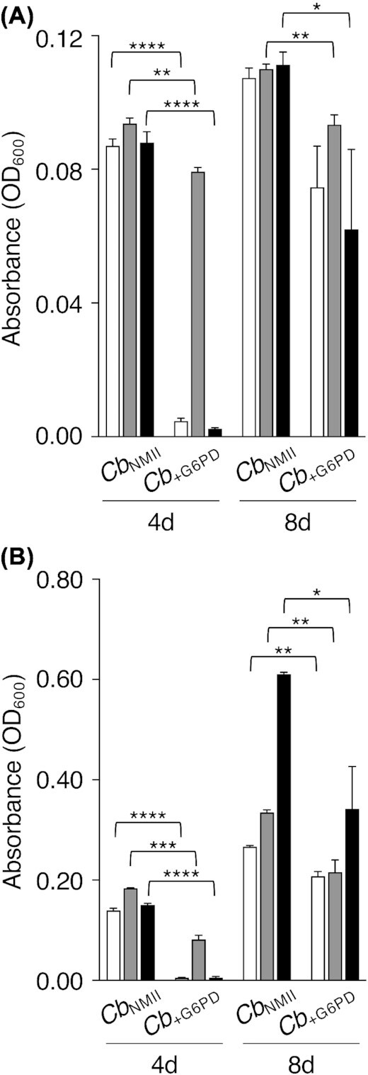
Ectopic expression of G6PD impairs C. burnetii fitness in the absence of glucose. The effect of complementing C. burnetii with zwf was tested under nutritionally controlled conditions. (A) Replication of the parental strain (CbNMII) and CbNMII::Lp-zwf (Cb+G6PD) was measured by analysis of OD600 in medium not supplemented with glucose (white), or media supplemented with either 5 mM glucose (gray) or glutamate (black). (B) Replication of CbNMII and Cb+G6PD was measured by analysis of OD600 using medium supplemented with the proteinogenic amino acid tryptophan but not glucose (white), or media supplemented with tryptophan and either 5 mM glucose (gray) or glutamate (black). Data represent the average of three independent experiments ± standard error of the mean (SEM). Asterisks indicate statistical significance (*P < 0.05; **P < 0.01; ***P < 0.001; ****P < 0.0001, unpaired Student's t-test).
As C. burnetii is a BIOP known to colonize different types of tissues, the effect of ectopic expression of G6PD was tested during C. burnetii intracellular replication in physiologically distinct cell lines (Fig. 2). Accordingly, murine macrophage-like J774A.1 and African green monkey kidney epithelial Vero cells cultured in medium containing 11 mM glucose were infected with the parental strain or CbNMII::Lp-zwf and bacterial loads determined by measuring genome equivalents (GEs) every 2 days over 8 days, essentially as described (Sanchez and Omsland 2020). In J774A.1 cells, the parental strain and CbNMII::Lp-zwf replicated with similar kinetics. During infection of Vero cells, CbNMII::Lp-zwf showed statistically significant reduced growth throughout the log phase. Infections performed with media containing 2.5 or 5 mM glucose did not result in exacerbated growth phenotypes for CbNMII::Lp-zwf, suggesting that even 2.5 mM glucose represents an excess of glucose or that the host cells adjust to glucose limitation, thus preventing a negative effect on the pathogen (data not shown). Overall, as seen under axenic conditions, expression of G6PD can impair C. burnetii intracellular replication.
Figure 2.
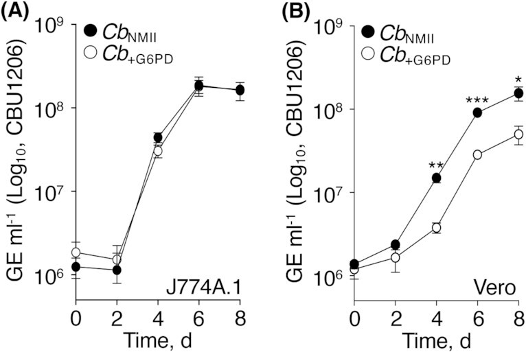
Ectopic expression of G6PD impairs C. burnetii fitness during intracellular replication. The effect of complementing C. burnetii with zwf was tested under intracellular replication in physiologically diverse cell lines. (A) Replication of the parental strain (CbNMII) and CbNMII::Lp-zwf (Cb+G6PD) in J774A.1 cells. (B) Replication of CbNMII and Cb+G6PD in Vero cells. Data points represent the average of three independent experiments ± SEM. Asterisks indicate statistical significance (*P < 0.05; **P < 0.01; ***P < 0.001, unpaired Student's t-test).
G6PD expression alters C. burnetii intracellular NADPH pools
To probe the mechanistic basis for why G6PD exhibits a negative impact on C. burnetii fitness under conditions of glucose limitation, intracellular pools of NADP+ and NADPH were measured in the parental strain and CbNMII::Lp-zwf cultured under different concentrations of glucose. As shown (Fig. 1), CbNMII::Lp-zwf does not develop by day 4 in medium not supplemented with glucose. However, near equivalent biomass production was observed by the parental strain and CbNMII::Lp-zwf in media containing 0.5 or 5 mM glucose (Fig. S2, Supporting Information). Notably, CbNMII::Lp-zwf replicated with higher variability under conditions of 0.5 and 1.5 (data not shown) mM glucose, consistent with suboptimal growth under glucose limitation. Therefore, 0.5 mM glucose represents a limiting glucose condition that supports generation of biomass for analysis by late exponential phase (i.e. day 4). When cultured in the presence of 0.5 mM glucose, CbNMII::Lp-zwf produced significantly more (∼50%) NADPH compared with the parental strain (Fig. 3A), consistent with expression of a functional G6PD enzyme. The (moderate) increase in NADPH produced by CbNMII::Lp-zwf is similar to that (∼30%) observed in E. coli upon induced expression of zwf (Sandoval, Arenas and Vásquez 2011). In medium containing 5 mM glucose, a condition where overall biomass production for the parental strain and CbNMII::Lp-zwf was similar and highly consistent between replicates (Fig. S2, Supporting Information), NADP+ and NADPH pools were equivalent between strains (Fig. 3B). Both strains exhibited low NADP+ pools in the presence of 5 mM glucose, with half the biological replicates testing below the limit of NADP+ detection.
Figure 3.
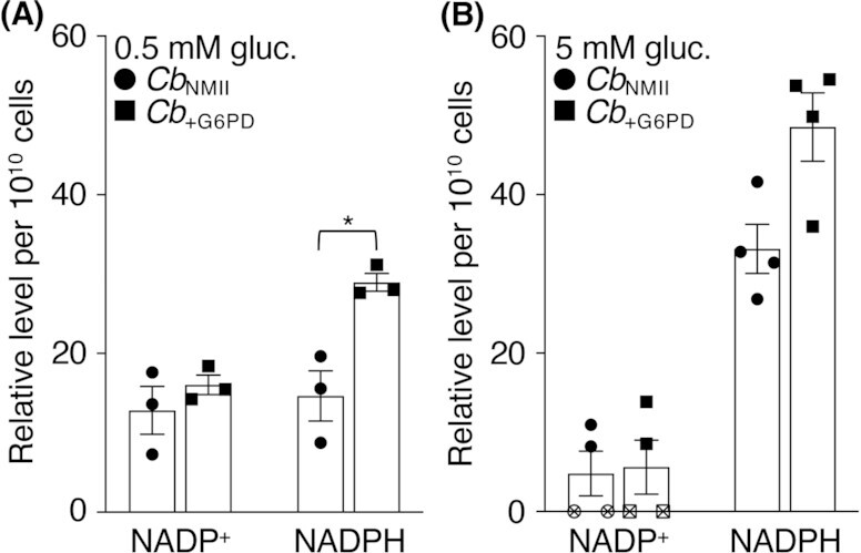
Coxiellaburnetii NADP+ and NADPH pools are affected by glucose availability. Intracellular pools of NADP+ and NADPH were measured in the parental strain (CbNMII) and CbNMII::Lp-zwf (Cb+G6PD) following 4 days of replication in media containing (A) 0.5 or (B) 5.0 mM glucose. Each symbol represents a biological replicate. Open symbols indicate samples for which cofactor levels were below the level of detection. The data represent relative averages of three to four independent experiments ± SEM. Asterisks indicate statistical significance (*P < 0.05, paired Student's t-test).
METHODS
Bacteria, cell lines and culture conditions
Coxiella burnetii NMII, clone 4 (RSA 439) was the parental strain used. The base medium used for bacterial culture was a modified ACCM-2 medium not supplemented with glucose. Bacteria were cultured with inocula normalized to 1 × 10⁶ GEs/ml under microaerobic (5% O2) conditions in the presence of 5% CO2. Air was displaced by nitrogen gas.
To complement C. burnetii with zwf, the gene was amplified from L. pneumophila subsp. pneumophila (ATCC 33152) DNA (LPG_RS02085, NC_002942) by PCR using the primers zwfLp-pJB_Forward (5′-CACTCCCTATCAGTGATAGAGAAAAGGATATATGCTCTTAAAAGCAGAATCGAAG-3′) and zwfLp-pJB_Reverse (5′-TATAATCACCGTCATGGTCTTTGTAGTCCCCTAAAATTATGAAACTGCATTGT-3′), cloned into pJB-CAT (Omsland et al. 2011) and C. burnetii transformed by electroporation according to standard protocols (Esquerra et al. 2017). Constitutive expression of zwf occurs under control of the low-medium strength promoter 1169P (Omsland et al. 2011). The resulting strain CbNMII::Lp-zwf was isolated by clonal isolation (Sanchez, Vallejo‐Esquerra and Omsland 2018) and characterized by PCR (Fig. S1, Supporting Information) to confirm successful transformation with pJB-CAT::Lp-zwf, and analysis of bacterial NADPH pools (Fig. 3).
Analysis of C. burnetii intracellular replication in J774A.1 (ATCC TIB-67) and Vero (ATCC CCL-81) cells was done essentially as described (Sanchez and Omsland 2020). Host cells were seeded in 12-well plates and infected with C. burnetii at a multiplicity of infection (MOI) of 5 via centrifugation (30 min at 400 × g, RT). Material from duplicate wells was used for quantification of GEs (Coleman et al. 2004) every 2 days.
Measurement of NADP+/NADPH
Bacterial NADP+/NADPH was measured using a commercial kit, NADP+/NADPH Quantification Kit (Sigma-Aldrich, St Louis, MO), with some modifications to the manufacturer's directions. Coxiella burnetii cytoplasmic extracts were prepared by pelleting bacteria from 4-day cultures, washing pellets in 0.4 ml of ice-cold ACCM-2 inorganic salts (Omsland et al. 2011), then resuspending pellets in 0.4 ml NADP+/NADPH extraction buffer. Samples were incubated for 10 min on ice before SDS was added at a final concentration of 0.02% (v/v), then incubated at RT for 5 min to aid lysis and inhibition of enzymatic activity in extracts. Samples were centrifuged to remove large debris (10 min at 20 000 × g, 4°C), before being split into two aliquots one of which was heated to 60°C for 30 min to decompose NADP+. Samples were stored at −20°C until analysis. Levels of NADP+ and NADPH were normalized to the number of bacterial cells as measured by GEs.
Statistical analysis
Analysis of statistical significance was done using Prism software (GraphPad Inc., San Diego, CA).
DISCUSSION
The canonical PPP has two branches: a reductive branch serving a critical function in nucleotide biosynthesis via generation of pentoses and an oxidative (ox) branch recognized as a major mechanism for regenerating NADPH. Because the C. burnetii genome is devoid of two genes that encode enzymes in the oxPPP, namely G6PD and 6-phosphogluconate dehydrogenase (6PGD), this pathogen has been proposed to metabolize under a suboptimal capacity to regenerate NADPH (Omsland et al. 2008; Omsland and Heinzen 2011). As in other bacteria, a critical role for nadB in synthesizing NAD+, required for generation of the reducing equivalents NADH and NADP+/NADPH, has been established for C. burnetii (Bitew et al. 2018). By ectopically expressing a gene encoding G6PD in C. burnetii, we sought to probe the physiological implications of increased capacity for NADPH regeneration in this pathogen. Generated data show that while expression of G6PD enhances NADPH biosynthesis, bacterial physiological fitness is severely compromised under glucose limitation.
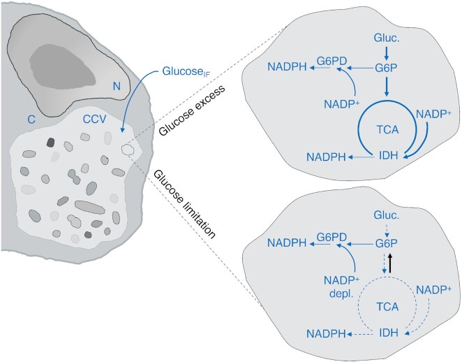
It is difficult to envision a specific mechanism for why enhanced NADPH biosynthesis would exert a negative effect on C. burnetii; however, G6PD-mediated depletion of limiting bacterial NADP+ and/or glucose-6P pools could reduce central metabolic flux (Fig. 4). This would be consistent with a loss of oxPPP capacity to ensure the availability of NADP+ for optimal flux through the tricarboxylic acid (TCA) cycle, which in C. burnetii includes an NADP+-dependent enzyme (see below). Optimal physiological fitness would thus be achieved by stabilizing central metabolism and TCA cycle activity. Because C. burnetii does not express a hexokinase, this pathogen relies on an alternative and possibly less efficient mechanism for phosphorylation of glucose. As G6PD activity is affected by concentrations of both substrate (i.e. glucose-6P) and coenzyme (NADP+) (Olavarría, Valdés and Cabrera 2012), expression of G6PD in C. burnetii could counteract other processes that depend on glucose-6P or NADP+. As observed with Zwf-1 of Pseudomonas fluorescence, bacterial adenosine triphosphate (ATP) pools can interfere with enzyme activity (Maleki, Maerk and Valla 2015) and prevent regeneration of NADPH under conditions where an energy source (e.g. glucose-6P) is available in excess. Alternatively, NADPH can inhibit synthesis of NADP+ from NAD+ via NAD kinase, as shown in Mycobacterium tuberculosis (Raffaelli et al. 2004). Potential interference of G6PD activity with NADP+ synthesis represents an alternative explanation for why C. burnetii does not benefit from expression of this enzyme. Because C. burnetii colonizes diverse host organisms, tissues and cells, expression of G6PD could negatively impact C. burnetii fitness under intracellular conditions where glucose is limited. Therefore, C. burnetii may have lost the oxPPP due to variable glucose availability within the CCV. Although data presented in Fig. 1 show an extended lag phase for CbNMII::Lp-zwf in the presence of glutamate, the possibility that another gluconeogenic carbon source (e.g. glycerol; Häuslein et al. 2017) could support growth of CbNMII::Lp-zwf as effectively as that observed with glucose cannot be excluded.
Figure 4.
Activity of G6PD as a source of metabolic imbalance in Coxiella. Expression of an NADP+-dependent isocitrate dehydrogenase (IDH) makes TCA cycle activity a mechanism for regeneration of NADPH in C. burnetii. The CCV is expected to be supplied with glucose via interstitial fluid (GlucoseIF). Under glucose excess, glycolysis and the TCA cycle run normally even with expression of G6PD. Bacterial NADP+ is present in excess allowing metabolic flux and bacterial replication. Under glucose limitation, glycolysis halts. Bacterial NADP+ (and any cellular reserve of glucose-6P) is depleted by G6PD, thus preventing central metabolic flux and replication until gluconeogenic activity restores sufficient levels of core metabolites to support production of biomass. N = host cell nucleus; C = host cell cytosol. The black arrow indicates gluconeogenesis.
Carbon source utilization in C. burnetii
Different carbon and/or energy sources have been proposed to serve as preferred substrates to fuel C. burnetii central metabolism. In 1964, Ormsbee and Peacock pointed to pyruvate as the principal energy source (Ormsbee and Peacock 1964), while later analyses of how different substrates affect pathogen membrane potential, maintenance of cytoplasmic pH and synthesis of ATP together pointed to glutamate as the preferred carbon and energy source for the pathogen (Hackstadt and Williams 1981a,b, 1983; Hackstadt 1983). In C. burnetii central carbon metabolism, glutamate is likely converted to α-ketoglutarate and funneled into the TCA cycle, thereby fueling gluconeogenesis. More recently, via genetic disruption of pckA (encoding PEPCK, the first committed step of gluconeogenesis), C. burnetii was shown to replicate efficiently when forced to utilize glucose to fuel biomass production (Esquerra et al. 2017). Isotopolog labeling in combination with metabolomic analysis has also pointed to promiscuous carbon source utilization in C. burnetii (Häuslein et al. 2017). Recent data has further revealed the ability of C. burnetii to import glucose via more than one transporter (Kuba et al. 2019). While current understanding suggests that utilization of glucose is important in C. burnetii central metabolism, the extent to which glucose is utilized by C. burnetii is obscured by apparent lack of a canonical mechanism to phosphorylate glucose.
Phosphorylation of glucose (in bacteria, typically performed by hexokinase) is critical for both oxidation of glucose via glycolysis and the oxPPP, of which G6PD is the initial enzyme. Despite apparent lack of a hexokinase gene in the C. burnetii genome (Seshadri et al. 2003), in 1962 Paretsky and colleagues described ‘hexokinase activity’ in C. burnetii cytoplasmic extracts via radiolabeling of glucose with 32P-carbamoyl phosphate (Paretsky, Consigli and Downs 1962). More recently, a phosphoenolpyruvate-based phosphotransferase system has also been suggested to function in C. burnetii glucose phosphorylation (Häuslein et al. 2017). A C. burnetii glucose 6-phosphatase (CBU1267, C. burnetii RSA493) has been proposed to function in pathogen glucose phosphorylation (Omsland and Heinzen 2011) but the apparent involvement of this enzyme in phosphatidylglycerol metabolism argues against a role in glucose phosphorylation (Stead, pers. comm.). While the biochemical mechanism for glucose phosphorylation in C. burnetii remains uncertain, the pathogen exhibits flexibility in the use of gluconeogenic versus glycolytic carbon substrates. The nutritional context of different host cells and tissues likely results in C. burnetii being exposed to environments with different availabilities of specific carbon sources. Thus, metabolic plasticity in central metabolism is a potentially critical virulence adaptation for C. burnetii that increases the likelihood of pathogen replication in nutritionally diverse environments. Selection against G6PD to optimize C. burnetii metabolic fitness under conditions of reduced glucose availability may represent a significant evolutionary adaptation of this pathogen.
First demonstrated experimentally in 1981 (Hackstadt and Williams 1981b), optimal C. burnetii metabolic activity is strictly dependent on moderately acidic pH. As observed with glutamate and glucose transport and catabolism (Hackstadt and Williams 1981b), extracellular pH influences the ability of C. burnetii to transport specific carbon sources and as such impacts which carbon source(s) the pathogen can use. More recently, it was demonstrated that C. burnetii manipulates CCV pH via secretion of type IV effector molecules, possibly to prevent activity of otherwise inhibitory proteolytic enzymes (Samanta et al. 2019). In addition to pH, a physiological dependence on CO2 has been established for C. burnetii (Esquerra et al. 2017) and correlates with activity of specific metabolic enzymes that require CO2 or bicarbonate (HCO3−). Carbamoyl phosphate is synthesized in part from HCO3−, generated enzymatically by carbonic anhydrase (CA). For C. burnetii, carbamoyl phosphate-dependent phosphorylation of glucose suggests CO2 metabolism may be a critical factor in regulating glucose metabolism. Notably, the C. burnetii CA (CBU0139, C. burnetii RSA493) is situated between the cell division-related genes ftsA and ftsQ (e.g. Mahone and Goley 2020), consistent with co-regulation of CA and genes directly related to cell division. Because glycolytic activity is directly related to generation of core metabolites required for biomass production, the sensing of CO2, a molecule produced by respiring animals, would be a clever trigger for C. burnetii replication upon infection of a new host.
The PPP and alternative strategies for NADPH regeneration in C. burnetii
Microbes rely on several enzymes for regeneration of NADPH, using NADP+ as a substrate (Spaans et al. 2015). In E. coli, a bacterium recognized for its incredible metabolic plasticity, NADPH is regenerated by five principal mechanisms (Lindner et al. 2018): G6PD and 6PGD of the oxPPP, NADP+-dependent malic enzyme and NADP+-dependent isocitrate dehydrogenase (IDH) of the TCA cycle, and the transhydrogenase PntAB. In comparison, of these canonical enzymes C. burnetii only encodes two, namely NADP+-dependent IDH (Nguyen et al. 1999) (CBU1200, C. burnetii RSA493) and PntAB (CBU1955/CBU1957, C. burnetii RSA493). The isotype of malic enzyme encoded by C. burnetii is annotated as the NAD+-dependent form (EC 1.1.1.38) (CBU0823, C. burnetii RSA493), not the NADP+-dependent form (EC 1.1.1.40) recognized as a source of NADPH. Of significance, the short-chain dehydrogenase SdrA (CBU1276, C. burnetii RSA493) was recently identified as a physiologically significant NADPH-regenerating enzyme in C. burnetii (Bitew et al. 2020). Predicted proteins with similarity to SdrA are found in other organisms, suggesting SdrA-dependent regeneration of NADPH is not unique to C. burnetii. Overall, IDH, PntAB and SdrA are the most likely sources of NADPH in C. burnetii. Under conditions of suboptimal TCA cycle activity, PntAB and/or SdrA may be critical for pathogen fitness.
Complementation of C. burnetii with G6PD resulted in a significant delay in pathogen replication under conditions of restricted glucose availability. Therefore, absence of oxPPP in C. burnetii is consistent with metabolic adaptation to environments with limiting glucose. Under such conditions, C. burnetii has the capacity to replicate near optimally using gluconeogenesis (Esquerra et al. 2017). Multiple amino acid auxotrophies prevent C. burnetii from generating biomass in the absence of amino acids. Of the gluconeogenic amino acids, C. burnetii has retained the capacity to synthesize glutamate (Sandoz et al. 2016), suggesting a selective pressure on glutamate generation under conditions when other (proteinogenic) amino acids are available for biomass production, but glucose is limiting.
General metabolic flux (e.g. glycolysis vs gluconeogenesis) and specific carbon source utilization has predictable effects on C. burnetii energetics. For the purpose of generating both core metabolites and conserve energy, glucose may be the most attractive carbon and energy source for this organism. Glucose is expected to be maintained at relatively stable concentration in the interstitial fluid of mammalian hosts, thus representing a reliable carbon source for C. burnetii assuming that the CCV is fed by fluid phase endocytosis during natural infection. Currently, there is no evidence to suggest that host cells infected with C. burnetii under natural conditions do not traffic compounds from the extracellular space into the CCV, as observed during infection of cultured cells (Heinzen et al. 1996).
Diversity in energy metabolism and PPP competence among intracellular pathogens
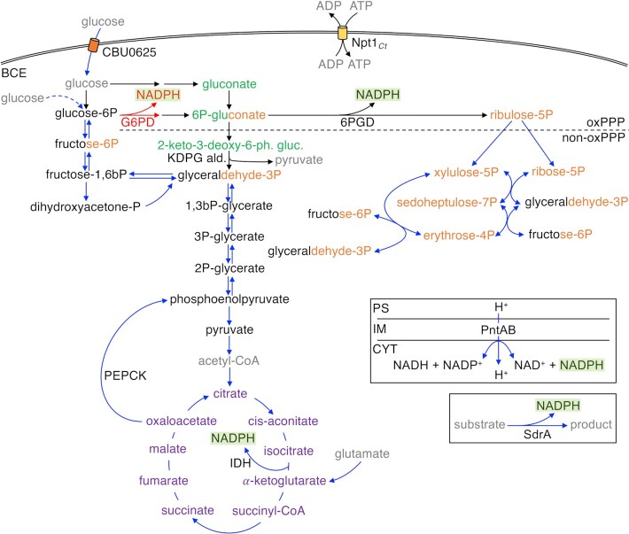
BOIPs and other bacterial pathogens that rely on intracellular parasitism as part of their virulence strategies show remarkable diversity in carbon and energy source utilization (Fig. 5). This includes bacteria of the genus Brucella, which have evolved to use different strategies for glucose metabolism. Notably, key species (e.g. B. abortus) share some pathogenic characteristics similar to those described for C. burnetii, including placental colonization in cattle and the ability to establish chronic infections. While pathogenic zoonotic Brucella spp. rely on the PPP for oxidation of glucose, species that group phylogenetically closer to free-living ancestors metabolize glucose via both the PPP and the Entner–Doudoroff (ED) pathway (Machelart et al. 2020), an alternative to the energetically favorable Embden–Meyerhof–Parnas (EMP) pathway (Flamholz et al. 2013). In comparison, L. pneumophila, a close phylogenetic relative of C. burnetii that also relies on intracellular parasitism as a virulence strategy, has retained a gene encoding glucokinase (EC 2.7.1.2) (Harada et al. 2010), an enzyme that is highly specific for glucose as a substrate, and thus similar to hexokinase (EC 2.7.1.1) with respect to glucose phosphorylation. Still, distribution of radiolabeled glutamate into several cellular components suggests that Legionella relies on amino acids as a significant carbon source (Tesh, Morse and Miller 1983). The lack of a canonical enzyme for glucose phosphorylation in C. burnetii distinguishes this bacterium from L. pneumophila, which metabolizes glucose via both the oxPPP and (primarily) the ED pathway (Schunder et al. 2014; Häuslein et al. 2016, 2017). In addition to absence of G6PD, C. burnetii does not encode the key ED-specific enzyme 6-phosphogluconate dehydratase (edd) (C. burnetii RSA493). The C. burnetii genome encodes a protein (CBU1277, C. burnetii RSA493) with 48% sequence identity at the amino acid level compared to the ED-specific enzyme 2-keto-3-deoxy-6-phosphogluconate aldolase (eda) (EC 4.1.2.14, E. coli K12), suggesting C. burnetii retains some enzymatic capacity of the ED pathway. Similar to C. burnetii, Francisella tularensis does not encode genes for a complete ED pathway or the oxPPP (Ziveri, Barel and Charbit 2017).
Figure 5.
Major components of central energy metabolism and NADPH regeneration in C. burnetii in comparison to other parasitic bacteria. Steps are indicated by black (canonical) or blue (C. burnetii) arrows. The broken blue line indicates a step for which the reaction mechanism in C. burnetii is uncertain. Red indicates reaction performed by G6PD and resulting NADPH in Cb+G6PD. NADPH from relevant reactions are highlighted in green. Intermediates related to major pathways are color coded: glycolysis/EMP and gluconeogenesis (black); ED pathway (green); PPP (orange); and TCA cycle (purple). Some intermediates relate to more than one pathway (dual coloring). Gray indicates components not related to a specific pathway. BCE indicates the bacterial cell envelope. oxPPP and non-oxPPP show separation between the two branches of the PPP. The reaction substrate(s) and product(s) for SdrA have not been determined. PntAB is coupled with the proton motive force. G6PD, glucose-6-phosphate dehydrogenase (zwf); 6PGD, 6-phosphogluconate dehydratase (edd); KDPG ald., 2-keto-3-deoxy-6-phosphogluconate aldolase (eda); PEPCK, phosphoenolpyruvate carboxykinase (pckA); PntAB, pyridine nucleotide transhydrogenase (pntA/pntB); IDH, isocitrate dehydrogenase (icd); and SdrA, short-chain dehydrogenase reductase A (sdrA). Boxes indicate reactions not directly related to the central metabolic network. PS, periplasmic space; IM, inner membrane; and CYT, cytoplasm.
Unlike C. burnetii, bacteria of the genus Chlamydia can behave as energy parasites. Curiously, the infectious elementary body (EB) and replicative reticulate body (RB) cell forms of Chlamydia trachomatis have been shown to rely on vastly different strategies for energy generation and acquisition (Omsland et al. 2012). The EB form does not appear to readily take up extracellular ATP but converts glucose-6P to ATP (Omsland et al. 2012). In contrast, the RB form scavenges ATP directly from the host (Tipples and McClarty 1993) via the Npt1Ct ADP/ATP translocase (Tjaden et al. 1999), yet may also take up and metabolize glucose. In addition to ATP, Npt1Ct functions in chlamydial NAD+ acquisition, critical in the absence of capacity to synthesize this cofactor (Fisher, Fernández and Maurelli 2013). While C. trachomatis does not appear capable of utilizing (nonphosphorylated) glucose, the bacterium promotes upregulation of host glucose transporters GLUT1 and GLUT3 during infection to support pathogen replication (Wang, Hybiske and Stephens 2017). The protein UhpC has been identified as a transporter for phosphorylated glucose in Chlamydia (Schwöppe, Winkler and Neuhaus 2002). Unlike C. burnetii, C. trachomatis (serovar L2) has an intact PPP (Iliffe‐Lee and McClarty 1999) and is thus expected to regenerate NADPH via the oxPPP. In contrast to Coxiella, Chlamydia, Legionella and Francisella, pathogens within the genus Rickettsia do not have genes for either glycolysis or the PPP (Driscoll et al. 2017). Consistent with absence of pathways for oxidation of glucose, Rickettsia prowazekii does not appear able to take up glucose or readily transport glucose phosphates (Winkler and Daugherty 1986). Similar to C. trachomatis, R. prowazekii can acquire extrabacterial ATP (Winkler 1976; Audia and Winkler 2006) and are thus energy parasites.
Outlook
Glucose is a molecule with unparalleled significance in central carbon metabolism. Therefore, deciphering the mechanism(s) behind C. burnetii glucose utilization has unquestionable significance in understanding the biology of this pathogen. The possibility that C. burnetii utilizes a novel approach to phosphorylate glucose makes the path for this research especially exciting. Moreover, questions remain as to why the C. burnetii central metabolic machinery has evolved away from canonical glucose phosphorylation (via hexokinase) and metabolism via the oxPPP. Loss of these otherwise well-conserved enzymes suggests their activity in C. burnetii impedes pathogen fitness. If this is indeed the case, beyond the test tube and cultured host cells as shown for G6PD in this study, understanding the underlying mechanisms would shed light on prominent anomalies in C. burnetii central metabolism and the evolutionary pressure(s) driving their existence.
While C. burnetii gene/genome content has been shown to be similar between pathogen isolates, their organization varies widely, including for genes related to central metabolism (Beare et al. 2009). For instance, the MFS superfamily of membrane transporters, which facilitate transport of small molecules including amino acids, exhibits heterogeneity among C. burnetii isolates (Beare et al. 2009). Because genome organization can affect expression of specific genes, it is possible that this observed diversity in genome organization represents a genuine virulence characteristic of C. burnetii. This is consistent with different C. burnetii isolates exhibiting a range of pathogenic potentials (Russell-Lodrigue et al. 2009). As differential gene expression may relate to isolate-specific capacity to metabolize various carbon and/or energy sources, it seems relevant to test potential links between isolate-specific central metabolic capacity and virulence. In both M. tuberculosis (Marrero et al. 2010) and F. tularensis (Brissac et al. 2015; Radlinski et al. 2018), gluconeogenic capacity is critical for optimal virulence in animal models. Determining the role of glycolysis and gluconeogenesis in C. burnetii virulence, including how such activities may impact the pathogenicity of different isolates, represents fundamental aspects of C. burnetii biology that have yet to be investigated.
In addition to how features endogenous to C. burnetii (e.g. gene content and genome structure) affect pathogen physiology, external conditions represent critical variables in this organism's physiology. This is in part due to the connection between the host extracellular environment (e.g. interstitial fluid) and the CCV via fluid-phase endocytosis (Heinzen et al. 1996). Development and use of physiological media that better reflect natural conditions for C. burnetii replication may be fruitful in this respect. For example, a Coxiella mutant unable to undergo gluconeogenesis (Cb∆pckA) exhibited an impairment in intracellular replication only when the concentration of glucose in the cell culture medium was reduced from 11 (i.e. the concentration of glucose in RPMI-1640) to 5 mM, a concentration of glucose more reflective of nondiabetic animals. For Salmonella enterica, pathogen metabolic processes can be masked under (host) cell culture conditions where nutrients (i.e. glucose) are in excess (Diacovich et al. 2016), a characteristic of commercial tissue culture media. Because the metabolism of BOIPs and their host cells is interconnected, effort must be placed on appropriately modeling a host's nutritional environment to allow for a physiologically relevant model to study the effect of nutrient availability on pathogen metabolism.
With the characteristic of transitioning between two physiologically distinct cell forms (i.e. the replicative large cell variant [LCV] and nonreplicative small cell variant [SCV]), the study of C. burnetii arguably poses challenges irrelevant to bacteria (e.g. E. coli) that have served as models for most developments in our understanding of bacterial physiology. As encouraged by Neidhardt (2006), researchers should be careful to assess bacterial (metabolic) activities and functions under conditions that facilitate reproducibility. For example, any intracellular or axenic analysis of C. burnetii growth that includes time points past the point of LCV-to-SCV transition will result in data representing the product of both LCV and SCV activity, or lack thereof. When possible, care should be taken to clearly specify and distinguish between growth phases, replication rates and cell forms. Key characteristics of C. burnetii replication have been described for both intracellular (e.g. Vero; Coleman et al. 2004) and axenic (e.g. ACCM [Omsland et al. 2009] vs ACCM-2 [Omsland et al. 2011; Sandoz et al. 2014]) conditions.
ACKNOWLEDGMENTS
We thank Drs Paul Beare and Bob Heinzen, Rocky Mountain Laboratories, NIAID, NIH, for plasmid pJB-CAT.
FUNDING
This work was supported by fellowships from the Poncin Scholarship Fund and the National Institutes of Health (1F31AI150167-01) to SES, and funds from Washington State University to AO.
Supplementary Material
Contributor Information
Savannah E Sanchez, Paul G. Allen School for Global Health, Washington State University, Pullman, WA 99164, USA; School of Molecular Biosciences, Washington State University, Pullman, WA 99164, USA.
Anders Omsland, Paul G. Allen School for Global Health, Washington State University, Pullman, WA 99164, USA.
AUTHOR CONTRIBUTIONS
SES and AO: experimental design, data analysis and manuscript preparation; SES: execution of experiments; AO: conception.
Conflict of interest
None declared.
REFERENCES
- Audia JP, Winkler HH. Study of the five Rickettsia prowazekii proteins annotated as ATP/ADP translocases (Tlc): only Tlc1 transports ATP/ADP, while Tlc4 and Tlc5 transport other ribonucleotides. J Bacteriol. 2006;188:6261–8. [DOI] [PMC free article] [PubMed] [Google Scholar]
- Baumgärtner W, Bachmann S. Histological and immunocytochemical characterization of Coxiella burnetii-associated lesions in the murine uterus and placenta. Infect Immun. 1992;60:5232–41. [DOI] [PMC free article] [PubMed] [Google Scholar]
- Beare PA, Gilk SD, Larson CLet al. Dot/Icm type IVB secretion system requirements for Coxiella burnetii growth in human macrophages. mBio. 2011;2:e00175–11. [DOI] [PMC free article] [PubMed] [Google Scholar]
- Beare PA, Unsworth N, Andoh Met al. Comparative genomics reveal extensive transposon-mediated genomic plasticity and diversity among potential effector proteins within the genus Coxiella. Infect Immun. 2009;77:642–56. [DOI] [PMC free article] [PubMed] [Google Scholar]
- Bitew MA, Hofmann J, Souza DPDet al. SdrA, an NADP(H)-regenerating enzyme, is crucial for Coxiella burnetii to resist oxidative stress and replicate intracellularly. Cell Microbiol. 2020;22:e13154. [DOI] [PubMed] [Google Scholar]
- Bitew MA, Khoo CA, Neha Net al. De novo NAD synthesis is required for intracellular replication of Coxiella burnetii, the causative agent of the neglected zoonotic disease Q fever. J Biol Chem. 2018;293:18636–45. [DOI] [PMC free article] [PubMed] [Google Scholar]
- Brissac T, Ziveri J, Ramond Eet al. Gluconeogenesis, an essential metabolic pathway for pathogenic Francisella: gluconeogenesis in Francisella virulence. Mol Microbiol. 2015;98:518–34. [DOI] [PubMed] [Google Scholar]
- Coleman SA, Fischer ER, Howe Det al. Temporal analysis of Coxiella burnetii morphological differentiation. J Bacteriol. 2004;186:7344–52. [DOI] [PMC free article] [PubMed] [Google Scholar]
- Diacovich L, Lorenzi L, Tomassetti Met al. The infectious intracellular lifestyle of Salmonella enterica relies on the adaptation to nutritional conditions within the Salmonella-containing vacuole. Virulence. 2016;8:975–92. [DOI] [PMC free article] [PubMed] [Google Scholar]
- Driscoll TP, Verhoeve VI, Guillotte MLet al. Wholly Rickettsia! Reconstructed metabolic profile of the quintessential bacterial parasite of eukaryotic cells. mBio. 2017;8:e00859–17. [DOI] [PMC free article] [PubMed] [Google Scholar]
- Eldin C, Mélenotte C, Mediannikov Oet al. From Q fever to Coxiella burnetii infection: a paradigm change. Clin Microbiol Rev. 2017;30:115–90. [DOI] [PMC free article] [PubMed] [Google Scholar]
- Esquerra EV, Yang H, Sanchez SEet al. Physicochemical and nutritional requirements for axenic replication suggest physiological basis for Coxiella burnetii niche restriction. Front Cell Infect Microbiol. 2017;7:190. [DOI] [PMC free article] [PubMed] [Google Scholar]
- Fisher DJ, Fernández RE, Maurelli AT. Chlamydia trachomatis transports NAD via the Npt1 ATP/ADP translocase. J Bacteriol. 2013;195:3381–6. [DOI] [PMC free article] [PubMed] [Google Scholar]
- Flamholz A, Noor E, Bar-Even Aet al. Glycolytic strategy as a tradeoff between energy yield and protein cost. Proc Natl Acad Sci USA. 2013;110:10039–44. [DOI] [PMC free article] [PubMed] [Google Scholar]
- Hackstadt T, Williams JC. Biochemical stratagem for obligate parasitism of eukaryotic cells by Coxiella burnetii. Proc Natl Acad Sci USA. 1981b;78:3240–4. [DOI] [PMC free article] [PubMed] [Google Scholar]
- Hackstadt T, Williams JC. pH dependence of the Coxiella burnetii glutamate transport system. J Bacteriol. 1983;154:598–603. [DOI] [PMC free article] [PubMed] [Google Scholar]
- Hackstadt T, Williams JC. Stability of the adenosine 5′-triphosphate pool in Coxiella burnetii: influence of pH and substrate. J Bacteriol. 1981a;148:419–25. [DOI] [PMC free article] [PubMed] [Google Scholar]
- Hackstadt T. Estimation of the cytoplasmic pH of Coxiella burnetii and effect of substrate oxidation on proton motive force. J Bacteriol. 1983;154:591–7. [DOI] [PMC free article] [PubMed] [Google Scholar]
- Harada E, Iida K-I, Shiota Set al. Glucose metabolism in Legionella pneumophila: dependence on the Entner–Doudoroff pathway and connection with intracellular bacterial growth. J Bacteriol. 2010;192:2892–9. [DOI] [PMC free article] [PubMed] [Google Scholar]
- Häuslein I, Cantet F, Reschke Set al. Multiple substrate usage of Coxiella burnetii to feed a bipartite metabolic network. Front Cell Infect Microbiol. 2017;7:285. [DOI] [PMC free article] [PubMed] [Google Scholar]
- Häuslein I, Manske C, Goebel Wet al. Pathway analysis using 13C-glycerol and other carbon tracers reveals a bipartite metabolism of Legionella pneumophila. Mol Microbiol. 2016;100:229–46. [DOI] [PubMed] [Google Scholar]
- Heinzen RA, Scidmore MA, Rockey DDet al. Differential interaction with endocytic and exocytic pathways distinguish parasitophorous vacuoles of Coxiella burnetii and Chlamydia trachomatis. Infect Immun. 1996;64:796–809. [DOI] [PMC free article] [PubMed] [Google Scholar]
- Iliffe-Lee ER, McClarty G. Glucose metabolism in Chlamydia trachomatis: the ‘energy parasite’ hypothesis revisited. Mol Microbiol. 1999;33:177–87. [DOI] [PubMed] [Google Scholar]
- Kohler LJ, Roy CR. Biogenesis of the lysosome-derived vacuole containing Coxiella burnetii. Microbes Infect. 2015;17:766–71. [DOI] [PMC free article] [PubMed] [Google Scholar]
- Kuba M, Neha N, Souza DPDet al. Coxiella burnetii utilizes both glutamate and glucose during infection with glucose uptake mediated by multiple transporters. Biochem J. 2019;476:BCJ20190504. [DOI] [PMC free article] [PubMed] [Google Scholar]
- Lindner SN, Ramirez LC, Krüsemann JLet al. NADPH-Auxotrophic E. coli: a sensor strain for testing in vivo regeneration of NADPH. ACS Synth Biol. 2018;7:2742–9. [DOI] [PubMed] [Google Scholar]
- Machelart A, Willemart K, Zúñiga-Ripa Aet al. Convergent evolution of zoonotic Brucella species toward the selective use of the pentose phosphate pathway. Proc Natl Acad Sci USA. 2020;117:26374–81. [DOI] [PMC free article] [PubMed] [Google Scholar]
- Mahone CR, Goley ED. Bacterial cell division at a glance. J Cell Sci. 2020;133:jcs237057. [DOI] [PMC free article] [PubMed] [Google Scholar]
- Maleki S, Maerk M, Valla S. Mutational analysis of glucose dehydrogenase and glucose-6-phosphate dehydrogenase genes in Pseudomonas fluorescence reveal their effects on growth and alginate production. Appl Environ Microbiol. 2015;81:3349–56. [DOI] [PMC free article] [PubMed] [Google Scholar]
- Marrero J, Rhee KY, Schnappinger Det al. Gluconeogenic carbon flow of tricarboxylic acid cycle intermediates is critical for Mycobacterium tuberculosis to establish and maintain infection. Proc Natl Acad Sci USA. 2010;107:9819–24. [DOI] [PMC free article] [PubMed] [Google Scholar]
- Neidhardt FC. Apples, oranges and unknown fruit. Nat Rev Microbiol. 2006;4:876. [DOI] [PubMed] [Google Scholar]
- Newton HJ, Kohler LJ, McDonough JAet al. A screen of Coxiella burnetii mutants reveals important roles for Dot/Icm effectors and host autophagy in vacuole biogenesis. PLoS Pathog. 2014;10:e1004286. [DOI] [PMC free article] [PubMed] [Google Scholar]
- Nguyen S, To H, Yamaguchi Tet al. Molecular cloning of an immunogenic and acid-induced isocitrate dehydrogenase gene from Coxiella burnetii. FEMS Microbiol Lett. 1999;175:101–6. [DOI] [PubMed] [Google Scholar]
- Olavarría K, Valdés D, Cabrera R. The cofactor preference of glucose-6-phosphate dehydrogenase from Escherichia coli-modeling the physiological production of reduced cofactors. FEBS J. 2012;279:2296–309. [DOI] [PubMed] [Google Scholar]
- Omsland A, Beare PA, Hill Jet al. Isolation from animal tissue and genetic transformation of Coxiella burnetii are facilitated by an improved axenic growth medium. Appl Environ Microbiol. 2011;77:3720–5. [DOI] [PMC free article] [PubMed] [Google Scholar]
- Omsland A, Cockrell DC, Fischer ERet al. Sustained axenic metabolic activity by the obligate intracellular bacterium Coxiella burnetii. J Bacteriol. 2008;190:3203–12. [DOI] [PMC free article] [PubMed] [Google Scholar]
- Omsland A, Cockrell DC, Howe Det al. Host cell-free growth of the Q fever bacterium Coxiella burnetii. Proc Natl Acad Sci USA. 2009;106:4430–4. [DOI] [PMC free article] [PubMed] [Google Scholar]
- Omsland A, Heinzen RA. Life on the outside: the rescue of Coxiella burnetii from its host cell. Annu Rev Microbiol. 2011;65:111–28. [DOI] [PubMed] [Google Scholar]
- Omsland A, Sager J, Nair Vet al. Developmental stage-specific metabolic and transcriptional activity of Chlamydia trachomatis in an axenic medium. Proc Natl Acad Sci USA. 2012;109:19781–5. [DOI] [PMC free article] [PubMed] [Google Scholar]
- Ormsbee RA, Peacock MG. Metabolic activity in Coxiella burnetii. J Bacteriol. 1964;88:1205–10. [DOI] [PMC free article] [PubMed] [Google Scholar]
- Paretsky D, Consigli RA, Downs CM. Studies on the physiology of rickettsiae III. Glucose phosphorylation and hexokinase activity in Coxiella burnetii. J Bacteriol. 1962;83:538–43. [DOI] [PMC free article] [PubMed] [Google Scholar]
- Radlinski LC, Brunton J, Steele Set al. Defining the metabolic pathways and host-derived carbon substrates required for Francisella tularensis intracellular growth. mBio. 2018;9:e01471–18. [DOI] [PMC free article] [PubMed] [Google Scholar]
- Raffaelli N, Finaurini L, Mazzola Fet al. Characterization of Mycobacterium tuberculosis NAD kinase: functional analysis of the full-length enzyme by site-directed mutagenesis. Biochemistry. 2004;43:7610–7. [DOI] [PubMed] [Google Scholar]
- Russell-Lodrigue KE, Andoh M, Poels MWJet al. Coxiella burnetii isolates cause genogroup-specific virulence in mouse and guinea pig models of acute Q fever. Infect Immun. 2009;77:5640–50. [DOI] [PMC free article] [PubMed] [Google Scholar]
- Samanta D, Clemente TM, Schuler BEet al. Coxiella burnetii type 4B secretion system-dependent manipulation of endolysosomal maturation is required for bacterial growth. PLoS Pathog. 2019;15:e1007855. [DOI] [PMC free article] [PubMed] [Google Scholar]
- Sanchez SE, Omsland A. Critical role for molecular iron in Coxiella burnetii replication and viability. mSphere. 2020;5:e00458–20. [DOI] [PMC free article] [PubMed] [Google Scholar]
- Sanchez SE, Vallejo-Esquerra E, Omsland A. Use of axenic culture tools to study Coxiella burnetii. Curr Protoc Microbiol. 2018;50:e52. [DOI] [PubMed] [Google Scholar]
- Sandoval JM, Arenas FA, Vásquez CC. Glucose-6-phosphate dehydrogenase protects Escherichia coli from tellurite-mediated oxidative stress. PLoS One. 2011;6:e25573. [DOI] [PMC free article] [PubMed] [Google Scholar]
- Sandoz KM, Beare PA, Cockrell DCet al. Complementation of arginine auxotrophy for genetic transformation of Coxiella burnetii by use of a defined axenic medium. Appl Environ Microbiol. 2016;82:3042–51. [DOI] [PMC free article] [PubMed] [Google Scholar]
- Sandoz KM, Sturdevant DE, Hansen Bet al. Developmental transitions of Coxiella burnetii grown in axenic media. J Microbiol Methods. 2014;96:104–10. [DOI] [PMC free article] [PubMed] [Google Scholar]
- Schunder E, Gillmaier N, Kutzner Eet al. Amino acid uptake and metabolism of Legionella pneumophila hosted by Acanthamoeba castellanii. J Biol Chem. 2014;289:21040–54. [DOI] [PMC free article] [PubMed] [Google Scholar]
- Schwöppe C, Winkler HH, Neuhaus HE. Properties of the glucose-6-phosphate transporter from Chlamydia pneumoniae (HPTcp) and the glucose-6-phosphate sensor from Escherichia coli (UhpC). J Bacteriol. 2002;184:2108–15. [DOI] [PMC free article] [PubMed] [Google Scholar]
- Seshadri R, Paulsen IT, Eisen JAet al. Complete genome sequence of the Q-fever pathogen Coxiella burnetii. Proc Natl Acad Sci USA. 2003;100:5455–60. [DOI] [PMC free article] [PubMed] [Google Scholar]
- Signs KA, Stobierski MG, Gandhi TN. Q fever cluster among raw milk drinkers in Michigan, 2011. Clin Infect Dis. 2012;55:1387–9. [DOI] [PubMed] [Google Scholar]
- Spaans SK, Weusthuis RA, Oost J van deret al. NADPH-generating systems in bacteria and archaea. Front Microbiol. 2015;6:742. [DOI] [PMC free article] [PubMed] [Google Scholar]
- Stephens RS, Kalman S, Lammel Cet al. Genome sequence of an obligate intracellular pathogen of humans: Chlamydia trachomatis. Science. 1998;282:754–9. [DOI] [PubMed] [Google Scholar]
- Tesh MJ, Morse SA, Miller RD. Intermediary metabolism in Legionella pneumophila: utilization of amino acids and other compounds as energy sources. J Bacteriol. 1983;154:1104–9. [DOI] [PMC free article] [PubMed] [Google Scholar]
- Tipples G, McClarty G. The obligate intracellular bacterium Chlamydia trachomatis is auxotrophic for three of the four ribonucleoside triphosphates. Mol Microbiol. 1993;8:1105–14. [DOI] [PubMed] [Google Scholar]
- Tjaden J, Winkler HH, Schwöppe Cet al. Two nucleotide transport proteins in Chlamydia trachomatis, one for net nucleoside triphosphate uptake and the other for transport of energy. J Bacteriol. 1999;181:1196–202. [DOI] [PMC free article] [PubMed] [Google Scholar]
- Voth DE, Heinzen RA. Lounging in a lysosome: the intracellular lifestyle of Coxiella burnetii. Cell Microbiol. 2007;9:829–40. [DOI] [PubMed] [Google Scholar]
- Wang X, Hybiske K, Stephens RS. Orchestration of the mammalian host cell glucose transporter proteins-1 and 3 by Chlamydiacontributes to intracellular growth and infectivity. Pathog Dis. 2017;75:ftx108. [DOI] [PMC free article] [PubMed] [Google Scholar]
- Winkler HH, Daugherty RM. Acquisition of glucose by Rickettsia prowazekii through the nucleotide intermediate uridine 5′-diphosphoglucose. J Bacteriol. 1986;167:805–8. [DOI] [PMC free article] [PubMed] [Google Scholar]
- Winkler HH. Rickettsial permeability. An ADP-ATP transport system. J Biol Chem. 1976;251:389–96. [PubMed] [Google Scholar]
- Ziveri J, Barel M, Charbit A. Importance of metabolic adaptations in Francisella pathogenesis. Front Cell Infect Microbiol. 2017;7:96. [DOI] [PMC free article] [PubMed] [Google Scholar]
Associated Data
This section collects any data citations, data availability statements, or supplementary materials included in this article.


