PLOS ONE logoLink to PLOS ONE
. 2021 Jul 22;16(7):e0240570. doi: 10.1371/journal.pone.0240570

Use of non-invasive intracranial pressure pulse waveform to monitor patients with End-Stage Renal Disease (ESRD)

Cristiane Rickli 1, Lais Daiene Cosmoski 1, Fábio André dos Santos 1, Gustavo Henrique Frigieri 2, Nicollas Nunes Rabelo 3, Adriana Menegat Schuinski 1, Sérgio Mascarenhas 2, José Carlos Rebuglio Vellosa 1,*
Editor: Patrick Barry Mark4
PMCID: PMC8297761  PMID: 34292964

Abstract

End-stage renal disease (ESRD) is treated mainly by hemodialysis, however, hemodialysis is associated with frequent complications, some of them involve the increased intracranial pressure. In this context, monitoring the intracranial pressure of these patients may lead to a better understanding of how intracranial pressure morphology varies with hemodialysis. This study aimed to follow-up patients with ESRD by monitoring intracranial pressure before and after hemodialysis sessions using a noninvasive method. We followed-up 42 patients with ESRD in hemodialysis, for six months. Noninvasive intracranial pressure monitoring data were obtained through analysis of intracranial pressure waveform morphology, this information was uploaded to Brain4care® cloud algorithm for analysis. The cloud automatically sends a report containing intracranial pressure parameters. In total, 4881 data points were collected during the six months of follow-up. The intracranial pressure parameters (time to peak and P2/P1 ratio) were significantly higher in predialysis when compared to postdialysis for the three weekly sessions and throughout the follow-up period (p<0.01) data showed general improvement in brain compliance after the hemodialysis session. Furthermore, intracranial pressure parameters were significantly higher in the first weekly hemodialysis session (p<0.05). In conclusion, there were significant differences between pre and postdialysis intracranial pressure in patients with ESRD on hemodialysis. Additionally, the pattern of the intracranial pressure alterations was consistent over time suggesting that hemodialysis can improve time to peak and P2/P1 ratio which may reflect in brain compliance.

Introduction

Chronic kidney disease (CKD), a leading cause of mortality and morbidity and a growing public health problem worldwide [1], is a complex disease that requires multiple treatment approaches [2].

Hemodialysis (HD) has become the predominant renal replacement therapy (RRT) in the world [3]. However, HD is associated with frequent complications, including hypotension and muscle cramps, in addition to postdialysis complaints of headache, fatigue, and inability to concentrate, which may significantly affect patients’ quality of life [4]. Mild signs and symptoms like headache, nausea, and muscle cramps are often attributed to volume depletion due to excessive ultrafiltration, but may represent a milder but not diagnosed form of dialysis disequilibrium syndrome (DDS) [5].

Even though maintenance HD has been a routine procedure for over 50 years, the exact mechanism of DDS remains poorly understood [6] and the syndrome manifests as neurologic symptoms and signs related to osmotic fluid shifts [5]. Cerebral edema and increased intracranial pressure (ICP) are the primary contributing factors to this syndrome and are the targets of therapy [6]. Neurologic manifestations progress sequentially as cerebral edema worsens and ICP rises and, if not promptly recognized and managed, can lead to coma and even death [7].

ICP monitoring could assist in the early diagnosis of DDS, but the methods in use are highly invasive, costly, and carry complication risks. However, a noninvasive method based on volumetric skull changes detected by a sensor has been developed [8]. This method allows for quick and safe access to ICP pulse waveform morphology, which is correlated with brain compliance [9].

This study aimed to follow-up patients with end-stage renal disease (ESRD) by monitoring ICP before and after HD sessions using a noninvasive method to assess ICP variations during HD treatment.

Materials and methods

Participants

This study was approved by Research Ethics Committee of the State University of Ponta Grossa/COEP-UEPG (process number: 1.834.627). It is a prospective longitudinal study of 42 patients aged ≥ 18 years with end-stage renal disease (ESRD) from a single RRT center who received HD periodically, three times per week with two one-day intervals and one two-day interval between sessions, for six months. HD session length ranged from three to four hours depending on the patient and his/her condition. The authors declare that they adhered to the Declaration of Helsinki. All participants received information about the study and provided written informed consent.

The clinical characteristics of participants including age, gender, underlying disease, start of treatment, and comorbidities were retrieved from the electronic medical records of the RRT center. The parameters mean arterial pressure (MAP) and interdialytic weight gain (IDWG) were obtained by consulting the notes of each hemodialysis session.

Intracranial pressure (ICP) and brain compliance monitoring

In total, 4881 data points were collected during the six months of follow-up. The noninvasive ICP monitoring equipment was provided by Brain4care® (São Paulo, SP, Brazil). This noninvasive method was validated by comparison with the invasive ICP monitoring method [8,10].

Predialysis monitoring sessions were done before the HD session in a private room with the patient seated in a chair similar to the one used in the HD session while ICP was monitored for 1 to 3 min. The patient should remain still during signal acquisition and the same procedure was performed after the HD session. Fig 1 shows a flowchart of how this search was conducted.

Fig 1. Flowchart of this research.

Fig 1

CKD: Chronic kidney disease; ESRD: End-stage renal disease; HD: Hemodialysis; ICP: Intracranial pressure.

Monro-Kellie doctrine states that the skull does not expand after the fontanels are closed. However, for the development of the ICP monitor, it was proven that the skulls, even those of adults, have volumetric changes, as a result of pressure variations and the noninvasive monitoring of ICP is based on the assessment of these changes in bone structure [11].

To perform the monitoring, a sensor that detects the micrometric deformations of the skull bones is attached to a plastic headband strapped around the patient’s head. The device filters, amplifies and digitizes the signal from the sensor before sending it to a computer [8].

Noninvasive ICP monitoring data were obtained through analysis of ICP waveform morphology. The ICP waveform is a modified blood pressure wave with three distinct peaks. The first peak (P1, or ‘percussive wave’) is the result of arterial pressure transmitted from the choroid plexus. The second peak (P2) signifies brain compliance and the last peak (P3) is the result of the aortic valve closure. Thus, under normal ICP conditions, the amplitude of the peaks is such that P1>P2>P3 [12,13]. However as brain compliance decreases and the ICP increases, the amplitude of the wave also increases and the P2 component of the wave exceeds P1 and P3 [14].

Following the ICP monitoring, the software saved the data to files that were later uploaded to Brain4care® for analysis. The result is a report containing the time to peak (TTP) and P2/P1 ratio. TTP was defined as the time at which the ICP curve reaches its tallest peak, either P1 or P2, starting from the start of the curve. The P2/P1 ratio assesses brain compliance and was defined as the ratio between the amplitudes of peaks P2 and P1 (R = AmpP2/AmpP1). Brain compliance is normal when R < 1.0 (P2<P1) and abnormal when R > 1.0 (P2>P1).

Statistical analysis

Clinical characteristics are expressed as mean ± standard deviation (SD) for continuous variables and relative frequency (%) for categorical variables. First, the mean ± SD of noninvasive ICP parameters (TTP and P2/P1 ratio) were calculated separately for the three weekly HD sessions (1st, 2nd, and 3rd) for each month of follow-up. The normality of the data (TTP and P2/P1 ratio) in each evaluation period at pre-dialysis and post-dialysis were assessed and confirmed with the Kolmogorov-Sminov test (p>0.05).

Predialysis and postdialysis noninvasive ICP parameters were compared using the Student’s t-test for paired samples. Besides, TTP and P2/P1 ratio were also compared between the three weekly HD sessions using repeated measures analysis of variance (ANOVA) and Tukey multiple comparison tests. For assessment of MAP and IDWG, volunteers were grouped and compared according to the dialysis session (1st, 2nd and 3rd) through analysis of variance (ANOVA) for repeated measures with Tukey’s post-test. As for MAP, the paired Student’s t test was used to compare the sessions. PAM and IDWG were correlated to TTP and the P2/P1 ratio through Person’s correlation. A p-value of <0.05 was considered statistically significant. All analyses were performed using SPSS version 17.0® for Windows (SPSS Inc., Chicago, IL, USA).

Results and discussion

Table 1 shows the baseline characteristics of the study participants.

Table 1. Clinical characteristics of the study patients*.

Clinical parameter Value
Age (years) 55.8 ± 16.5
Age ≥ 60 years (%) 50.0
Female gender (%) 45.2
CKD cause (%)
    Undetermined 23.8
    Multifactorial 11.9
    diabetic nephropathy 11.9
    chronic glomerulonephritis 11.9
    polycystic kidney disease 9.5
    hypertensive nephrosclerosis 4.8
    Other 26.2
Comorbidities (%)
    systemic arterial hypertension 64.3
    diabetes mellitus 21.4
Mean HD session length (min) 220.0 ± 23.9
HD session length ≥ 240 min (%) 52.4
Mean HD time (years) 4.8 ± 4.8

*Sample size: 42 patients with CKD on regular hemodialysis.

CKD, chronic kidney disease; HD, hemodialysis.

Values are mean ± standard deviation or relative frequency (%).

The results of the intracranial pressure (ICP) pulse waveform monitorings are shown in Fig 2. The parameters TTP and P2/P1 ratio were generally higher in the predialysis moment compared to the postdialysis moment. This change was repeated over time and a statistically significant difference was demonstrated in all evaluated sessions and months (p<0.01, paired Student’s t-test).

Fig 2. Predialysis and postdialysis noninvasive intracranial pressure (ICP) parameters at the three weekly hemodialysis sessions over the six months of follow-up.

Fig 2

A) Time to peak (TTP). B) P2/P1 ratio. Differences between predialysis and postdialysis intracranial pressure parameters were analyzed by paired Student’s t-test (*p<0.01). Differences in TTP and P2/P1 ratio between the three weekly hemodialysis sessions were analyzed by repeated-measures ANOVA but were not significant (p>0.05). Data are presented as Mean ± SD.

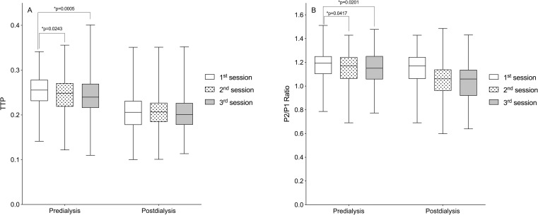
Still, in Fig 2, a second analysis comparing ICP parameters between the three weekly hemodialysis sessions over the six months of follow-up showed no significant differences in predialysis and postdialysis measurements. However, when all monitorings from each HD session (1st, 2nd, and 3rd weekly session) were pooled, there were significant differences in predialysis TTP and P2/P1 ratio with higher non-invasive ICP parameter values in the first session of the week (Fig 3) (p<0.05, repeated-measures ANOVA followed by the Tukey’s post-hoc test).

Fig 3. Comparison of predialysis and postdialysis noninvasive intracranial pressure (ICP) parameters according to the hemodialysis sessions over the six months of follow-up.

Fig 3

(A) Time to peak (TTP). (B) P2/P1 ratio. Box-plots illustrate the median and interquartile range of the noninvasive intracranial pressure parameters at the three weekly hemodialysis sessions (1st, 2nd, and 3rd) over the six months of follow-up (*p<0.05, repeated-measures ANOVA followed by the Tukey’s post-hoc test).

The main finding of this study was the significant difference detected between predialysis and postdialysis noninvasive ICP parameters (TTP and P2/P1 ratio) over the six months of follow-up (Fig 2). These parameters obtained through analysis of ICP waveform morphology. According to Nucci et al. (2016), changes in ICP waveform morphology can reflect changes in ICP whereas ICP wave morphological analysis can in turn predict the ICP measurements of invasive methods [15].

Considering that in general the parameters of non-invasive ICP were higher in the predialysis moment, it is suggested that the removal of fluids promoted by HD may be beneficial in improving cerebral compliance of patients with ESRD. Nevertheless, HD is associated with frequent complications, some of them involve the increased intracranial pressure, such as DDS [6].

Intradialytic hypotension is the commonest complication among HD patients [1620] and may precede DDS. The symptoms like headache, nausea, and muscle cramps experienced by some patients of the current study during follow-up may represent a milder but not diagnosed spectrum of DDS [5]. Our understanding of the pathophysiology of DDS has improved since its initial description and it is now evident from animal and human studies that DDS is associated with the development of cerebral edema and increased ICP [21,22].

At first DDS was believed to occur only in patients with acute kidney injury when hemodialysis was first initiated, but it has also been reported in patients with CKD [7,23].

According to Castro (2001), DDS can be prevented in patients with very high plasma urea levels by performing low-efficiency dialysis sessions of brief duration, reducing the interdialytic interval, and adding hypertonic solutions such as mannitol in the dialysate, which contribute to reduce cerebral edema [24].

In the current study, we showed that noninvasive ICP parameters (TTP and P2/P1 ratio) were higher in the first HD session of the week (Fig 3). Foley et al. (2011) found that in patients receiving maintenance HD, adverse events including all-cause mortality, myocardial infarction, and hospital admissions occurred more frequently on the day after the long interdialytic interval (1st weekly session) than on other days. We believe that this long (two-day) interdialytic interval contributes to higher interdialytic weight gain (IDWG) and consequently, changes in ICP [25].

Studies using transcranial doppler ultrasonography have evaluated the effects of HD on cerebral hemodynamics and concluded that there is a decrease in CBF after the procedure [26,27]. CBF can influence ICP, just as ICP can influence CBF. An elevated blood flow triggers a response of the self-regulation mechanism of the cerebral circulation that promotes vasodilation, increasing the cerebral blood volume, which consequently increases the ICP. In contrast, when the ICP rises, for any other reason, the cerebral perfusion pressure decreases, which hinders blood circulation and reduces CBF [28].

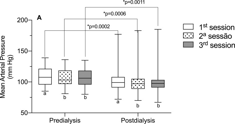
Considering the role of MAP in brain self-regulation, as it directly influences CBF [29], we evaluated this parameter of the volunteers in this study (Fig 4), however, there was no correlation between MAP and TTP and the P2/P1 ratio. However, the data is interesting because it shows that, in the 1st weekly HD session, MAP is higher, in relation to the 2nd and 3rd session of the week. This may have occurred, due to the greater fluid overload that occurs in the HD session two days’ time gap It has also been observed that PAM data, as well as PIC data, are higher in the pre-dialysis moment, in comparison with the post-dialysis.

Fig 4. Mean arterial pressure in the different hemodialysis sessions of the week.

Fig 4

Significant statistical differences were observed for the mean arterial pressure (MAP) parameter when comparing it with the different hemodialysis sessions of the week (1st, 2nd, and 3rd). Different letters, significant differences between the different sessions in the pre- and post-dialysis moments (p<0.05, ANOVA for repeated measures and Tukey’s post-test).

IDWG is the result of salt and water intake between two HD sessions and is influenced by several factors. It is recommended that IDWG does not exceed 4.5% of ‘dry body weight’ [30]. Recently, it has been reported that patients with IDWG ≥ 5.7% and 4%, respectively, are at an elevated risk for mortality and increased risk for fluid-overload hospitalization [31].

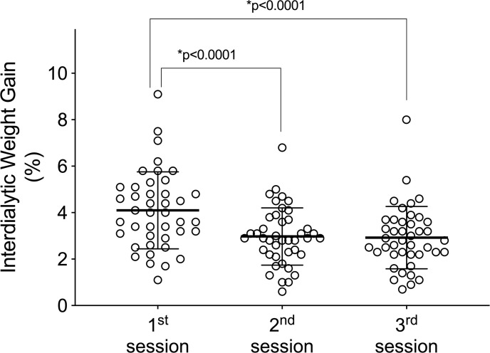
The volunteers in this study had significantly higher IDWG in the first HD session of the week (Fig 5). We hypothesize that patients with an IDWG ≥ 4% have worse brain compliance and greater chances of complications, because ICP is derived from cerebral blood and parenchyma and cerebrospinal fluid (CSF) circulatory dynamics, an increase in any of these components (blood, CSF, or parenchyma) may increase ICP [32,33], however, when correlating the PIC parameters (TTP and P2/P1 ratio) with the IDWG, no significant correlation was observed (p>0.05).

Fig 5. Interdialytic weight gain in the different hemodialysis sessions of the week.

Fig 5

Significant statistical differences were observed for the interdialytic weight gain parameter when comparing it with the different hemodialysis sessions of the week (1st, 2nd, and 3rd) (p <0.05, ANOVA for repeated measures and Tukey’s post-test).

A higher IDWG is associated with complications including higher predialysis blood pressure [34,35], intradialytic hypotension as a result of rapid fluid removal during the HD session [36], and increased mortality [37,38], and it may also be related to changes in ICP. Ipema et al. (2016) highlighted the importance of personalized advice on fluid and sodium restriction in HD patients [39].

This work was able to demonstrate through a non-invasive method that changes in the ICP of patients undergoing hemodialysis occur, and that these changes are repeated over the months. It is suggested that hemodialysis can improve the parameters of ICP that reflect brain compliance, however, future studies are warranted that examine the causes of ICP alterations, especially considering that prolonged ICP elevation is associated with poor neurocognitive outcomes [40]. Besides that, we show that the noninvasive ICP parameters TTP and P2/P1 ratio were higher in the first weekly HD session than in the second and third sessions, which may happen as a function of the time gap between the last and the first session of the week, which results in a greater accumulation of liquids (Fig 5).

As previously exposed, the noninvasive method used in this study was validated by comparison with the invasive ICP monitoring method [8,10] and has been used in the study of several situations, physiological and pathological, involving the central nervous system [4145]. Based on this context, the routine uses of non-invasive ICP monitoring in RRT centers could contribute to the clinical evaluation of patients with ESRD.

Conclusions

Through this unprecedented study, using a non-invasive method, it was possible to understand how the ICP of patients with ESRD behaves. Results of ICP wave morphology analysis of patients with ESRD followed-up for six months by noninvasive ICP monitoring revealed significant differences between predialysis and postdialysis ICP parameters. Also, the pattern of ICP alterations was consistent throughout the study.

Limitations of the study

Due to the dynamics used to carry out this study, it was not possible to detect complications and correlate them to the ICP. New studies that evaluate patients individually and for a longer time in each dialysis session could explain this issue.

Supporting information

S1 File. Average TTP values and P2/P1 ratio in the six-month follow-up.

Mean values of TTP values and P2/P1 ratio in the six months of follow-up, pre- and post-dialysis, of the 42 volunteers included in the study.

(XLSX)

Data Availability

All relevant data are within the manuscript and its Supporting Information files.

Funding Statement

The Braincare Desenvolvimento e Inovação Tecnológica S.A. provided the equipment free of charge for this study. CR declares Scholarship Funding from CAPES - Coordenação de Aperfeiçoamento de Pessoal de Nível Superior – Finance Code 001. This financing did not role in study design, data collection and analysis, decision to publish, or preparation of the manuscript. GHF declares personal fees as employee (Research Coordinator) from Braincare Desenvolvimento e Inovação Tecnológica S.A., during the conduct of the study; In addition, GHF has a patent US9826934B2 issued, and a patent US9993170B1 issued. The funder (Braincare Desenvolvimento e Inovação Tecnológica S.A.) only provided support in the form of salaries for author GHF, but did not have any additional role in the study design, data collection and analysis, decision to publish, or preparation of the manuscript. GHF contributed as a researcher with Conceptualization, Methodology, Software, Visualization, Writing – review & editing, as described in the ‘author contributions’ section. This commercial affiliation does not alter our adherence to PLOS ONE policies on sharing data and materials. NNR declares personal fees as medical consultant from Braincare Desenvolvimento e Inovação Tecnológica S.A., during the conduct of the study. The funder (Braincare Desenvolvimento e Inovação Tecnológica S.A.) only provided support in the form of consultant fee for author NNR, but did not have any additional role in the study design, data collection and analysis, decision to publish, or preparation of the manuscript. NNR contributed as a researcher with Methodology, Visualization, Writing – review & editing, as described in the ‘author contributions’ section. This commercial affiliation does not alter our adherence to PLOS ONE policies on sharing data and materials.

References

  • 1.Provenzano M, Mancuso C, Garofalo C, De Nicola L, Andreucci M. Temporal variation of Chronic Kidney Disease’s epidemiology. 2019;36: 0–13. [PubMed] [Google Scholar]
  • 2.Bastos MG, Bregman R, Kirsztajn GM. Doença renal crônica: frequente e grave, mas também prevenível e tratável. Rev Assoc Med Bras. 2010;56: 248–253. doi: 10.1590/s0104-42302010000200028 [DOI] [PubMed] [Google Scholar]
  • 3.Liew A. Perspectives in renal replacement therapy: Haemodialysis. Nephrology. 2018;23: 95–99. doi: 10.1111/nep.13449 [DOI] [PubMed] [Google Scholar]
  • 4.Denhaerynck K, De Geest S, Manhaeve D, Dobbels F, Garzoni D, Nolte C. Prevalence and consequences of nonadherence to hemodialysis regimens. Am J Crit Care. 2007;16: 222–236. [PubMed] [Google Scholar]
  • 5.Mistry K. Dialysis disequilibrium syndrome prevention and management. Int J Nephrol Renovasc Dis. 2019;12: 69–77. doi: 10.2147/IJNRD.S165925 [DOI] [PMC free article] [PubMed] [Google Scholar]
  • 6.Zepeda-orozco D, Quigley R. Dialysis disequilibrium syndrome. 2012;27: 2205–2211. doi: 10.1007/s00467-012-2199-4 [DOI] [PMC free article] [PubMed] [Google Scholar]
  • 7.Dalia T, Tuffaha AM. Dialysis disequilibrium syndrome leading to sudden brain death in a chronic hemodialysis patient. Hemodial Int. 2018;22: E39–E44. doi: 10.1111/hdi.12635 [DOI] [PubMed] [Google Scholar]
  • 8.Cabella B, Vilela GHF, Mascarenhas S, Czosnyka M, Smielewski P, Dias C, et al. Validation of a New Noninvasive Intracranial Pressure Monitoring Method by Direct Comparison with an Invasive Technique. 2016;122: 93–96. doi: 10.1007/978-3-319-22533-3_18 [DOI] [PubMed] [Google Scholar]
  • 9.Germon K. Interpretation of ICP pulse waves to determine intracerebral compliance. The Journal of neuroscience nursing: journal of the American Association of Neuroscience Nurses. 1988;20: 344–351. doi: 10.1097/01376517-198812000-00004 [DOI] [PubMed] [Google Scholar]
  • 10.Frigieri G, Andrade RAP, Dias C, Spavieri DL, Brunelli R, Cardim DA, et al. Analysis of a non-invasive intracranial pressure monitoring method in patients with traumatic brain injury. Acta Neurochir Suppl. 2018;126: 107–110. doi: 10.1007/978-3-319-65798-1_23 [DOI] [PubMed] [Google Scholar]
  • 11.Mascarenhas S, Vilela G, Carlotti C, Damiano L, Seluque W, Colli B, et al. The new ICP minimally invasive method shows that the Monro-Kellie doctrine is not valid. Acta Neurochir Suppl. 2012;114: 117–120. doi: 10.1007/978-3-7091-0956-4_21 [DOI] [PubMed] [Google Scholar]
  • 12.Adams JP, McKinlay J, Bell D. Neurocritical Care: A Guide to Practical Management. J Intensive Care Soc. 2010;11: 215–215. doi: 10.1177/175114371001100326 [DOI] [Google Scholar]
  • 13.Cardoso ER, Rowan JO, Galbraith S. Analysis of the cerebrospinal fluid pulse wave in intracranial pressure. J Neurosurg. 1983;59: 817–821. doi: 10.3171/jns.1983.59.5.0817 [DOI] [PubMed] [Google Scholar]
  • 14.Avezaat C, Van Eijndhoven J, Wyper D. Cerebrospinal fluid pulse pressure and intracranial volume-pressure relationships. J Neurol Neurosurg Psychiatry. 1979;42: 687–700. doi: 10.1136/jnnp.42.8.687 [DOI] [PMC free article] [PubMed] [Google Scholar]
  • 15.Nucci CG, De Bonis P, Mangiola A, Santini P, Sciandrone M, Risi A, et al. Intracranial pressure wave morphological classification: automated analysis and clinical validation. Acta Neurochir (Wien). 2016;158: 581–588. doi: 10.1007/s00701-015-2672-5 [DOI] [PubMed] [Google Scholar]
  • 16.Davenport A. Balancing risks: blood pressure targets, intradialytic hypotension, and ischemic brain injury. Semin Dial. 2014;27: 13–15. doi: 10.1111/sdi.12153 [DOI] [PubMed] [Google Scholar]
  • 17.Flythe JE, Xue H, Lynch KE, Curhan GC, Brunelli SM. Association of mortality risk with various definitions of intradialytic hypotension. J Am Soc Nephrol. 2015;26: 724–734. doi: 10.1681/ASN.2014020222 [DOI] [PMC free article] [PubMed] [Google Scholar]
  • 18.Mc Causland FR, Claggett B, Sabbisetti VS, Jarolim P, Waikar SS. Hypertonic Mannitol for the Prevention of Intradialytic Hypotension: A Randomized Controlled Trial. Am J Kidney Dis. 2019;74: 483–490. doi: 10.1053/j.ajkd.2019.03.415 [DOI] [PMC free article] [PubMed] [Google Scholar]
  • 19.Sands JJ, Usvyat LA, Sullivan T, Segal JH, Zabetakis P, Kotanko P, et al. Intradialytic hypotension: Frequency, sources of variation and correlation with clinical outcome. Hemodial Int. 2014;18: 415–422. doi: 10.1111/hdi.12138 [DOI] [PubMed] [Google Scholar]
  • 20.Da Silva GL, Thomé EG. Complicações do procedimento hemodialítico em pacientes com insuficiência renal aguda: intervenções de enfermagem. Revista gaúcha de enfermagem. 2009;30: 33–39. [PubMed] [Google Scholar]
  • 21.Lund A, Damholt MB, Strange DG, Kelsen J, Møller-Sørensen H, Møller K. Increased Intracranial Pressure during Hemodialysis in a Patient with Anoxic Brain Injury. Case Reports Crit Care. 2017; 2017: 1–4. doi: 10.1155/2017/5378928 [DOI] [PMC free article] [PubMed] [Google Scholar]
  • 22.Krane NK. Intracranial Pressure Measurement in a Patient Undergoing Hemodialysis and Peritoneal Dialysis. Am J Kidney Dis. 1989;13: 336–339. doi: 10.1016/s0272-6386(89)80042-3 [DOI] [PubMed] [Google Scholar]
  • 23.Bagshaw SM, Peets AD, Hameed M, Boiteau PJE, Laupland KB, Doig CJ. Dialysis Disequilibrium Syndrome: Brain death following hemodialysis for metabolic acidosis and acute renal failure–A case report. BMC Nephrol. 2004;5: 1–5. doi: 10.1186/1471-2369-5-1 [DOI] [PMC free article] [PubMed] [Google Scholar]
  • 24.Castro MCM De. Actualização em dialise—complicações em diálise. J Bras Nefrol. 2001;23: 108–113. [Google Scholar]
  • 25.Foley RN, Gilbertson DT, Murray T, Collins AJ. Long Interdialytic Interval and Mortality among Patients Receiving Hemodialysis. N Engl J Med. 2011;365: 1099–1107. doi: 10.1056/NEJMoa1103313 [DOI] [PubMed] [Google Scholar]
  • 26.Hata R, Matsumoto M, Handa N, Terakawa H, Sugitani Y, Kamada T. Effects of hemodialysis on cerebral circulation evaluated by transcranial doppler ultrasonography. Stroke. 1994;25: 408–412. doi: 10.1161/01.str.25.2.408 [DOI] [PubMed] [Google Scholar]
  • 27.Skinner H, Mackaness C, Bedforth N, Mahajanl R. Cerebral haemodynamics in patients with chronic renal failure: Effects of haemodialysis. British Journal of Anaesthesia. 2005;94: 203–205. doi: 10.1093/bja/aei016 [DOI] [PubMed] [Google Scholar]
  • 28.Reivich M. Arterial PCO2 and Cerebral Hemodynamics. The American journal of physiology. 1964;206: 25–35. doi: 10.1152/ajplegacy.1964.206.1.25 [DOI] [PubMed] [Google Scholar]
  • 29.Koizumi MS. Monitorização da pressão intracraniana. Rev. Esc. Enf. USP, São Paulo. 1981;75: 147–154. doi: 10.1590/0080-6234198101500200147 [DOI] [PubMed] [Google Scholar]
  • 30.Fouque D, Vennegoor M, Ter Wee P, Wanner C, Basci A, Canaud B, et al. EBPG guideline on nutrition. Nephrol Dial Transplant. 2007;22: ii45–ii87. doi: 10.1093/ndt/gfm020 [DOI] [PubMed] [Google Scholar]
  • 31.Wong MMY, McCullough KP, Bieber BA, Bommer J, Hecking M, Levin NW, et al. Interdialytic Weight Gain: Trends, Predictors, and Associated Outcomes in the International Dialysis Outcomes and Practice Patterns Study (DOPPS). Am J Kidney Dis. 2017;69: 367–379. doi: 10.1053/j.ajkd.2016.08.030 [DOI] [PubMed] [Google Scholar]
  • 32.Czosnyka M, Pickard JD. Monitoring and interpretation of intracranial pressure. J Neurol Neurosurg Psychiatry. 2004;75: 813–821. doi: 10.1136/jnnp.2003.033126 [DOI] [PMC free article] [PubMed] [Google Scholar]
  • 33.Elixmann IM, Hansinger J, Goffin C, Antes S, Radermacher K, Leonhardt S. Single pulse analysis of intracranial pressure for a hydrocephalus implant. Proc Annu Int Conf IEEE Eng Med Biol Soc EMBS. 2012;2012: 3939–3942. doi: 10.1109/EMBC.2012.6346828 [DOI] [PubMed] [Google Scholar]
  • 34.Nerbass FB, Morais JG, Santos RG dos, Krüger TS, Koene TT, Filho HA da L. Factors related to interdialytic weight gain in hemodialysis patients. J Bras Nefrol. 2011;33: 300–305. 10.1590/S0101-28002011000300005. [DOI] [PubMed] [Google Scholar]
  • 35.Kuipers J, Usvyat LA, Oosterhuis JK, Dasselaar JJ, De Jong PE, Westerhuis R, et al. Variability of predialytic, intradialytic, and postdialytic blood pressures in the course of a week: A study of Dutch and US maintenance hemodialysis patients. Am J Kidney Dis. 2013;62: 779–788. doi: 10.1053/j.ajkd.2013.03.034 [DOI] [PubMed] [Google Scholar]
  • 36.Lai CT, Wu CJ, Chen HH, Pan CF, Chiang CL, Chang CY, et al. Absolute interdialytic weight gain is more important than percent weight gain for intradialytic hypotension in heavy patients. Nephrology. 2012;17: 230–236. doi: 10.1111/j.1440-1797.2011.01542.x [DOI] [PubMed] [Google Scholar]
  • 37.Flythe JE, Curhan GC, Brunelli SM. Disentangling the Ultrafiltration rate-mortality association: The respective roles of session length and weight gain. Clin J Am Soc Nephrol. 2013;8: 1151–1161. doi: 10.2215/CJN.09460912 [DOI] [PMC free article] [PubMed] [Google Scholar]
  • 38.Sarkar SR, Kotanko P, Levin NW. Interdialytic weight gain: Implications in hemodialysis patients. Semin Dial. 2006;19: 429–433. doi: 10.1111/j.1525-139X.2006.00199_1.x [DOI] [PubMed] [Google Scholar]
  • 39.Ipema KJR, Kuipers J, Westerhuis R, Gaillard CAJM, Van Der Schans CP, Krijnen WP, et al. Causes and Consequences of Interdialytic weight gain. Kidney Blood Press Res. 2016;41: 710–720. doi: 10.1159/000450560 [DOI] [PubMed] [Google Scholar]
  • 40.Steiner LA, Andrews PJD. Monitoring the injured brain: ICP and CBF. Br J Anaesth. 2006;97: 26–38. doi: 10.1093/bja/ael110 [DOI] [PubMed] [Google Scholar]
  • 41.Ballestero MFM, Frigieri G, Cabella BCT, de Oliveira SM, de Oliveira RS. Prediction of intracranial hypertension through noninvasive intracranial pressure waveform analysis in pediatric hydrocephalus. Child’s Nerv Syst. 2017;33: 1517–1524. doi: 10.1007/s00381-017-3475-1 [DOI] [PubMed] [Google Scholar]
  • 42.Bollela VR, Frigieri G, Vilar FC, Spavieri DL, Tallarico FJ, Tallarico GM, et al. Noninvasive intracranial pressure monitoring for HIV-associated cryptococcal meningitis. Brazilian J Med Biol Res. 2017;50: 10–14. doi: 10.1590/1414-431X20176392 [DOI] [PMC free article] [PubMed] [Google Scholar]
  • 43.Rochetti Bezerra TA, Spavieri Júnior DL, Frigieri G, Brunell R, de Oliveira SM. In-flight analysis of intracranial pressure in pilots undergoing variation in Gz. Aeronaut Aerosp Open Access J. 2018;2: 126–131. doi: 10.15406/aaoaj.2018.02.00042 [DOI] [Google Scholar]
  • 44.Cardim DA, Frigieri GH, Cabella BCT, Malheiros JM, Cardim AC, Wang CC, et al. Characterization of intracranial pressure behavior in chronic epileptic animals: A preliminary study. Acta Neurochirurgica, Supplementum. 2016;122: 329–333. doi: 10.1007/978-3-319-22533-3_65 [DOI] [PubMed] [Google Scholar]
  • 45.Cardim DA, do Val da Silva RA, Cardim AC, Cabella BCT, Frigieri GH, de Sousa Torres CV, et al. Characterization of ICP behavior in an experimental model of hemorrhagic stroke in rats. Acta Neurochirurgica, Supplementum. 2016;122: 121–124. doi: 10.1007/978-3-319-22533-3_24 [DOI] [PubMed] [Google Scholar]

Decision Letter 0

Patrick Barry Mark

12 Jan 2021

PONE-D-20-29529

Use of non-invasive intracranial pressure pulse waveform to monitor patients with End-Stage Renal Disease (ESRD)

PLOS ONE

Dear Professor Vellosa,

Thank you for submitting your manuscript to PLOS ONE. After careful consideration, we feel that it has merit but does not fully meet PLOS ONE’s publication criteria as it currently stands. Therefore, we invite you to submit a revised version of the manuscript that addresses the points raised during the review process.

Please submit your revised manuscript by 11/4/21. If you will need more time than this to complete your revisions, please reply to this message or contact the journal office at plosone@plos.org. When you're ready to submit your revision, log on to https://www.editorialmanager.com/pone/ and select the 'Submissions Needing Revision' folder to locate your manuscript file.

Please include the following items when submitting your revised manuscript:

  • A rebuttal letter that responds to each point raised by the academic editor and reviewer(s). You should upload this letter as a separate file labeled 'Response to Reviewers'.

  • A marked-up copy of your manuscript that highlights changes made to the original version. You should upload this as a separate file labeled 'Revised Manuscript with Track Changes'.

  • An unmarked version of your revised paper without tracked changes. You should upload this as a separate file labeled 'Manuscript'.

If you would like to make changes to your financial disclosure, please include your updated statement in your cover letter. Guidelines for resubmitting your figure files are available below the reviewer comments at the end of this letter.

If applicable, we recommend that you deposit your laboratory protocols in protocols.io to enhance the reproducibility of your results. Protocols.io assigns your protocol its own identifier (DOI) so that it can be cited independently in the future. For instructions see: http://journals.plos.org/plosone/s/submission-guidelines#loc-laboratory-protocols

We look forward to receiving your revised manuscript.

Kind regards,

Patrick Barry Mark

Academic Editor

PLOS ONE

Journal Requirements:

When submitting your revision, we need you to address these additional requirements.

1. Please ensure that your manuscript meets PLOS ONE's style requirements, including those for file naming. The PLOS ONE style templates can be found at

https://journals.plos.org/plosone/s/file?id=wjVg/PLOSOne_formatting_sample_main_body.pdf and

https://journals.plos.org/plosone/s/file?id=ba62/PLOSOne_formatting_sample_title_authors_affiliations.pdf

2.Thank you for stating the following in the Competing Interests section:

"I have read the journal's policy and the authors of this manuscript have the following competing interests:

•    CR declares no competing interests/ has nothing to disclose.

•    LDC declares no competing interests/ has nothing to disclose.

•    FAS declares no competing interests/ has nothing to disclose.

•    GHF declares personal fees as employee (Research Coordinator) from Braincare Desenvolvimento e Inovação Tecnológica S.A., during the conduct of the study; In addition, GHF has a patent US9826934B2 issued, and a patent US9993170B1 issued.

•    NNR declares personal fees as medical consultant from Braincare Desenvolvimento e Inovação Tecnológica S.A., during the conduct of the study

•    AMS declares no competing interests/ has nothing to disclose.

•    SM declares he has a patent US9826934B2 issued, and a patent US9993170B1 issued.

•    JCRV declares no competing interests/ has nothing to disclose."

We note that one or more of the authors are employed by a commercial company: Braincare Desenvolvimento e Inovação Tecnológica S.A.

a) Please provide an amended Funding Statement declaring this commercial affiliation, as well as a statement regarding the Role of Funders in your study. If the funding organization did not play a role in the study design, data collection and analysis, decision to publish, or preparation of the manuscript and only provided financial support in the form of authors' salaries and/or research materials, please review your statements relating to the author contributions, and ensure you have specifically and accurately indicated the role(s) that these authors had in your study. You can update author roles in the Author Contributions section of the online submission form.

Please also include the following statement within your amended Funding Statement.

“The funder provided support in the form of salaries for authors [insert relevant initials], but did not have any additional role in the study design, data collection and analysis, decision to publish, or preparation of the manuscript. The specific roles of these authors are articulated in the ‘author contributions’ section.”

If your commercial affiliation did play a role in your study, please state and explain this role within your updated Funding Statement.

b) Please also provide an updated Competing Interests Statement declaring this commercial affiliation along with any other relevant declarations relating to employment, consultancy, patents, products in development, or marketed products, etc.  

c) We note that you have a patent relating to material pertinent to this article. Please provide an amended statement of Competing Interests to declare this patent (with details including name and number), along with any other relevant declarations relating to employment, consultancy, patents, products in development or modified products etc.

Within your Competing Interests Statement, please confirm that this commercial affiliation does not alter your adherence to all PLOS ONE policies on sharing data and materials by including the following statement: "This does not alter our adherence to  PLOS ONE policies on sharing data and materials.” (as detailed online in our guide for authors http://journals.plos.org/plosone/s/competing-interests) . If this adherence statement is not accurate and  there are restrictions on sharing of data and/or materials, please state these. Please note that we cannot proceed with consideration of your article until this information has been declared.

Please include both an updated Funding Statement and Competing Interests Statement in your cover letter. We will change the online submission form on your behalf.

Please know it is PLOS ONE policy for corresponding authors to declare, on behalf of all authors, all potential competing interests for the purposes of transparency. PLOS defines a competing interest as anything that interferes with, or could reasonably be perceived as interfering with, the full and objective presentation, peer review, editorial decision-making, or publication of research or non-research articles submitted to one of the journals. Competing interests can be financial or non-financial, professional, or personal. Competing interests can arise in relationship to an organization or another person. Please follow this link to our website for more details on competing interests: http://journals.plos.org/plosone/s/competing-interests

Additional Editor Comments:

This is an interesting paper highlighting a novel technique for monitoring intracranial pressure in dialysis. Overall I thought it was well done and whilst there was some divergence of opinion in the reviewers, I thought it would merit revision and look forward to have a looking at another version.

[Note: HTML markup is below. Please do not edit.]

Reviewers' comments:

Reviewer's Responses to Questions

Comments to the Author

1. Is the manuscript technically sound, and do the data support the conclusions?

The manuscript must describe a technically sound piece of scientific research with data that supports the conclusions. Experiments must have been conducted rigorously, with appropriate controls, replication, and sample sizes. The conclusions must be drawn appropriately based on the data presented.

Reviewer #1: Yes

Reviewer #2: No

**********

2. Has the statistical analysis been performed appropriately and rigorously?

Reviewer #1: Yes

Reviewer #2: No

**********

3. Have the authors made all data underlying the findings in their manuscript fully available?

The PLOS Data policy requires authors to make all data underlying the findings described in their manuscript fully available without restriction, with rare exception (please refer to the Data Availability Statement in the manuscript PDF file). The data should be provided as part of the manuscript or its supporting information, or deposited to a public repository. For example, in addition to summary statistics, the data points behind means, medians and variance measures should be available. If there are restrictions on publicly sharing data—e.g. participant privacy or use of data from a third party—those must be specified.

Reviewer #1: No

Reviewer #2: Yes

**********

4. Is the manuscript presented in an intelligible fashion and written in standard English?

PLOS ONE does not copyedit accepted manuscripts, so the language in submitted articles must be clear, correct, and unambiguous. Any typographical or grammatical errors should be corrected at revision, so please note any specific errors here.

Reviewer #1: Yes

Reviewer #2: Yes

**********

5. Review Comments to the Author

Please use the space provided to explain your answers to the questions above. You may also include additional comments for the author, including concerns about dual publication, research ethics, or publication ethics. (Please upload your review as an attachment if it exceeds 20,000 characters)

Reviewer #1: In the manuscript, authors present data from a study assessing the “Use of non-invasive intracranial pressure pulse waveform to monitor patients with End-Stage Renal Disease”. The study is in general of interest and provides some new insights, especially concerning the application of a non-invasive device for ICP measurement. Nevertheless, there are some open questions and thus will not provide full impact for the readership:

Minor issues:

• The data are nicely presented and, I guess, “behave” as expected. Seen differences are like data known for "normal" blood pressure. So, it may reflect just the same phenomenon (removal of fluid) as well? Blood pressure is increasing from end of dialysis session till next start (thus higher at beginning of week). This is all nicely described in the discussion as well. Do you have data on ultrafiltration volume and intradialytic weight gain? These data would be of interest to be included in your analyses. Rate of change from pre- to post-dialytic session could be directly related to UFV and IDWG. Do you have blood pressure data as well? Would be interesting to see the “same” behaviour in these data as well. Please comment.

• Methods for normality testing: Have you checked normality using QQ-plots (or other means to visualize data) or used any formal tests? Just reporting checking of skewness and kurtosis is in my opinion not common in the medical domain (although might be correct).

• Table 1: Are you sure all data are normally distributed? E.g. Are you sure mean HD session length is normally distributed, if 52.4% of the sessions are longer than 240 minutes and the mean is 220 mins? What about the mean HD time? If looking at the SD, I assume they are not normally distributed, thus use of mean and SD are not correct.

• Figure 2 and Figure 3: Are you sure the captions are correct? In Figure 2, I cannot see any comparison of sessions. In Figure 3, I cannot see any data “over the six months of FU”. Furthermore, in Figure 3 it says, “ANOVA is used for comparing data over six months” Please comment.

• Figure 3B, is it correct that for post-dialysis, there are no significant differences? Especially between session 1 and session 2, the difference seems to be “large”.

• In the discussion, you mention that TTP and P2/P1 ratio were higher in the first session compared to the second and third. You argue that it “may happen as a function of the time gap between the last and the first session of the week.” I guess it is not the time, but the fluid overload, thus IDWG.

Reviewer #2: In this study the authors non-invasively recorded intracranial pressure in patients with end-stage renal disease (ESRD) receiving hemodialysis by using the Brain4care device. The authors concluded that intracranial pressure parameters (time to peak and P1/P2) were higher before dialysis compared to after dialysis and that these differences were significantly elevated in the first session relative to the subsequent dialysis sessions. The authors indicated that the rationale for doing this study is based on the fact that some ESRD patients on dialysis may progress into dialysis disequilibrium syndrome suffering of cerebral edema and high intracranial pressure. Although this phenomenon is rare in clinical practice, the authors suggest that the presence of cramps, headaches, fatigue and inability to concentrate after dialysis may be mild manifestations of the dialysis disequilibrium syndrome.

This reviewer has several concerns and comments with this investigation:

1) Although the concept of this work is interesting, I am concerned with the validation of the non-invasive device (Brain4care®) used for measuring intracranial pressure in humans. The authors report that this device measures volumetric changes of the skull in adults by simply applying the sensor through a plastic band around the head and apparently, there is no need for calibration. This appears to be a very simple device in its utilization, but its validation is a concern, particularly when most studies of validation were performed in animal models. Moreover, it seems that the two studies that have addressed validation have been performed by the same group and it appears that there has not been any further independent validation. In addition, data on the device reliability and accuracy in humans is not provided and in consequence, extrapolating a relationship between this device and any gold standard from animals into humans without a complete assessment of reliability may be an important source of study bias.

2) The description of the methodology is succinct and there is no information on the technical details of the measurements. Also, it is not clear what the 4881 data-points from the 42 subjects represent. From this number, I speculate that 116 data points were obtained per subject along the 6-month period at a rate of 3 times sessions per week. Then, we should expect that only 1.61 data-points per patient and session were accomplished. Is the rate of these data points per patient and session sufficient and reliable to provide confidence in these measurements? Unfortunately, without any clear assessment of the reproducibility of the technique any interpretation becomes speculative.

3) It is not clear to this reviewer what would be the contribution of extracranial sources to these measurements and whether this could be a source of artifacts to the readings.

4) Few investigations have used transcranial Doppler from the middle cerebral artery to determine the effects of dialysis on the cerebral circulation. These studies have reported that mean flow velocities remain high before dialysis, decline significantly during dialysis, and stay lower in the post-dialysis period. What is interesting from these studies is that the reduction in flow velocity negatively correlates with the ultrafiltrate volumes, amount of fluid removed and the loss of weight after hemodialysis. Moreover, the post-dialysis reduction in mean flow velocity after 12 months of continued dialysis correlated significantly with the patients’ lower global and executive functions and with progression of their white matter hyperintensities with MRI. It seems then, that these studies are opposite the conclusions achieved by the current study which indicates that intracranial pressure remains high before dialysis. In my opinion, these hemodynamic changes of cerebral blood flow before, during and after dialysis should be discussed in the context of the authors’ findings. [See Stroke 1994;25:408-412; J Am Soc Nephrol 2019;30:147-158.

5) Figure 2 shows the statistical significance of 18 paired comparisons between pre-dialysis and post-dialysis through the use of individual paired student t-tests. In my opinion, these multiple comparisons require an adjustment in the alpha value due to the number of comparisons. Statistical advice is suggested. In addition, Figure 3 displays the comparisons of all measurements through the 6-month period and the authors conclude that session 1 was significantly higher than 2 and 3. Unfortunately, with the large standard deviations and without information on the variability of the measurements, it is difficult to believe that there was a significant difference.

6) An important parameter that would help to understand these changes is systemic blood pressure. However, there was no attempt to document this information. Data on blood pressure may be particularly important as they reported that 64% of the patients were classified with systemic arterial hypertension.

7) In my opinion, most of these hemodynamic changes occur during the dialysis session. If such device is demonstrated to be a valid surrogate of the intracranial pressure, intra-dialytic recordings would be more interesting in order to determine the impact of dialysis on the brain dynamics.

**********

6. PLOS authors have the option to publish the peer review history of their article (what does this mean?). If published, this will include your full peer review and any attached files.

If you choose “no”, your identity will remain anonymous but your review may still be made public.

Do you want your identity to be public for this peer review? For information about this choice, including consent withdrawal, please see our Privacy Policy.

Reviewer #1: No

Reviewer #2: No

[NOTE: If reviewer comments were submitted as an attachment file, they will be attached to this email and accessible via the submission site. Please log into your account, locate the manuscript record, and check for the action link "View Attachments". If this link does not appear, there are no attachment files.]

While revising your submission, please upload your figure files to the Preflight Analysis and Conversion Engine (PACE) digital diagnostic tool, https://pacev2.apexcovantage.com/. PACE helps ensure that figures meet PLOS requirements. To use PACE, you must first register as a user. Registration is free. Then, login and navigate to the UPLOAD tab, where you will find detailed instructions on how to use the tool. If you encounter any issues or have any questions when using PACE, please email PLOS at figures@plos.org. Please note that Supporting Information files do not need this step.

PLoS One. 2021 Jul 22;16(7):e0240570. doi: 10.1371/journal.pone.0240570.r002

Author response to Decision Letter 0


17 May 2021

Title: Use of non-invasive intracranial pressure pulse waveform to monitor patients with End-Stage Renal Disease (ESRD)

Patrick Barry Mark

Academic Editor

PLOS ONE

Answers to Reviewer Comments

Dear reviewers,

Thank you so much for the valuable comments and critics. Your considerations have certainly provided us with a valuable opportunity to improve our work. Below, you will find an itemized response to each of your concerns:

Reviewer 1:

• The data are nicely presented and, I guess, “behave” as expected. Seen differences are like data known for "normal" blood pressure. So, it may reflect just the same phenomenon (removal of fluid) as well? Blood pressure is increasing from end of dialysis session till next start (thus higher at beginning of week). This is all nicely described in the discussion as well. Do you have data on ultrafiltration volume and intradialytic weight gain? These data would be of interest to be included in your analyses. Rate of change from pre- to post-dialytic session could be directly related to UFV and IDWG. Do you have blood pressure data as well? Would be interesting to see the “same” behaviour in these data as well. Please comment.

Dear reviewer, we value your suggestion and include data on blood pressure and IDWG. The data were included in the manuscript as figure 4 and figure 5. Ultrafiltration data were not obtained. The data on blood pressure and IDWG were correlated to the parameters of ICP (TTP and P2/P1 ratio), however, there was no significant correlation (Person’s correlation).

• Methods for normality testing: Have you checked normality using QQ-plots (or other means to visualize data) or used any formal tests? Just reporting checking of skewness and kurtosis is in my opinion not common in the medical domain (although might be correct).

Dear reviewer, we appreciate your suggestion, and we have modified the method for evaluating the normality of the data. We apply a more formal test (Kolmogorov-Smirnov).

The information has been changed in the manuscript (Material and Methods section).

Below you can see the table with the details of the normality tests (not included in the manuscript).

It can be seen that the points are very approximate to the straight line, so we can assume that the data have distributions that are similar to a normal curve.

The normality of data can be analyzed descriptively through histograms, box-plots, Q-Q Plots, and skewness and kurtosis coefficients, respectively, the degree of deviation or skewness from the symmetry and flatness of the distribution. In addition to descriptive methods, hypothesis tests assess normality, such as the Kolmogorov-Smirnov and Shapiro-Wilks tests. However, it is essential to note that these tests are extremely rigorous and easily reject the hypothesis of normality. Therefore, we should be cautious and not base our decision only on these tests' descriptive levels (p-values). There is no simple relationship between relative power and sample size and no clear rationale for the frequently cited threshold of 30 – 50 patients per group, indicating acceptability of parametric statistics (Vickers 2005).

1. Vickers AJ. Parametric versus non-parametric statistics in the analysis of randomized trials with non-normally distributed data. BMC Med Res Methodol. 2005 Nov 3;5:35. doi: 10.1186/1471-2288-5-35. PMID: 16269081; PMCID: PMC1310536.

• Table 1: Are you sure all data are normally distributed? E.g. Are you sure mean HD session length is normally distributed, if 52.4% of the sessions are longer than 240 minutes and the mean is 220 mins? What about the mean HD time? If looking at the SD, I assume they are not normally distributed, thus use of mean and SD are not correct.

Dear reviewer, Table 1 presents the data concerning the subjects' characteristics included in this clinical trial. We did not perform inferential statistics with the data shown in Table 1. Therefore, the normality of the data presented in Table 1 does not influence our study's results and conclusions.

• Figure 2 and Figure 3: Are you sure the captions are correct? In Figure 2, I cannot see any comparison of sessions. In Figure 3, I cannot see any data “over the six months of FU.” Furthermore, in Figure 3 it says, “ANOVA is used for comparing data over six months” Please comment.

In figure 2, we consider the two parameters (A. TTP and B. P2/P1 ratio); We do not include statistical significance values in the figure since we did not find significant differences between the evaluation periods. Therefore the indication of the statistic in the face of non-significant differences is not necessary. However, if the Reviewer/Editor considers this information relevant, we can include it in the manuscript.

In figure 3, we show the data regarding the follow-up during the six months of the study. For the data analysis, the means were obtained for the first, second, and third hemodialysis sessions during the six months of the research. Thus, the data from the sessions were obtained during the entire follow-up period (six months).

Analysis of Variance (ANOVA) is a common and robust statistical test that you can use to compare the mean scores collected from different conditions or groups in an experiment.

A repeated-measures (or within-participants) test is what you use when you want to compare the performance of the same group of participants in different experimental moments. That is, when the same participants take part in all of the conditions in your study.

• Figure 3B, is it correct that for post-dialysis, there are no significant differences? Especially between session 1 and session 2, the difference seems to be “large”.

The reviewer has raised an important point in Figure 3B. We have checked the data, and there are no significant differences between the predialysis and post-dialysis times. Figure 3B the y-axis shows a maximum value for P2/P1 ratio of 1.7, and the lower limit of the graph starts at 0.5. This way, the visual difference in the figure becomes more evident. Therefore, Figure 3B was re-edited with the y-axis starting at zero.

• In the discussion, you mention that TTP and P2/P1 ratio were higher in the first session compared to the second and third. You argue that it “may happen as a function of the time gap between the last and the first session of the week.” I guess it is not the time, but the fluid overload, thus IDWG.

Dear reviewer, your note is correct and that is what we wanted to suggest. The sentence was rewritten in the manuscript.

Reviewer 2:

1) Although the concept of this work is interesting, I am concerned with the validation of the non-invasive device (Brain4care®) used for measuring intracranial pressure in humans. The authors report that this device measures volumetric changes of the skull in adults by simply applying the sensor through a plastic band around the head and apparently, there is no need for calibration. This appears to be a very simple device in its utilization, but its validation is a concern, particularly when most studies of validation were performed in animal models. Moreover, it seems that the two studies that have addressed validation have been performed by the same group and it appears that there has not been any further independent validation. In addition, data on the device reliability and accuracy in humans is not provided and in consequence, extrapolating a relationship between this device and any gold standard from animals into humans without a complete assessment of reliability may be an important source of study bias.

Dear reviewer, in the discussion session, we cite studies that assessed non-invasive ICP in humans by the method used in this study, such as Ballestero MFM, Frigieri G, Cabella BCT, de Oliveira SM, de Oliveira RS. Prediction of intracranial hypertension through noninvasive intracranial pressure waveform analysis in pediatric hydrocephalus. Child’s Nerv Syst. 2017; 33: 1517–1524. In addition, the method in question is registered with the National Health Surveillance Agency (registration number 81157910004) and is present in reference hospitals such as Hospital Sírio Libanês, Hospital Nove de Julho, Beneficiência Portuguesa – BP and Neuresp Neurologia Especializada. This study did not aim to correlate the Brain4care method with the gold standard, not least because in practice the invasive method is not routinely used in renal patients. The method used in this study provides a unique strategy for evaluating patients who do not have any information on brain compliance.

2) The description of the methodology is succinct and there is no information on the technical details of the measurements. Also, it is not clear what the 4881 data-points from the 42 subjects represent. From this number, I speculate that 116 data points were obtained per subject along the 6-month period at a rate of 3 times sessions per week. Then, we should expect that only 1.61 data-points per patient and session were accomplished. Is the rate of these data points per patient and session sufficient and reliable to provide confidence in these measurements? Unfortunately, without any clear assessment of the reproducibility of the technique any interpretation becomes speculative.

To work with the large volume of data collected, the TTP values and the P2 / P1 ratio of the different sessions (1st, 2nd and 3rd) were initially averaged for each month of follow-up (file sent in .xlm format). In addition, within the 6 months that each volunteer was followed up, there were eventually days when the volunteer's brain compliance was not monitored (due to not accepting on a certain day, not feeling well or missing the hemodialysis session).

3) It is not clear to this reviewer what would be the contribution of extracranial sources to these measurements and whether this could be a source of artifacts to the readings.

Dear reviewer, the extracranial sources that could generate artifacts refer to the patient's posture (sitting, lying or standing) and movement during monitoring. As for posture, patients were always monitored in the same way in the pre- and post-dialysis moments, sitting on their own hemodialysis chairs, in addition, the sensor was placed on the same side of the head at both times (pre- and post-dialysis). dialysis). Regarding the patient's movement during monitoring, the volunteers were instructed to remain immobile during the procedure, however, if movement occurred, the acquisition was totally modified and visible, as it is an extremely sensitive method that captures cranial microalterations. In this sense, the period that the patient moved was removed from the analysis and the volunteer was monitored for a longer time to compensate for the excluded period.

4) Few investigations have used transcranial Doppler from the middle cerebral artery to determine the effects of dialysis on the cerebral circulation. These studies have reported that mean flow velocities remain high before dialysis, decline significantly during dialysis, and stay lower in the post-dialysis period. What is interesting from these studies is that the reduction in flow velocity negatively correlates with the ultrafiltrate volumes, amount of fluid removed and the loss of weight after hemodialysis. Moreover, the post-dialysis reduction in mean flow velocity after 12 months of continued dialysis correlated significantly with the patients’ lower global and executive functions and with progression of their white matter hyperintensities with MRI. It seems then, that these studies are opposite the conclusions achieved by the current study which indicates that intracranial pressure remains high before dialysis. In my opinion, these hemodynamic changes of cerebral blood flow before, during and after dialysis should be discussed in the context of the authors’ findings. [See Stroke 1994;25:408-412; J Am Soc Nephrol 2019;30:147-158.

We would like to thank you the suggestion, we adjusted the text in the discussion session.

5) Figure 2 shows the statistical significance of 18 paired comparisons between predialysis and post-dialysis through the use of individual paired student t-tests. In my opinion, these multiple comparisons require an adjustment in the alpha value due to the number of comparisons. Statistical advice is suggested. In addition, Figure 3 displays the comparisons of all measurements through the 6-month period and the authors conclude that session 1 was significantly higher than 2 and 3. Unfortunately, with the large standard deviations and without information on the variability of the measurements, it is difficult to believe that there was a significant difference.

In Figure 2, the paired t-test was applied considering predialysis and postdialysis in each evaluated period. Thus, for each pairwise comparison, the use of the paired t-test is adequate. We did not perform multiple comparisons with the paired t-test. The Bonferroni correction for alpha adjustment would be applied if multiple comparisons were made. A second analysis involving the different periods within the same group; we employed the ANOVA test for repeated measures for this purpose.

The reviewer has raised an important point in Figure 3B. We have checked the data, and there are no significant differences between the predialysis and post-dialysis times. Figure 3B the y-axis shows a maximum value for P2/P1 ratio of 1.7, and the lower limit of the graph starts at 0.5. This way, the visual difference in the figure becomes more evident. Therefore, Figure 3B was re-edited with the y-axis starting at zero.

In Table 2, we can see the distribution of the data. The descriptive statistics show a low coefficient of variation. The mean and median values are very similar. The standard deviation and interquartile range present low values. The skewness and kurtosis coefficients are between -1 and +1, indicating that the data demonstrate an approximately normal distribution.

In figure 2 (below), we can see the frequency histograms and Q-Q Plot. The figure supports the normal distribution of the analyzed data. The histograms show a distribution very similar to a normal distribution. The Q-Q Plot has the vast majority of points fitted to a straight line, indicating a normal distribution approximation.

Therefore, this information reinforces the reliability of the statistical analysis performed.

In Table 3, we can see the distribution of the data from the P2/P1 ratio. The results are similar to those observed in Table 2. The descriptive statistics show a low coefficient of variation. The mean and median values are very similar. The standard deviation and interquartile range present low values. The skewness and kurtosis coefficients are between -1 and +1, indicating that the data suggest an approximately normal distribution.

In figure 3 (below), we can see the frequency histograms and Q-Q Plot. The figure supports the normal distribution of the analyzed data. The histograms show a distribution very similar to a normal distribution. The Q-Q Plot has the vast majority of points fitted to a straight line, indicating a normal distribution approximation.

Therefore, this information reinforces the reliability of the statistical analysis performed.

6) An important parameter that would help to understand these changes is systemic blood pressure. However, there was no attempt to document this information. Data on blood pressure may be particularly important as they reported that 64% of the patients were classified with systemic arterial hypertension.

Dear reviewer, we value your suggestion and include data on blood pressure. The data were included in the manuscript as figure 4. The data on blood pressure were correlated to the parameters of ICP (TTP and P2/P1 ratio), however, there was no significant correlation (Person’s correlation).

7) In my opinion, most of these hemodynamic changes occur during the dialysis session. If such device is demonstrated to be a valid surrogate of the intracranial pressure, intra-dialytic recordings would be more interesting in order to determine the impact of dialysis on the brain dynamics.

Dear reviewer, your collocations are interesting, however, in this study the objective was to evaluate the effect of hemodialysis on cerebral compliance and for that, the patients were monitored for a few minutes before and after dialysis. In this study, it was not feasible to monitor the patient during part or all the hemodialysis session for the following reasons: 42 volunteers were monitored for a period of 6 months; we believe that we would have patients drop out if, in order to participate in the study, they had to be monitored in all their sessions for a "long" period. In this study, it was not feasible to keep a patient connected to the equipment for a long period, due to the number of equipment and researchers available to carry out the evaluations. However, this study is the first to clearly demonstrate the effect of hemodialysis on brain compliance and most importantly, it was not occasional changes, but reproducible over the months. It is expected and positive that the results of this study will awaken many other questions to be answered. Future studies, which continuously monitor hemodialysis sessions of patients with end-stage renal disease and acute kidney disease, could assist in establishing the application of the non-invasive method for assessing intracranial pressure in these situations.

Attachment

Submitted filename: Response to Reviewers.pdf

Decision Letter 1

Patrick Barry Mark

27 May 2021

Use of non-invasive intracranial pressure pulse waveform to monitor patients with End-Stage Renal Disease (ESRD)

PONE-D-20-29529R1

Dear Dr. Vellosa,

We’re pleased to inform you that your manuscript has been judged scientifically suitable for publication and will be formally accepted for publication once it meets all outstanding technical requirements.

Within one week, you’ll receive an e-mail detailing the required amendments. When these have been addressed, you’ll receive a formal acceptance letter and your manuscript will be scheduled for publication.

An invoice for payment will follow shortly after the formal acceptance. To ensure an efficient process, please log into Editorial Manager at http://www.editorialmanager.com/pone/, click the 'Update My Information' link at the top of the page, and double check that your user information is up-to-date. If you have any billing related questions, please contact our Author Billing department directly at authorbilling@plos.org.

If your institution or institutions have a press office, please notify them about your upcoming paper to help maximize its impact. If they’ll be preparing press materials, please inform our press team as soon as possible -- no later than 48 hours after receiving the formal acceptance. Your manuscript will remain under strict press embargo until 2 pm Eastern Time on the date of publication. For more information, please contact onepress@plos.org.

Kind regards,

Patrick Barry Mark

Academic Editor

PLOS ONE

Additional Editor Comments (optional):

Thank you for revising this interesting study

Reviewers' comments:

Acceptance letter

Patrick Barry Mark

14 Jul 2021

PONE-D-20-29529R1

Use of non-invasive intracranial pressure pulse waveform to monitor patients with End-Stage Renal Disease (ESRD)

Dear Dr. Vellosa:

I'm pleased to inform you that your manuscript has been deemed suitable for publication in PLOS ONE. Congratulations! Your manuscript is now with our production department.

If your institution or institutions have a press office, please let them know about your upcoming paper now to help maximize its impact. If they'll be preparing press materials, please inform our press team within the next 48 hours. Your manuscript will remain under strict press embargo until 2 pm Eastern Time on the date of publication. For more information please contact onepress@plos.org.

If we can help with anything else, please email us at plosone@plos.org.

Thank you for submitting your work to PLOS ONE and supporting open access.

Kind regards,

PLOS ONE Editorial Office Staff

on behalf of

Prof Patrick Barry Mark

Academic Editor

PLOS ONE

Associated Data

    This section collects any data citations, data availability statements, or supplementary materials included in this article.

    Supplementary Materials

    S1 File. Average TTP values and P2/P1 ratio in the six-month follow-up.

    Mean values of TTP values and P2/P1 ratio in the six months of follow-up, pre- and post-dialysis, of the 42 volunteers included in the study.

    (XLSX)

    Attachment

    Submitted filename: Response to Reviewers.pdf

    Data Availability Statement

    All relevant data are within the manuscript and its Supporting Information files.


    Articles from PLoS ONE are provided here courtesy of PLOS

    RESOURCES