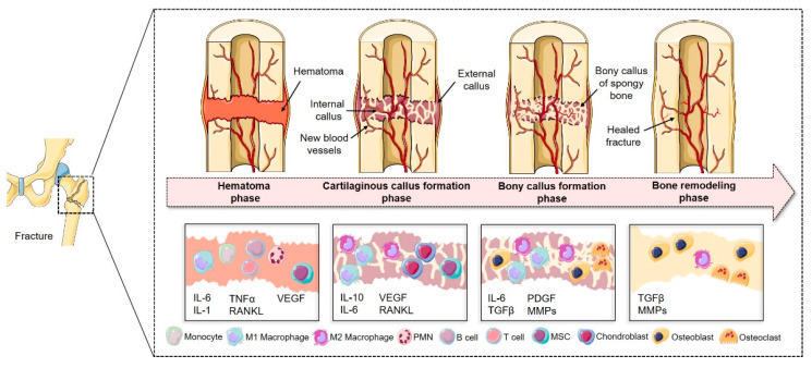
Figure 1.
Schematic description of the four phases of fracture healing: The first phase is characterized by the formation of the fracture hematoma and a local inflammation. Immune cells, such as peripheral multinucleated cells (PMN), T- and B-cells, monocytes and MSCs, are activated and recruited towards the fracture gap via autocrine and paracrine pathways (e.g., by the release of cytokines such as interleukin (IL-1), IL-6 or tumor necrosis factor (TNFα)). Activation of, for instance, vascular endothelial growth factor (VEGF) also paves the way for revascularization in this early phase. In the following phase, chondroprogenitor cells differentiate into chondroblasts and start to build an early fibrocartilaginous bridging area, while angiogenic processes are also upheld. The third phase is characterized by endochondral ossification, thereby substituting cartilage with primitive bone tissue. In the last phase bone structure and function is completely restored by the constrict interplay of bone-forming and bone-resorbing cells. Figure was modified from Servier Medical Art, licensed under a Creative Common Attribution 3.0 Generic License.

