Intravitreal injections of anti-vascular endothelial growth factor (anti-VEGF) agents are the mainstay treatment for several retinal diseases including neovascular age related macular degeneration and macular oedema due to retinal vascular and inflammatory diseases. This procedure is the most common procedure done in Ophthalmology. Intravitreal aflibercept (Eylea®, Bayer Inc) was approved for use in the National Health Service (NHS) since 2012. Traditionally, it is dispensed as a vial containing 100 μl, equivalent to 4 mg aflibercept injected in the eye using a 1 ml syringe. More recently, in a step towards standardizing the dose delivery, aflibercept received its market authorization for use in a pre-filled syringe (PFS) from April, 2020. As per the Summary of Product Characteristics (SmPC) one PFS contains 90 μl, equivalent to 3.6 mg aflibercept [1]. This provides a usable amount to deliver a single dose of 50 μl containing 2 mg aflibercept. Potential side-effects of anti-VEGF therapies are rare, but ocular adverse effects such as immediate and sustained rise in intraocular pressure (IOP), intraocular inflammation, vitreous haemorrhage, retinal tears, endophthalmitis, development of cataracts and have been noted [2, 3].
The safety profile of aflibercept was determined by eight phase III studies encompassing all major indications [4, 5]. Serious ocular adverse reactions in the study eye related to the injection procedure occurred in less than 1 in 1,900 intravitreal aflibercept injections. Of the most commonly reported adverse reactions defined as in at least 5% of patients, intraocular pressure increase was noted in 8% patients [6]. Comparatively, there is limited data on the safety profile of PFS. The change in intraocular pressure following intravitreal injection with PFS aflibercept is of particular interest as it offers regulated and more controlled volume of drug injected. The aim of this study was to compare post injection IOP change between the traditional mode of injection from vial using 1 ml syringe versus PFS.
In this study, we included intravitreal aflibercept injections performed for various clinical indications in Moorfields Eye Hospital between January 1st and October 20th, 2020. Moorfields Eye Hospital adopted the use of PFS aflibercept in May 2020. From the electronic database, we recorded the pre-and post IOP of 748 consecutive patients injected with PFS with 565 historical group of patients who were administered Aflibercept from the vial. Our protocol suggests that IOP is only recorded at least 15 min after the intravitreal injection.
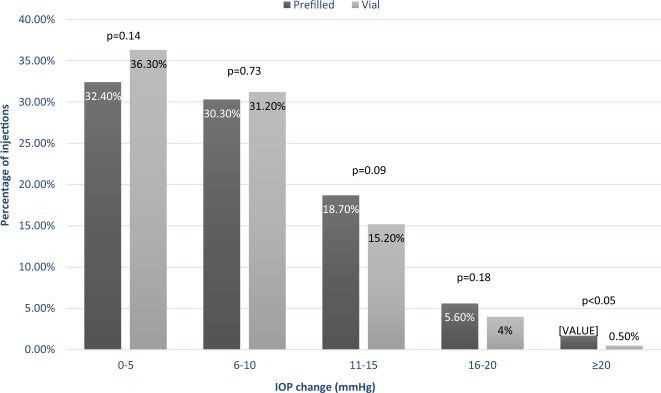
Baseline IOPs were similar between the two groups with mean IOP being 13.7 mm Hg (SD 4.4) in the PFS group and 13.9 mm Hg (SD 4.3) in the vial group. We found a significant mean IOP rise in the PFS group compared to the vial group but with very large standard deviation in the PFS group. Therefore, we categorised post-injection IOP rise into 5 groups of 5 mmHg increase in ascending order (Fig. 1). The majority of patients had IOP increase of 10 mmHg or more in both groups with no significant difference between groups (62.7% of the control group and 67.5% of the PFS group). However, an increase in IOP rise of 20 mm Hg or more was recorded in a small number of patients more frequently in the PFS arm (n = 13) than in the vial arm (n = 3). This difference was statistically significant (p value < 0.05).
Fig. 1. Change of IOP shown in 5 groups of ascending IOP values.
The PFS treated arm is demonstrated in dark grey and the vial treated arm is demonstrated in light grey. Non significant difference between the two arms was observed in the 0–5, 6–10, 11–15, and 16–20 mmHg rise groups. However, there was a statistically significant difference in IOP rise between the two treatment arms in the ≥20 mmHg increase group.
We reviewed the records of those that had post-injection IOP rise of 20 mmHg or more in the PFS group. Three had previous diagnosis of glaucoma (1 ocular hypertension, 1 primary open angle glaucoma and 1 primary angle closure suspect). The mean number of injections in the PFS treatment arm was 23.8 injections (SD 17.7, range: 1–60) until the IOP spike. The patients were switched to vial in subsequent visits because it was postulated that the IOP rise may be related to the PFS injection procedure.
All the three injections in the vial group that noted a substantial IOP rise were from 3 different patients and all of them had a previous diagnosis of primary open angle glaucoma.
Our study demonstrates that a small proportion of patients may experience a rise in IOP post PFS aflibercept injection. Recently, Gallagher and colleagues published a case series of transient central retinal artery occlusions following PFS aflibercept injections. They analysed the volume expressed from 1 ml syringe versus the PFS and reported that the PFS expressed a greater range of volumes than the 1 ml syringe with 21% of PFS repeats expressing 0.07 ml or more, highlighting that the IOP rise may be due to an increase in volume injected due to errors in positioning of the syringe plunger [7]. The error in volume expressed is the product of linear error in plunger alignment and the internal diameter of the syringe. As the internal diameter of a PFS syringe is almost twice as wide as the 1 ml syringe, a unit error in plunger alignment will lead to almost a twofold volume error in PFS as compared to a 1 ml syringe.
Pallikaris et al. demonstrated a linear relationship between IOP rise and volume injected [8]. Friedenwald has defined ocular rigidity as a measure of the resistance that the eyeball offered to a change in intraocular pressure [9]. Therefore, eyes with glaucoma, documented to have the lowest ocular rigidity, afford least resistance to IOP rise due to change in volume. Thus, the margin of error while using a PFS is low and a minimal parallax error can lead to a large increase in IOP post-injection.
This study highlights the need for training in the delivery of aflibercept loaded PFS especially in terms of the correct alignment of the plunger to the mark on the syringe that defines the standard volume to be injected. All injectors should also be aware of the effect of using a larger syringe. It is recommended that post-injection IOP checks are done in all patients injected with aflibercept PFS until such time as the injecting community gets used to delivering standardised volumes from the PFS. Extra caution is required when delivering PFS in patients with glaucoma and close monitoring and management of IOP is recommended after each aflibercept PFS is delivered.
Author contributions
RH was involved in interpreting results, contributed to writing the report, provided feedback on the report. EK was involved in designing the review protocol, writing the protocol and report, analyzing data and interpreting results. SC was involved in designing the review protocol, writing the protocol and report, analyzing data and interpreting results. SF was involved in conducting the search, contributed to data extraction and provided feedback on the report. SS was involved in designing the review protocol, interpreting results, contributed to writing the report.
Compliance with ethical standards
Conflict of interest
SS has received research grants and attended advisory board meetings of Bayer, Allergan, Novartis, Roche, Boehringer Ingleheim, Optos. SS has attended advisory board meetings of Heidelberg Engineering, Oxurion, Apellis and Oculis. RH reports personal fees from Bayer, Novartis, Allergan and Ellex. SC is funded by the GCRF UKRI (MR/P207881/1). All other authors declare no conflict of interest.
Footnotes
Publisher’s note Springer Nature remains neutral with regard to jurisdictional claims in published maps and institutional affiliations.
References
- 1.Eylea 40 mg/ml solution for injection in pre-filled syringe. 2020. Available from: https://www.medicines.org.uk/emc/product/11273/smpc.
- 2.Schauwvlieghe AM, Dijkman G, Hooymans JM, Verbraak FD, Hoyng CB, Dijkgraaf MG, et al. Comparing the effectiveness of bevacizumab to ranibizumab in patients with exudative age-related macular degeneration. The BRAMD study. PLoS One. 2016;11:e0153052. doi: 10.1371/journal.pone.0153052. [DOI] [PMC free article] [PubMed] [Google Scholar]
- 3.Matušková V, Balcar VJ, Khan NA, Bonczek O, Ewerlingová L, Zeman T, et al. CD36 gene is associated with intraocular pressure elevation after intravitreal application of anti-VEGF agents in patients with age-related macular degeneration: Implications for the safety of the therapy. Ophthalmic Genet. 2018;39:4–10. doi: 10.1080/13816810.2017.1326508. [DOI] [PubMed] [Google Scholar]
- 4.Kitchens JW, Do DV, Boyer DS, Thompson D, Gibson A, Saroj N, et al. Comprehensive review of ocular and systemic safety events with intravitreal aflibercept injection in randomized controlled trials. Ophthalmology. 2016;123:1511–20. doi: 10.1016/j.ophtha.2016.02.046. [DOI] [PubMed] [Google Scholar]
- 5.Ikuno Y, Ohno-Matsui K, Wong TY, Korobelnik JF, Vitti R, Li T, et al. Intravitreal aflibercept injection in patients with myopic choroidal neovascularization: the MYRROR Study. Ophthalmology. 2015;122:1220–7. doi: 10.1016/j.ophtha.2015.01.025. [DOI] [PubMed] [Google Scholar]
- 6.Eylea 40mg/ml solution for injection in a vial. 2020. Available from: https://www.medicines.org.uk/emc/product/2879/smpc#gref.
- 7.Gallagher K, Raghuram AR, Williams GS, Davies N. Pre-filled aflibercept syringes-variability in expressed fluid volumes and a case series of transient central retinal artery occlusions. Eye. 2020. 10.1038/s41433-020-01211-4. [DOI] [PMC free article] [PubMed]
- 8.Pallikaris IG, Kymionis GD, Ginis HS, Kounis GA, Tsilimbaris MK. Ocular rigidity in living human eyes. Invest Ophthalmol Vis Sci. 2005;46:409–14. doi: 10.1167/iovs.04-0162. [DOI] [PubMed] [Google Scholar]
- 9.Wang J, Freeman EE, Descovich D, Harasymowycz PJ, Kamdeu Fansi A, Li G, et al. Estimation of ocular rigidity in glaucoma using ocular pulse amplitude and pulsatile choroidal blood flow. Invest Ophthalmol Vis Sci. 2013;54:1706–11. doi: 10.1167/iovs.12-9841. [DOI] [PubMed] [Google Scholar]


