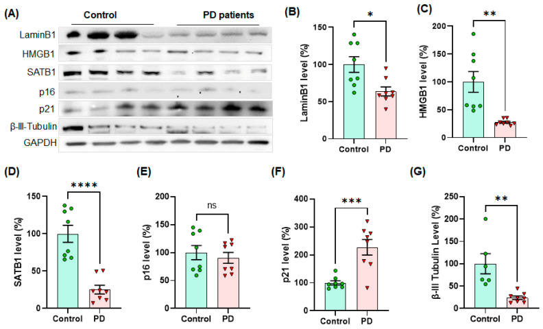
Figure 7.
The levels of cellular senescence markers, such as Lamin B1, HMGB1 and SATB1, were significantly lower in human PD SNpc than age- and gender-matched control tissues, whereas the level of p21 was higher in human PD post-mortem SNpc (n = 8/group). In Western blots, the examples of senescence markers are displayed in quadruplets per group (A). The quantified levels of Lamin B1 (B), HMGB1 (C), SATB1 (D), p16 (E), p21 (F) and β-III-tubulin (G) were statistically analyzed in unpaired Student’s t-test for significance. The band intensity of GAPDH was normalized and displayed as relative band intensities (100% for age-matched controls, n = 8/group) in mean ± SEM. *: p < 0.05, **: p < 0.01, ***: p < 0.001 and ****: p < 0.0001. ns: not significant.

