



Abstract
Mitral annular calcification (MAC) is a chronic, degenerative condition of the fibrous mitral annulus, which may transform to liquefaction necrosis MAC, a rare variant of caseous MAC. We present a series of experiences, showing the varying manifestations of caseous MAC according to multimodal imaging. (Level of Difficulty: Intermediate.)
Key Words: cardiac magnetic resonance imaging, caseous mitral annular calcification, computed tomography, liquefaction necrosis
Abbreviations and Acronyms: CMAC, caseous mitral annular calcification; CMR, cardiac magnetic resonance; CT, computed tomography; MAC, mitral annular calcification; TAVI, transcatheter aortic valve intervention
Graphical abstract

Caseous mitral annular calcification (CMAC) is a rare variant of degenerative mitral annular calcification (MAC). It primarily affects older patients with hypertension, with an echocardiographic prevalence of 0.6% of all MACs and an overall prevalence of up to 0.07% in the general population (1,2). Due to the general benign prognosis, conservative management of this lesion is performed in most cases. However, CMACs may grow large in size and infiltrate adjacent territories such as the myocardium. CMAC rarely was linked to severe mitral valve dysfunction, transient aortic outflow tract obstruction, embolization, heart block, or constrictive pericarditis (3). Furthermore, echocardiographic as well as computed tomography (CT) observations suggest a dynamic course of the condition, with conversion processes from MAC to CMAC and vice versa. Some of these cases are associated with histories of chronic kidney disease and hemodialysis treatment (1,4,5).
Learning Objectives
-
•
To familiarize with the clinical entity of CMACs.
-
•
To train imaging characteristics of CMACs in different modalities.
-
•
To recognize atypical imaging presentations of CMACs.
Differentiation of a CMAC from other cardiac masses attached to the mitral annulus may be challenging due to its variable imaging characteristics depending on its stage of evolution. Using only a single imaging modality such as echocardiography is often not sufficient for a clear diagnosis. Therefore, a multimodal imaging approach is normally used; that is, echocardiography, cardiac CT imaging, and cardiac magnetic resonance (CMR). Echocardiography as first-line modality assesses the mass as well as the functional significance of the CMAC. On both transthoracic echocardiography and in particular transesophageal echocardiography, a CMAC can be recognized as a well-defined, echo-dense mass with central echolucency surrounded by a calcified envelope at the posterior periannular region of the mitral valve (6). In addition, cardiac CT imaging confirms this calcified nature of CMACs, revealing a variable hyperdense mass, with a central hypodense content and peripheral calcifications without enhancement after contrast agent administration (7). CMR provides the best tissue characterization and may exclude other entities. CMR usually visualizes a solid mass with low-signal intensity in both T1- and T2-weighted sequences, reflecting its calcium content. First-pass perfusion sequence reveals no contrast enhancement, whereas late gadolinium enhancement depicts a peripheral rim of enhancement (8).
Peripheral calcifications and avascularity are key features in CMACs. These features can be used to differentiate CMACs from other mass-like lesions involving the atrioventricular grooves but lacking calcifications, such as myxoma, papillary fibroelastoma, myocardial abscess, infective endocarditis and vegetations, lipomatous hypertrophy, or dilated coronary sinus (9). They can also be used to differentiate from lesions that are well vascularized, such as myxoma, hemangioma, dilated coronary sinus or left circumflex artery aneurysm, and enlarged lymph nodes. Distinctions here can easily be accomplished by using color Doppler or contrast enhancement in CT imaging or CMR. However, myocardial abscess within the annular region with an echo-dense appearance and systolic blood flow by color Doppler can closely resemble a CMAC, which may explain the first descriptions of CMACs as a “sterile myocardial abscess“ (1,9).
The interior of a CMAC is composed of a liquefied mixture of calcium, cholesterol, and fatty acids, which explains the central echolucency on transthoracic echocardiography/transesophageal echocardiography and the central hypodensity in CT imaging (6); it is therefore also known as a “toothpaste-like” tumor among surgeons because of its similar consistency. Imagers should be familiar with this rare entity because these lesions have various clinical implications and may even simulate tumors. Due to the increasing use of CT and CMR imaging, CMAC may be more frequently encountered today in clinical practice. Thus, the purpose of our case series was to describe the versatile spectrum of imaging characteristics of CMACs, emphasizing the value of multimodal imaging.
Case 1: Dynamic Evolution and Atypical Location of a CMAC
A 76-year-old man with history of a biological aortic valve replacement and bypass surgery 11 years earlier was presented for evaluation before valve-in-valve transcatheter aortic valve intervention (TAVI). Cardiac CT imaging revealed a heavily calcified posterior mitral annulus with a large, atypically located isolated caseous calcification in its continuation to the aortic outflow tract at the aorto-mitral continuity. This represents an exceptionally rare CMAC location. Moreover, exophytic lesions at the aortic outflow tract may lead to complications during positioning and expansion of the prosthesis as well as to post-procedural device deformation. These lesions are associated with an increased risk of aortic root rupture during the TAVI procedure (3).
Furthermore, by evaluating former CT images, we were able to reconstruct the dynamic nature of a degenerative CMAC over a period of 19 years: initially, the anterior and posterior mitral annulus showed minimal calcifications (Figure 1, year 2001) that slowly but gradually progressed to a lumpy appearance. However, 10 years later, a small caseous part developed at the anterior annulus calcification (Figure 1, year 2011). Three years later, at TAVI-planning CT imaging, as discussed earlier, another increase of the lesion at the anterior mitral annulus was visualized, together with a continued significant decrease in its density (Figure 1, year 2014). However, the calcified part at the posterior annulus remained stable. Another 2 years later, after the TAVI procedure, the anterior CMAC consisted of a complete liquefaction necrosis surrounded by a delicate, partially calcified rim (Figure 1, year 2016). Current CT images from 2020 reveal the anterior part of this CMAC lesion to be reduced to small residues (Figure 1, year 2020), whereas the posterior annulus shows an increased calcified mass with small areas of central liquefaction.
Figure 1.
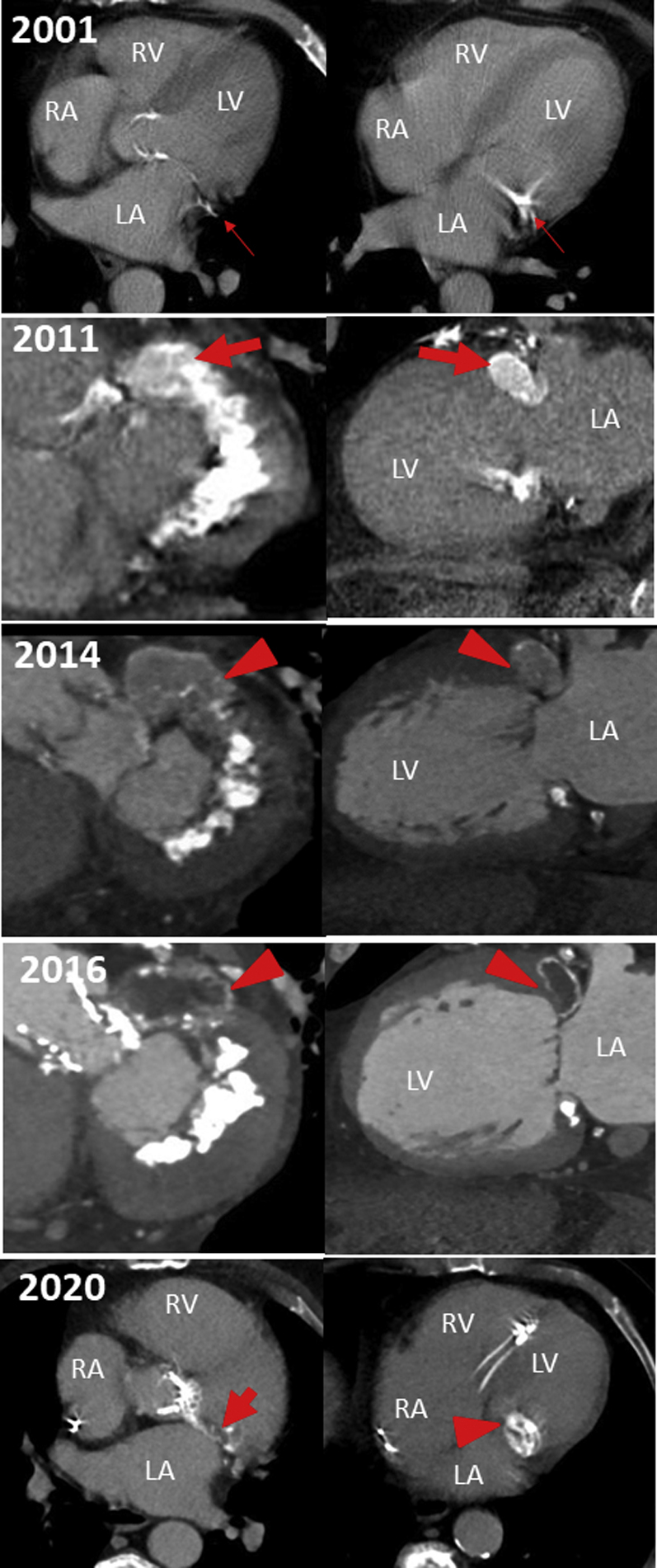
Dynamic Evolution and Atypical Location of a Caseous Mitral Annular Calcification
LA = left atrium; LV = left ventricle; RA = right atrium; RV = right ventricle.
This case contributes to the accumulating evidence regarding the dynamic progression from MAC to CMAC as well as its remission from CMAC to MAC. As this case shows, even a temporally and morphologically diverse progress of individual parts of the lesion seems to be possible.
Because this disease is associated with complications after transcatheter mitral and aortic valve interventions and mitral valve surgery, its clinical implications are of high importance (10, 11, 12, 13, 14).
Case 2: Atypical Mobile CMAC Mimicking a Valvular Mass—Differential Diagnosis
Transthoracic and subsequent transesophageal echocardiography of a 78-year-old woman with hypertension and a medical history of cerebral emboli and non–ST-segment elevation myocardial infarction revealed a shelf-like, partially echo-dense mass beneath the P2 segment of the mitral leaflet but not clearly distinguishable from the posterior mitral valve leaflet (Figures 2A to 2C, Video 1). Due to its mobility (Videos 1 and 2), its heteroechogenicity, and its location at the mitral leaflet, the possible differential diagnosis included: 1) valvular mass tumor such as papillary fibroelastoma; 2) old, organized vegetation or thrombus; or 3) atypical, mobile CMAC.
Figure 2.

Atypical Mobile Caseous Mitral Annular Calcification Mimicking a Valvular Mass
(A) Apical 4-chamber view of transesophageal echocardiography. (B) Apical 4-chamber tissue Doppler in transesophageal echocardiography. (C) Apical 4-chamber view of transthoracic 3D echocardiography. (D) Biphasic iodine contrast-enhanced electrocardiographically gated computed tomography (CT). (E) Basal short-axis slice of late gadolinium enhancement cardiac magnetic resonance imaging.
AV = aortic valve; other abbreviations as in Figure 1.
A biphasic iodine contrast-enhanced electrocardiographically gated CT scan revealed a subtotal calcified mass located at the junction of the atrioventricular groove and posterior mitral leaflet (Figure 2D). Because the lesion did not exhibit contrast enhancement in both CT and CMR late gadolinium imaging (Figure 2E), tumors such as myxoma or papillary fibroelastoma could be firmly excluded; this allowed us to diagnose degenerative CMAC with a small central liquefaction component.
This case highlights the advantage of a multimodal imaging approach to clearly differentiate CMAC from other lesions.
Case 3: CMAC at CMR Imaging
The incidental finding during adenosine stress CMR of a 45-year-old man with a history of hemodialysis treatment and chronic myocardial infarction was a large, mass-like lesion of mitral annulus adjacent to the P1 and P2 segment of the posterior leaflet. Due to its CMR signal characteristics (i.e., general T2 hypointense signal [Figure 3A], a weak heterogeneous T1 hypointensity [Figure 3B], central hypointensity at cine steady-state free precession sequence with a brighter border [Figure 3C] evidencing a broad rim of late gadolinium enhancement [Figure 3D]), the differential diagnosis was an atypically located fibroma (usually characterized by homogenous late enhancement) or a CMAC with a prominent fibrous capsule. A subsequent electrocardiographically gated CT scan revealed a centrally hypodense mass with irregular calcified borders (Figure 3E) in continuity to a roughly calcified chronic myocardial scar (Figure 3F) at the territory of the circumflex artery. This allowed a safe diagnosis of a CMAC with a liquefied necrotic core surrounded by a prominent capsule with an inner fibrotic layer and an outer calcified layer. Imagers, however, may be more familiar with the appearances of CMAC at CT scans, which are usually used to plan transcatheter valve interventions and to clarify echocardiographically unclear valve masses. This case familiarizes imagers and clinicians with the typical imaging features of a CMAC in CMR.
Figure 3.

Caseous Mitral Annular Calcification at Cardiac Magnetic Resonance Imaging
(A) Basal short-axis slice of a T2-weighted turbo-spin-echo cardiac magnetic resonance imaging sequence. (B) Basal short-axis slice of a T1-weighted turbo-spin-echo cardiac magnetic resonance imaging sequence. (C) 4-chamber view of a cine steady-state free precession cardiac magnetic resonance imaging sequence. (D) 4-chamber view of a late gadolinium enhancement cardiac magnetic resonance imaging sequence. (E) Iodine contrast-enhanced electrocardiographically gated computed tomography (CT). (F) Contrast-free electrocardiographically gated CT.
Conclusions
CMACs exhibit heterogeneous imaging characteristics, mainly depending on their stage of evolution. Differentiation of other intracardiac masses is therefore challenging. However, using a multimodal imaging approach, precise diagnosis can be accomplished. Imagers should be attentive to this disease due to its various clinical implications, including those of rapidly evolving transcatheter valve procedures.
Author Disclosures
The authors have reported that they have no relationships relevant to the contents of this paper to disclose.
Footnotes
The authors attest they are in compliance with human studies committees and animal welfare regulations of the authors’ institutions and Food and Drug Administration guidelines, including patient consent where appropriate. For more information, visit the Author Center.
Appendix
For supplemental videos, please see the online version of this paper.
Appendix
Transesophageal echocardiography shows a shelf-like, partially echo-dense, mobile mass at the junction of the atrioventricular groove and the P2 segment of the mitral leaflet.
Three-dimensional transesophageal echocardiography evaluation of the mitral valve reveals the mass-like lesion at the P2 segment of the mitral leaflet.
References
- 1.Deluca G., Correale M., Ieva R., Del Salvatore B., Gramenzi S., Di Biase M. The incidence and clinical course of caseous calcification of the mitral annulus: a prospective echocardiographic study. J Am Soc Echocardiogr. 2008;21:828–833. doi: 10.1016/j.echo.2007.12.004. [DOI] [PubMed] [Google Scholar]
- 2.Pomerance A. Pathological and clinical study of calcification of the mitral valve ring. J Clin Pathol. 1970;23:354–361. doi: 10.1136/jcp.23.4.354. [DOI] [PMC free article] [PubMed] [Google Scholar]
- 3.Motwani M., Fairbairn T.A., Jogiya R., Greenwood J.P., Plein S. Caseous calcification of the mitral valve complicated by embolization, mitral regurgitation, and pericardial constriction. Eur Heart J Cardiovasc Imaging. 2012;13:792. doi: 10.1093/ehjci/jer180. [DOI] [PubMed] [Google Scholar]
- 4.Asselbergs F.W., Mozaffarian D., Katz R. Association of renal function with cardiac calcifications in older adults: the Cardiovascular Health Study. Nephrol Dial Transplant. 2009;24:834–840. doi: 10.1093/ndt/gfn544. [DOI] [PMC free article] [PubMed] [Google Scholar]
- 5.Koito H., Nakamura C., Suzuki J., Takahashi H., Iwasaka T. Reduced size of liquefaction necrosis of mitral annular calcification in chronic renal failure by using low calcium concentration hemodialysis. Jpn Circ J. 1999;63:490–492. doi: 10.1253/jcj.63.490. [DOI] [PubMed] [Google Scholar]
- 6.Harpaz D., Auerbach I., Vered Z., Motro M., Tobar A., Rosenblatt S. Caseous calcification of the mitral annulus: a neglected, unrecognized diagnosis. J Am Soc Echocardiogr. 2001;14:825–831. doi: 10.1067/mje.2001.111877. [DOI] [PubMed] [Google Scholar]
- 7.Vanovermeire O.M., Duerinckx A.J., Duncan D.A., Russell W.G. Caseous calcification of the mitral annulus imaged with 64-slice multidetector CT and magnetic resonance imaging. Int J Cardiovasc Imaging. 2006;22:553–559. doi: 10.1007/s10554-005-9069-0. [DOI] [PubMed] [Google Scholar]
- 8.Monti L., Renifilo E., Profili M., Balzarini L. Cardiovascular magnetic resonance features of caseous calcification of the mitral annulus. J Cardiovasc Magn Reson. 2008;10:25. doi: 10.1186/1532-429X-10-25. [DOI] [PMC free article] [PubMed] [Google Scholar]
- 9.McKernan N.P., Culp W.C., Jr., Knight W.L., Johnston W.E., Sniecinski R.M., Lazar H.L. CASE 2—2012 intraoperative diagnosis and management of caseous calcification of the mitral annulus. J Cardiothorac Vasc Anesth. 2012;26:327–332. doi: 10.1053/j.jvca.2011.07.015. [DOI] [PubMed] [Google Scholar]
- 10.Barbanti M., Yang T.H., Rodes Cabau J. Anatomical and procedural features associated with aortic root rupture during balloon-expandable transcatheter aortic valve replacement. Circulation. 2013;128:244–253. doi: 10.1161/CIRCULATIONAHA.113.002947. [DOI] [PubMed] [Google Scholar]
- 11.Boerlage-Van Dijk K., Kooiman K.M., Yong Z.Y. Predictors and permanency of cardiac conduction disorders and necessity of pacing after transcatheter aortic valve implantation. Pacing Clin Electrophysiol. 2014;37:1520–1529. doi: 10.1111/pace.12460. [DOI] [PubMed] [Google Scholar]
- 12.Durst R., Avelar E., McCarty D. Outcome and improvement predictors of mitral regurgitation after transcatheter aortic valve implantation. J Heart Valve Dis. 2011;20:272–281. [PubMed] [Google Scholar]
- 13.Fusini L., Ghulam Ali S., Tamborini G. Prevalence of calcification of the mitral valve annulus in patients undergoing surgical repair of mitral valve prolapse. Am J Cardiol. 2014;113:1867–1873. doi: 10.1016/j.amjcard.2014.03.013. [DOI] [PubMed] [Google Scholar]
- 14.Okada Y. Surgical management of mitral annular calcification. Gen Thorac Cardiovasc Surg. 2013;61:619–625. doi: 10.1007/s11748-013-0207-7. [DOI] [PubMed] [Google Scholar]
Associated Data
This section collects any data citations, data availability statements, or supplementary materials included in this article.
Supplementary Materials
Transesophageal echocardiography shows a shelf-like, partially echo-dense, mobile mass at the junction of the atrioventricular groove and the P2 segment of the mitral leaflet.
Three-dimensional transesophageal echocardiography evaluation of the mitral valve reveals the mass-like lesion at the P2 segment of the mitral leaflet.