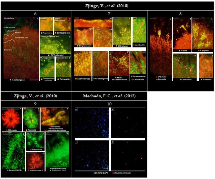
Figure 4.
Panel of the gathered images. (6) Localization of the most abundant species in subgingival biofilms. (6A) Overview of the four layers of subgingival biofilm. Actinomyces sp. (green), bacteria (red), and eukaryotic cells (large green cells on top). (6B) Spirochaetes (yellow) outside the biofilm, without clear organization. (6C) Detail of Synergistetes (yellow) in the top layer near PMN’s (Polymorphonuclear leukocytes) (green). (6D) Presence of the Cytophaga-Flavobacterium-Bacteroides cluster (CFB-cluster) (yellow) in the top and intermediate layer. (6E) Fusobacterium nucleatum in the intermediate layer. (6F) Tannerella sp. (yellow) in the intermediate layer. Each panel is double-stained with probe EUB338 labeled with FITC (Fluorescein isothiocyanate) or Cy3 (Cyanine 3). Bars indicate 10 µm. (7) Localization of the most abundant species in supragingival biofilms. (7A1–C) The second layer. (7D–G) Basal layer. (7A1–A3) Streptococcus sp. disposed of in different ways on the second layer. (7B) Cells from the CFB-cluster stained in the top layer of the biofilm. (7C) Lactobacillus sp. (red) through the top layer. (7D) On the basal layer, Actinomyces sp. cells (yellow). (7E) Actinomyces sp. (green) and chains of cocci. (7F) Colonies of Streptococcus sp. (yellow) all-around yeast cells (green) and bacteria unidentified (red). (7G) Streptococcus sp. (green) growing closely to Lactobacillus sp. (red). Black holes might be channels through the biofilm. Panels (A–C,E,F) are double stained with probe EUB338 labeled with FITC or Cy3. Bars indicate 10 µm. (8) Localization of presumptive species associated with periodontitis in subgingival biofilms. (8A) Colonization of the subgingival biofilm by the CFB-cluster species (red) and Prevotella sp. (yellow). Since Prevotella sp. are part of the CFB-cluster of bacteria, cells appear in yellow. (8B) Top of the biofilm with a micro-colony of Parvimonas micra (yellow). (8C,D) Micro-colonies of Porphyromonas gingivalis (yellow) and Porphyromonas endodontalis (yellow) in the top layer, respectively. (8E) Micro-colonies of Prevotella intermedia in the top layer. Panels B, C, D, and E are double stained with probe EUB338 labeled with FITC or Cy3. Bars indicate 10 µm. (9) Bacterial aggregates detected in both sub- and supragingival plaque. (9A) Filamentous cells from the CFB-cluster in the fourth layer of the subgingival plaque. (9B) Tannerella sp. (yellow) in a test-tube brush. (9C) Test-tube brush with Lactobacillus sp. (red rods) as central structures. Fusobacterium nucleatum (green) and CFB-cluster filaments (red), morphologically identical to Tannerella forsythia, perpendicularly radiating around lactobacilli. (9D) Test tube brush stained with the eubacterial probe. (9E) Synergistetes group A species forming aggregates solely with themselves. (9F) Streptococcus sp. (green) aggregation around a central cell (not stained) in supragingival plaque. (9G) Supragingival plaque with Streptococcus sp. (green cocci) adhering to a central axis of yeast cells or hyphae, such as Candida albicans (green hyphae). Bars indicate 10 µm. (10a,c) Plaque specimen stained for total bacterial cells with DAPI (4′, 6-diamino-2-phenylindole) in pregnant and non-pregnant women, respectively. (10b,d) Plaque specimen stained for Prevotella intermedia (with probe Pint649) in pregnant and non-pregnant women, respectively. Bars indicate 20 µm. All images were reprinted and adapted with the publisher’s permission.

