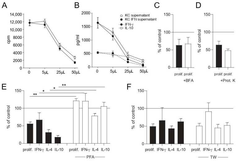
Figure 5.
Keratinocytes inhibit T cell effector functions through the production of soluble factor/s. Cell-free culture supernatants of keratinocyte- or IFN-γ-prestimulated keratinocyte cultures (KC IFN) were obtained and used in different volumes in co-culture experiments. Proliferation (A) and cytokine production (B) were measured after 48 h by 3H-thymidine incorporation and ELISA of co-culture supernatants, respectively. Unstimulated keratinocytes were fixed with PFA (E) or treated with brefeldin A (BFA) to block the Golgi apparatus (C) prior to co-culture with nickel-specific T cell clones and dendritic cells. Supernatants of unstimulated keratinocytes were treated for 30 min with proteinase K (Prot. K) (D) prior to co-culture with nickel-specific T cell clones and dendritic cells. Keratinocytes were separated from nickel-specific T cells and dendritic cells via a transwell chamber (TW) (F). To rule out any effects from the keratinocyte medium, the medium was added to every control stimulation in the same volume as was used for the keratinocyte supernatant experiments. ((A) results of one representative nickel-specific Th1 clone; (E) clones = 8, n = 16; (C,D) clones = 4, n = 8; (F) clones = 5, n = 14; *: p < 0.05; **: p < 0.01).

