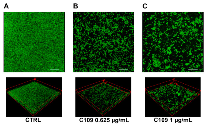
Figure 5.
CLSM images of S. aureus ATCC 25923 biofilms grown in a Lab-Tek II Chamber Slide. Pictures were taken with an overall magnification of 400×. Cells were grown overnight at 37 °C in TSB + 1% glucose with no C109 (CTRL, (A)), 0.625 μg/mL of C109 (B), or 1 μg/mL of C109 (C). Eighty planes at equal distances along the Z-axis of the biofilm were imaged by CLSM. These 2D images were stacked to reconstruct the 3D biofilm image. Scale bar represents 28 μm.

