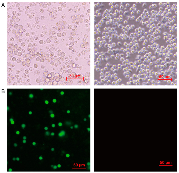
Figure 1.
Observation of PAM cells after ASFV infection. (A) Typical CPEs caused by ASFV were shown by optical microscope hours 72 h post infection. (B) PAM cells were infected by ASFV at MOI of 1. At 24 h post infection, the expression of ASFV encoding protein p30 in PAM cells was detected by indirect immunofluorescence. The left was ASFV-infected PAM cells visual field, the right was mock-infected PAM cells visual field. The red bars in graphs were on behalf of 50 μm.

