Abstract
Objective:
To study the airflow, transmission, and clearance of aerosols in the clinical spaces of a hospital ward that had been used to care for patients with coronavirus disease 2019 (COVID-19) and to examine the impact of portable air cleaners on aerosol clearance.
Design:
Observational study.
Setting:
A single ward of a tertiary-care public hospital in Melbourne, Australia.
Intervention:
Glycerin-based aerosol was used as a surrogate for respiratory aerosols. The transmission of aerosols from a single patient room into corridors and a nurses’ station in the ward was measured. The rate of clearance of aerosols was measured over time from the patient room, nurses’ station and ward corridors with and without air cleaners [ie, portable high-efficiency particulate air (HEPA) filters].
Results:
Aerosols rapidly travelled from the patient room into other parts of the ward. Air cleaners were effective in increasing the clearance of aerosols from the air in clinical spaces and reducing their spread to other areas. With 2 small domestic air cleaners in a single patient room of a hospital ward, 99% of aerosols could be cleared within 5.5 minutes.
Conclusions:
Air cleaners may be useful in clinical spaces to help reduce the risk of acquisition of respiratory viruses that are transmitted via aerosols. They are easy to deploy and are likely to be cost-effective in a variety of healthcare settings.
Coronavirus disease 2019 (COVID-19) presents a major global health challenge, with extraordinary clinical, societal and economic impacts. Although initially there may have been controversy on the role of airborne transmission, most authorities now suggest that transmission of severe acute respiratory coronavirus virus 2 (SARS-CoV-2) can occur via contact, droplet and/or airborne routes depending on the circumstances.1–8 The different mechanisms of transmission likely arise due to variation in the size of respiratory particles generated, which depends on a number of factors including, but not limited to, the site of primary infection (lower vs upper respiratory tract), exposure to aerosol-generating procedures (eg, nebulizer use), or aerosol-generating behaviors of the patient (eg, coughing, shouting, or singing).9–16 The context is also likely to be very important because, for example, airborne transmission may be more likely to occur where there are several infected people (sources) in a confined space with a limited clearance of aerosolized particles due to poor ventilation.17–19
In many countries, an increased risk of infection with SARS-CoV-2 has been reported among frontline healthcare workers. At the Royal Melbourne Hospital, 271 healthcare workers acquired COVID-19 infection, and the consequent epidemiologic and genomic investigation suggested that most of these cases were healthcare acquired.20 Clinicians observed that transmission to healthcare workers seemed to be more common when the number of concurrent COVID-19 infected patients in a given ward was high, and in association with particular behaviors (eg, shouting or persistent cough). Staff without patient contact were also noted to become infected, especially if they spent prolonged times in the ward (namely corridors and nurses’ stations) but not necessarily in the patient rooms. The high rate of staff infection raised concern about possible aerosol transmission, so staff within the wards were required to wear N95/P2 masks rather than surgical masks in mid-July ahead of state and national guidance. A group of multidisciplinary researchers from engineering, aerosol science, virology, infection prevention, and infectious diseases and respiratory medicine was convened to discuss what was known about aerosol transmission of SARS-CoV-2 in July 2020 and met weekly thereafter. As new evidence emerged to support that hypothesis, the focus quickly moved to considering what mitigation strategies could be used in a clinical space to reduce risks for staff, including the value of portable air cleaners.21–29 In this study, we traced airflow and the movement of aerosolized particles within a ward where known COVID-19 patients had been cared for and transmission to staff had occurred to document the effectiveness of air cleaners in reducing airborne particle concentrations.
Methods
Setting
The Royal Melbourne Hospital is a university-affiliated, tertiary-care hospital with 550 acute-care beds and 150 subacute care (rehabilitation, geriatric medicine) inpatient beds. The hospital has cared for the largest number of inpatients with COVID-19 in Australia to date, namely 525 inpatients, with a peak of 99 concurrent inpatients in August 2020. The infectious diseases ward has 14 Class N negative-pressure rooms with anterooms. In 2020, as the number of concurrent inpatients with COVID-19 infection climbed and the capacity of the infectious diseases ward was exceeded, 5 other medical and surgical wards were progressively converted to COVID-19 wards. All staff entering the wards were required to wear PPE (ie, long-sleeved gowns, gloves, eye protection via full face shield, and N95/P2 mask) including while in patient rooms, corridors, and nurses’ stations. Many patients with COVID-19 who were cared for in these wards were elderly and required high levels of nursing care. Due to patient frailty, the doors to patient rooms sometimes stayed open to provide appropriate supervision.
In December 2020, the research team had full access to an empty ward that had previously been used to care for COVID-19 patients (and where staff acquisitions had occurred) to conduct in situ experiments assessing airflow and aerosol clearance. This ward has a long central corridor and 11 rooms, which usually accommodate 25 patient beds (4 single rooms with en suite bathrooms and 7 three-bed shared rooms, each with a shared en suite bathroom). When the ward was occupied, patients were predominantly cared for with 1 patient per room and the peak concurrent number of patients in that ward was 15. The nurses’ station is situated halfway along the corridor, directly opposite 2 single rooms. No rooms in this ward have negative pressure, and the ward has its own closed, ducted heating, ventilation, and air conditioning (HVAC) system, which delivers 12 air changes per hour. No windows in the ward can be opened, and the return air vent for the whole ward is above the single entrance and exit point to the ward (just inside the door to the ward). Rooms all have doors with a small gap at the bottom (∼5 cm) to allow air egress. One of the single-patient rooms with a room floor space of 12.8 m2 and volume of ∼37 m3 was selected for the study. The corridor outside the room was ∼2 m wide, and the patient room was directly opposite the open nurses’ station, which had a front desk with entrance spaces on either side.
Study design
In this intervention study, we investigated the air flows of aerosols from a room used for the care of patients with COVID-19 to the corridor and the nursing station in that clinical ward. The space with a glycerin-based smoke and the pathways were video recorded, and changes in aerosol levels over time in the room and adjacent spaces were monitored. The effect of portable air cleaners on the rate of clearance of air particles in these areas was also examined.
Intervention
For each experiment, glycerine-based aerosol smoke with a mean aerosol size of 1 µm was injected for 15 seconds into the patient room to flood the air space with smoke. The aerosol size distribution of the glycerin-based smoke was measured to confirm that the particle sizes of this smoke roughly approximated the sizes of SARS-CoV-2 infectious aerosols measured in the air of hospitals elsewhere (<1 µm). This information was used as a surrogate for replicate respiratory aerosol movement and airborne transmission.30 This type of smoke testing forms part of the indoor air assessment industry standard accepted processes for assessing airflows. Once injected, smoke was permitted to mix and visibly fill all corners of the room, which took ∼30 seconds. The visible pathways of travel of the smoke within the patient room, corridor, and nurses’ station were then observed and video recorded.
The sensors used in this experiment to measure aerosols were the TSI DustTrak DRX 8533 (called DRX) and the TSI DustTrak II 8530 (called DRII). The DustTrak sensors measured aerosol concentration using a combination of particle cloud and single-particle detection to measure the mass concentration of aerosols per unit volume. The DustTrak’s sensors were both fitted with an identical 2.5-µm inlet that only allowed aerosols 2.5 µm or smaller to pass through. The DRII was placed inside the patient room (or corridor), and the DRX was placed in the nurses’ station to determine the amount of aerosol that could move through the corridor or across into the nurses’ station. Both devices were calibrated by the manufacturer before the study. Although the DRX also has the ability to detect masses in different aerosol-size bins, for this study we used only the common mass observation of aerosols <2.5 µm in size for both the DRX and DRII. Side-by-side and zero calibrations assured us that the DRX and the DRII provided identical observations under the same conditions.
The air cleaners used were domestic appliances (Samsung AX5500K) equipped with H13 HEPA filters capable of filtering 99.97% of particles at a clean air delivery rate of 467 m3 per hour on the highest fan speed setting. Based on laboratory testing, for the size of the hospital room and the capacity of the air cleaner selected, 2 air cleaners were placed along the bedside and at the foot of the bed.
Measurements
Measurements were taken concurrently in the single-patient room and at the nurses’ station at 10-second intervals until the aerosols cleared. This process was reported as the normalized and absolute aerosol concentration decay over time.
Intervention tests evaluated the following 4 aspects:
(1) Effect of installation of portable air cleaners in the patient room vs usual HVAC on aerosols within and external to the room,
(2) Effect of patient room door open vs door closed on aerosols outside the room,
(3) Effect of installation of variable number of portable air cleaners in the corridor on aerosols in the corridor, and
(4) Effect of installation of different barriers to enclose the nurse’s station on aerosols in within the nurses’ station.
Outcomes
The effects of each intervention were assessed as changes in the clearance rate of aerosols from the space. For example, the time taken to clear the patient room and the nurses’ station from aerosols under existing HVAC settings at an air change rate of 12 times per hour was compared to the time taken when air cleaners were placed in the patient room. The time taken to clear the aerosols from the patient room and the nurses’ station opposite was measured when the patient room had the door open and when the door was closed, for comparison. The impact of additional protections for the nurses’ station included when the nurses’ station was protected by an air cleaner barrier or a plastic ZipWall. An air-cleaner barrier consisted of 3 air cleaners lined up 1 m apart directly in front of the desk. The ZipWall is a clear plastic barrier that was erected in front of the desk, sealed at the ceiling and walls on either side, with a magnetized self-closing door for people to enter and exit the space.
Measurements of aerosol clearance were also taken in the corridor of the ward, which was ∼50 m long and 2 m wide. The corridor was flooded with smoke and the rates of clearance of aerosols were compared with different numbers of air cleaners positioned along its length to identify the optimum number of air cleaners for that space.
Results
At baseline, the air in the patient room had negligible demonstrable aerosols, and after smoke flooding was applied, the entire room was rapidly and visibly filled with smoke. During the tests with the patient room door closed, within the first minute after the room was flooded with smoke, smoke escaped under the gap at the bottom of the door and moved along the corridor toward the return air vent at the entrance of the ward. When the door to the patient room was opened, smoke immediately moved out to the corridor and travelled along the corridor to the return air vent.
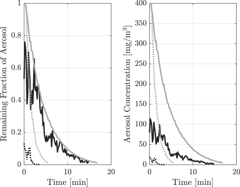
Using measurements within the patient room, at usual HVAC settings and with a closed door, it took 16 minutes for the aerosols in the patient room to clear back down to 1% of the baseline maximum measurable by the instruments. The concentration of aerosols concurrently reaching the nurses’ station even when the patient room door was closed were high and decreased at the same rate as aerosols inside the room. The relative amount of aerosols in the nurses’ station compared to the patient room was highly variable representing 25%–50% of the room’s concentration with the door closed using 2 air cleaners. When 2 air cleaners were placed in the patient room with the door closed or open, the room cleared of 99% of all aerosols in 5.5 minutes (a 67% reduction compared with no air cleaners). The smoke at the nurses’ station cleared even more quickly, in <3 minutes (Fig. 1). Having the bathroom door open with exhaust fan running made a negligible difference to the clearance time (∼50 seconds, and within the variability of the observations).
Fig. 1.
The effect of no air cleaners versus 2 air cleaners on aerosol clearance and transmission of aerosols within a patient room with the door closed. The left image shows the values normalized to the saturation value of the sensor whereas right shows the measured value. Note. The grey solid line indicates measures taken within the standard patient room. Black solid line indicates measures taken at nurses’ station. The grey dotted line indicates measures taken within the patient room with 2 air cleaners running. The black dotted line indicates measures taken at the nurses’ station when the 2 air cleaners were in the patient room.
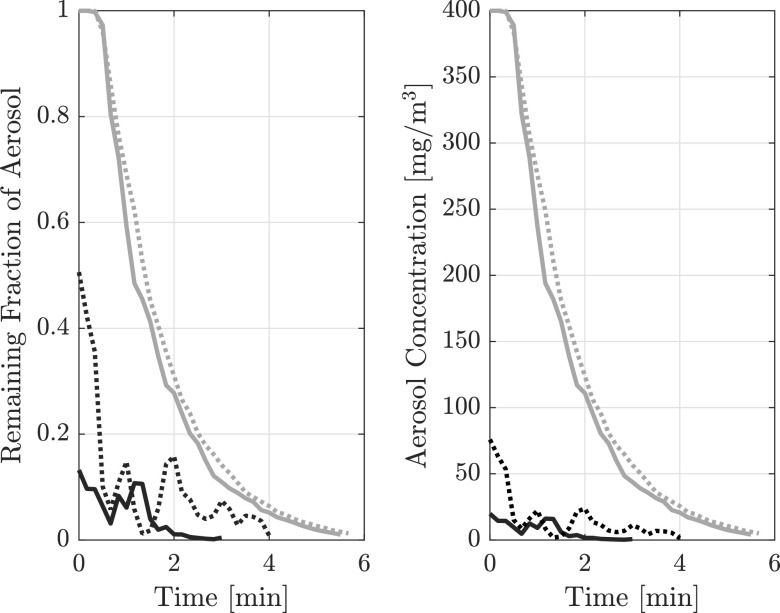
When the door to the patient room was open, high levels of aerosols had crossed the corridor and entered the nurses’ station at baseline measurement. When the air cleaners were used in the patient room, the aerosols cleared from the nurses’ station in 4 minutes. When the door to the patient room was closed and air cleaners were used in the patient room, very low levels of aerosol were detectable at the nurses’ station at baseline, and 99% of these cleared within ∼2 minutes (Fig. 2).
Fig. 2.
The effect of open vs closed door to patient room on aerosol clearance and transmission of aerosols. Left image shows the values normalized to the saturation value of the sensor whereas right shows the measured value. The measurements were taken with 2 air cleaners in the patient room. Note. The grey solid line indicates measures taken within the patient room with door to the corridor closed. The black solid line indicates measures taken at the nurses’ station (NS) when the door was closed. The grey dotted line indicates measures taken within the patient room with door to the corridor open. Black dotted line indicates measures taken at the nurses’ station (NS) when the door was open.
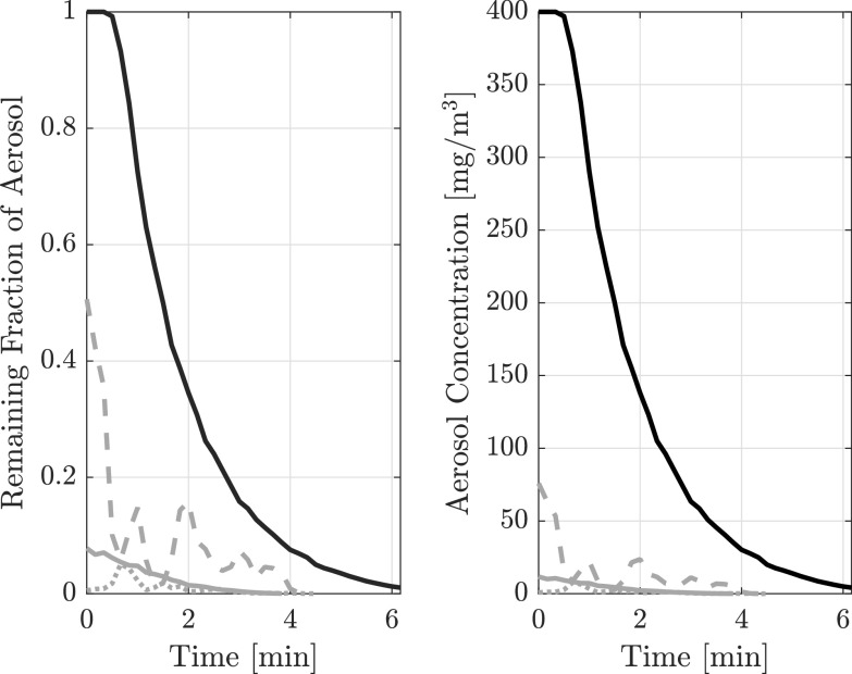
Two different barriers were tested to determine the most effective way to provide protection to the nurses’ station. When the air-cleaner barrier was used, negligible smoke particles from the patient room were measured at the nurses’ station (Fig. 3). Similarly, the ZipWall substantially reduced the ability of the aerosolized smoke particles to enter the nurses’ station (Fig. 3).
Fig. 3.
Comparing different interventions at the nurses’ station on aerosol clearance and transmission of aerosols. Left image shows the values normalized to the saturation value of the sensor whereas right shows the measured value. The measurements were taken with 2 air cleaners in the patient room. Note. The black solid line indicates measures taken within the patient room. The grey solid line indicates measures taken at the nurses’ station (NS) with Zipwall present. The grey dashed line indicates measures taken at NS without barrier. The grey dotted line indicates measures taken at NS with air cleaner barrier present (3 air cleaners in front of desk).
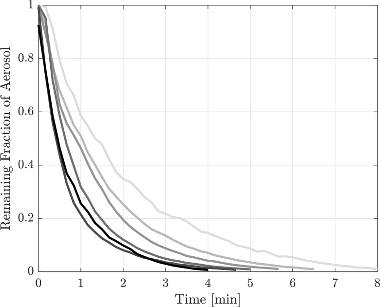
When the corridor was flooded with smoke, between 4 and 12 air cleaners were evenly spaced along its length. Adding extra air cleaners did not increase the clearance much beyond the clearance achieved with 8 air cleaners, which was 99% in 5 minutes (Fig. 4).
Fig. 4.
Rate of clearance of aerosolized smoke particles in the corridor with differing numbers of air cleaners. Some sources also called these “portable HEPA filters.” Line color goes from light to dark for cases with progressively more air cleaners, with 0, 4, 6, 8, 10, and 12 evenly spaced air cleaners considered in the corridor.
Discussion
In this study, we demonstrated that the existing ward HVAC system alone was quite poor at clearing a patient room of aerosols. Our results suggest that commercially available air cleaners may have a role in clearing aerosolized particles that may contain respiratory viruses, such as SARS-CoV-2, in clinical environments. The actual 99% clearance of aerosols from the air in patient rooms using existing HVAC alone at 12 air exchanges per hour was quite slow. Aside from isolation rooms, HVAC systems in hospitals and other places are designed for comfort rather than infection control. Notably, air exchange rates of 2 would not be uncommon in an office environment. In an Australian hospital environment, 6 air exchanges per hour is the standard, and 10 air exchanges per hour has been suggested for wards managing COVID-19 patients. These data demonstrate that the relationship between reported air exchanges and actual aerosol clearance from rooms is not predictable, probably because it does not account for flow recirculation regions or other air flow anomalies. Theoretically, to clean 99% of the air of a well-mixed room in <10 minutes would require the same air exchange equivalent to air exchanges of 30 per hour.31 For context, typical operating rooms have 20 air exchanges per hour,32,33and some have recommended waiting for 5 full air exchanges (18 minutes for 99.3% clearance) before staff can enter without airborne precautions where necessary.34 It is impossible to achieve >30 air exchanges with the typical hospital ward HVAC systems alone, but it is not difficult with air cleaners.
Our data suggest that any air that enters a patient room needs to travel to reach a return air vent. In some cases, return air vents are placed in the patient room or in the en suite bathroom. In the ward we studied, the position of the return air vent meant that air was leaving the patient room and traveling along the corridor. It is important to understand where and how air exits a patient room so that the path of least resistance for air to travel from the patient to the return air vent can be considered when patients are placed on a ward. On the ward studied, the preference is now to utilize rooms close to that air return for higher-risk patients (eg, earlier in their illness, actively coughing) where possible.
Our findings also confirmed that the doors to the patient rooms should be kept closed wherever possible to protect areas outside the recognized patient zone because the air and the aerosols carried in it may still travel. We also showed that air cleaners in the patient room limit how much aerosol is likely to escape into the corridor or nurses’ station, which likely provides protection for staff. Air cleaners placed along the corridor should be considered (in addition to cleaners in the patient room) if the ward was filled with patients infected with SARS-CoV-2 to help clear the air of potentially infected aerosols that may have escaped from the patient’s room. Finally, a physical barrier, such as a ZipWall, may provide additional protection for the staff if they spend time at a workstation.
Importantly, this work used a glycerin-based aerosol as a proxy for respiratory aerosols that contain live virus. Extrapolations must be made from other studies in which viral RNA or culturable virus has been identified in aerosols from clinical spaces caring for people with SARS-CoV-2 infection.26,27,29,35 Nevertheless, we have demonstrated that aerosols the same size as respiratory particles can travel long distances. Viral transmission via aerosols is likely to be very variable, both between patients and at different times in a given patient’s infection course. Transmission and may also be affected by other factors such as relative humidity and temperature.36 We hypothesize that if multiple patients are in a given confined space, each producing some respiratory particles, then the density of infectious aerosols in the air may become high enough to put staff at increased risk of virus acquisition via aerosols, even in corridors or nurses’ stations of dedicated COVID-19 wards. Given that the air cleaners remove aerosols from the air before it leaves the patient room, it is likely that they would help protect staff inside and outside patient rooms. These air cleaners are a relatively low cost, readily implementable mitigation strategy that should be considered in clinical spaces. Our findings may also have implications outside healthcare institutions that warrant further investigation.
In conclusion, despite exceeding recommended air exchange rates for a hospital, the HVAC alone did not effectively remove aerosols from the clinical space in a timely matter. In addition, depending on the location of the return air duct, the existing HVAC system promoted the dispersal of aerosols beyond the patient room. However, we were able to demonstrate that relatively low-cost air cleaners could dramatically increase the clearance rate of aerosols. Air in clinical spaces does travel, and with it any respiratory aerosols containing potentially infectious virus. An understanding of directional airflow is important to help limit the risk to staff and will likely be different in different spaces. Air cleaners enabled the air to be cleaned locally, providing protection equivalent to >30 air exchanges per hour to staff in common areas, far exceeding the current best-practice guidelines for hospital indoor air-ventilation rates. Based on our findings, the hospital studied has adopted air cleaners for use in rooms of patients with suspected or confirmed COVID-19, with appropriate policies, procedures, and training to ensure their safe and effective use.
Acknowledgments
Supplementary material
For supplementary material accompanying this paper visit http://dx.doi.org/10.1017/ice.2021.284.
click here to view supplementary material
Financial support
The work was funded by The Royal Melbourne Hospital.
Conflicts of interest
All authors report no conflicts of interest relevant to this article.
References
- 1.Transmission of SARS-CoV-2: implications for infection prevention precautions. World Health Organization website. https://www.who.int/news-room/commentaries/detail/transmission-of-sars-cov-2-implications-for-infection-prevention-precautions. Accessed March 27, 2021.
- 2. Shiu EY, Leung NH, Cowling BJ. Controversy around airborne versus droplet transmission of respiratory viruses: implication for infection prevention. Curr Op Infect Dis 2019;32:372–379. [DOI] [PubMed] [Google Scholar]
- 3. Morawska L, Cao J. Airborne transmission of SARS-CoV-2: the world should face the reality. Environ Intern 2020;139:105730. [DOI] [PMC free article] [PubMed] [Google Scholar]
- 4. Tang S, Mao Y, Jones RM, et al. Aerosol transmission of SARS-CoV-2? Evidence, prevention, and control. Environ Intern 2020;144:106039. [DOI] [PMC free article] [PubMed] [Google Scholar]
- 5. Dancer SJ, Tang JW, Marr LC, Miller S, Morawska L, Jimenez JL. Putting a balance on the aerosolization debate around SARS-CoV-2. J Hosp Infect 2020;105:569–570. [DOI] [PMC free article] [PubMed] [Google Scholar]
- 6. Tang JW, Bahnfleth WP, Bluyssen PM, et al. Dismantling myths on the airborne transmission of severe acute respiratory syndrome coronavirus (SARS-CoV-2). J Hosp Infect 2021;110:89–96. [DOI] [PMC free article] [PubMed] [Google Scholar]
- 7. Asadi S, Bouvier N, Wexler AS, Ristenpart WD. The coronavirus pandemic and aerosols: Does COVID-19 transmit via expiratory particles? Aerosol Sci Technol 2020. doi: 10.1080/02786826.2020.1749229. [DOI] [PMC free article] [PubMed] [Google Scholar]
- 8. Morawska L, Milton DK. It is time to address airborne transmission of coronavirus disease 2019 (COVID-19). Clin Infect Dis 2020;71:2311–2313. [DOI] [PMC free article] [PubMed] [Google Scholar]
- 9. Fennelly KP. Particle sizes of infectious aerosols: implications for infection control. Lancet Resp Med 2020;8:914–924. [DOI] [PMC free article] [PubMed] [Google Scholar]
- 10. Somsen GA, van Rijn C, Kooij S, Bem RA, Bonn D. Small droplet aerosols in poorly ventilated spaces and SARS-CoV-2 transmission. Lancet Resp Med 2020;8:658–659. [DOI] [PMC free article] [PubMed] [Google Scholar]
- 11. Bahl P, de Silva C, Bhattacharjee S, et al. Droplets and aerosols generated by singing and the risk of coronavirus disease 2019 for choirs. Clin Infect Dis 2020;ciaa1241. [DOI] [PMC free article] [PubMed] [Google Scholar]
- 12. Meyerowitz EA, Richterman A, Gandhi RT, Sax PE. Transmission of SARS-CoV-2: a review of viral, host, and environmental factors. Ann Intern Med 2021;174:69–79. [DOI] [PMC free article] [PubMed] [Google Scholar]
- 13. Edwards DA, Ausiello D, Langer R, et al. Exhaled aerosol increases with COVID-19 infection, and risk factors of disease symptom severity. Proc Nat Acad Sci 2021;118:e2021830118. [DOI] [PMC free article] [PubMed] [Google Scholar]
- 14. Mürbe D, Kriegel M, Lange J, Schumann L, Hartmann A, Fleischer M. Aerosol emission of adolescents voices during speaking, singing and shouting. Plos One 2021;16:e0246819. [DOI] [PMC free article] [PubMed] [Google Scholar]
- 15. Echternach M, Gantner S, Peters G, et al. Impulse dispersion of aerosols during singing and speaking: a potential COVID-19 transmission pathway. Am J Resp Crit Care Med 2020;202:1584–1587. [DOI] [PMC free article] [PubMed] [Google Scholar]
- 16. Morawska L, Johnson G, Ristovski Z, et al. Size distribution and sites of origin of droplets expelled from the human respiratory tract during expiratory activities. J Aero Sci 2009;40:256–269. [Google Scholar]
- 17. Bourouiba L. Turbulent gas clouds and respiratory pathogen emissions: potential implications for reducing transmission of COVID-19. JAMA 2020;323:1837–1838. [DOI] [PubMed] [Google Scholar]
- 18. Hamner L. High SARS-CoV-2 attack rate following exposure at a choir practice—Skagit County, Washington, March 2020. Morbid Mortal Wkly Rept 2020;69:606–610. [DOI] [PubMed] [Google Scholar]
- 19. Morawska L, Tang JW, Bahnfleth W, et al. How can airborne transmission of COVID-19 indoors be minimised? Environ Int 2020;142:105832. [DOI] [PMC free article] [PubMed] [Google Scholar]
- 20. Buising KL, Williamson D, Cowie BC, et al. A hospital-wide response to multiple outbreaks of COVID-19 in health care workers: lessons learned from the field. Med J Aust 2021;214:101–104. [DOI] [PMC free article] [PubMed] [Google Scholar]
- 21. Islam MS, Rahman KM, Sun Y, et al. Current knowledge of COVID-19 and infection prevention and control strategies in healthcare settings: a global analysis. Infect Cont Hosp Epi 2020;41:1196–1206. [DOI] [PMC free article] [PubMed] [Google Scholar]
- 22. Goldman E. Exaggerated risk of transmission of COVID-19 by fomites. Lancet Infect Dis 2020;20:892–893. [DOI] [PMC free article] [PubMed] [Google Scholar]
- 23. Liu Y, Ning Z, Chen Y, et al. Aerodynamic analysis of SARS-CoV-2 in two Wuhan hospitals. Nature 2020;582:557–560. [DOI] [PubMed] [Google Scholar]
- 24. Setti L, Passarini F, De Gennaro G, et al. Airborne transmission route of COVID-19: why 2 meters/6 feet of interpersonal distance could not be enough. Int J Env Res Pub Health 2020;17:2932. [DOI] [PMC free article] [PubMed] [Google Scholar]
- 25. Sommerstein R, Fux CA, Vuichard-Gysin D, et al. Risk of SARS-CoV-2 transmission by aerosols, the rational use of masks, and protection of healthcare workers from COVID-19. Antimicr Resist Infect Cont 2020;9:100. [DOI] [PMC free article] [PubMed] [Google Scholar]
- 26. Guo Z-D, Wang Z-Y, Zhang S-F, et al. Aerosol and surface distribution of severe acute respiratory syndrome coronavirus 2 in hospital wards, Wuhan, China, 2020. Emerg Infect Dis 2020; 26:1586. [DOI] [PMC free article] [PubMed] [Google Scholar]
- 27. Chia PY, Coleman KK, Tan YK, et al. Detection of air and surface contamination by SARS-CoV-2 in hospital rooms of infected patients. Nature Comm 2020;11:1–7. [DOI] [PMC free article] [PubMed] [Google Scholar]
- 28. Santarpia JL, Rivera DN, Herrera VL, et al. Aerosol and surface contamination of SARS-CoV-2 observed in quarantine and isolation care. Scient Rep 2020;10:12732. [DOI] [PMC free article] [PubMed] [Google Scholar]
- 29. Lednicky JA, Lauzard M, Fan ZH, et al. Viable SARS-CoV-2 in the air of a hospital room with COVID-19 patients. Int J Infect Dis 2020;100:476–482. [DOI] [PMC free article] [PubMed] [Google Scholar]
- 30. Birgand G, Peiffer-Smadja N, Fournier S, Kerneis S, Lescure F-X, Lucet J-C. Assessment of air contamination by SARS-CoV-2 in hospital settings. JAMA Network Open 2020;3:e2033232-e. [DOI] [PMC free article] [PubMed] [Google Scholar]
- 31.Healthcare Infection Control Practices Advisory Committee (HICPAC): Guidelines for environmental infection control in healthcare facilities, 2003. Centers for Disease Control and Prevention website. http://www.cdc.gov/hicpac/pdf/guidelines/eic_in_hcf_03.pdf Publishrd 2003. Accessed March 26, 2021.
- 32.Design guidelines for hospitals and day procedure centres. Part E - Building services and environmental design. 2004. The Victorian Department of Health and Human Services website. http://www.healthdesign.com.au/vic.dghdp/. Accessed March 20, 2021.
- 33. Cook T, Harrop-Griffiths W. Aerosol clearance times to better communicate safety after aerosol-generating procedures. Anaesthesia 2020;75:1122–1123. [DOI] [PubMed] [Google Scholar]
- 34. Cook T, El-Boghdadly K, McGuire B, McNarry A, Patel A, Higgs A. Consensus guidelines for managing the airway in patients with COVID-19. Anaesthesia 2020;75:785–799. [DOI] [PMC free article] [PubMed] [Google Scholar]
- 35. Zhou L, Yao M, Zhang X, et al. Breath-, air-, and surface-borne SARS-CoV-2 in hospitals. J Aerosol Sci 2021;152:105693. [DOI] [PMC free article] [PubMed] [Google Scholar]
- 36. Memarzadeh F. Literature review of the effect of temperature and humidity on viruses. ASHRAE Transact 2012;118:1049–1060. [Google Scholar]
Associated Data
This section collects any data citations, data availability statements, or supplementary materials included in this article.
Supplementary Materials
For supplementary material accompanying this paper visit http://dx.doi.org/10.1017/ice.2021.284.
click here to view supplementary material


