Description
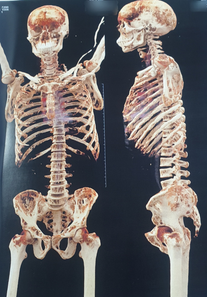
A 36-year-old gentleman of Indian origin presented with bone pains and difficulty in walking of around 1-year duration. There was no history of fractures, renal stones or acid peptic disease or any psychiatric disturbance. He looked emaciated with significant proximal muscle wasting. Neck examination revealed a 3 cm nodule on the left side of the neck that moved with deglutition. There were no bony swellings or deformities. Serum chemistries showed hypercalcaemia (16 mg/dL, N: 8.8–10.6), hypophosphataemia (2.1 mg/dL, N: 2.5–4.5), hyperphosphatasia (1767 IU/L, N: 32–120) and elevated parathormone (PTH) 2831 pg/mL, N: 12–88). Renal and liver functions were normal. Serum prolactin and thyroid function tests were also within normal limits. A diagnosis of primary hyperparathyroidism was made and CT was done with three-dimensional reconstruction (figure 1), which showed numerous lytic lesions of varying sizes in both flat (iliac crests, ribs, skull) and long bones (femur, humerus).
Figure 1.

Three-dimensional CT reconstructive image of the skeleton illustrating moth-eaten appearance of skull, ribs, vertebrae, pelvis, humerus and femur.
The pathological counterpart of these lytic lesions is called osteitis fibrosa cystica (OFC) or Von Recklinghausen disease of bone. Technetium sestamibi nuclear imaging showed uptake corresponding to the clinically felt nodule, which was confirmed by CT scan and ultrasonogram. He underwent neck exploration and removal of the parathyroid tumour. Other parathyroid glands were normal. Histopathology showed parathyroid tumour with capsular breach and high mitotic index confirming the diagnosis of a parathyroid carcinoma. Postoperatively, he had profound hypocalcaemia, worsening of hypophosphataemia and increasing alkaline phosphate levels suggesting hungry bone syndrome. The latter complication is seen in the postoperative setting of hyperparathyroidism, especially patients with extensive skeletal involvement, as in our case. This was managed with repletion of vitamin D stores (cholecalciferol), stiff doses of calcium carbonate, active vitamin D (initially). He gradually improved with significant relief in bony pains and was doing well on follow-up.
The classical radiological features of OFC, which includes salt and pepper appearance of skull, distal clavicle tapering, subperiosteal bone resorption, bone cysts and brown tumours, are rarely seen, mostly because of the paradigm shift in the disease presentation from symptomatic to asymptomatic hypercalcaemia owing to routine biochemical screening of serum calcium.1 However, when compared with its benign counterpart, it is still commonly seen in 40%–70% of parathyroid carcinomas.2 Overproduction of PTH leads to increased osteoclastic resorption, causing cortical bony destruction and fibrous cyst formation. The closest differential to these cystic lytic lesions is a multicentric giant cell tumour, which is indistinguishable on imaging and histology but calcium profile can help with the diagnosis. Parathyroidectomy has been shown to completely cure these lytic lesions.3 However, there is a risk of hungry bone syndrome in cases of prolonged untreated hyperparathyroidism, high bone turnover and extensive bone involvement, which should be promptly recognised and treated with large doses of calcium supplementation.
Another highlight in this case is the diagnosis of parathyroid carcinoma, which is very rare, accounting to <1% of primary hyperparathyroidism. Few clinical and biochemical features that point towards parathyroid carcinoma include : serum calcium levels>14 mg/dL with symptoms, PTH levels>3–10 times the upper limit of normal, renal and skeletal involvement at diagnosis, very high level of alkaline phosphatase and a palpable neck mass,2 4 (as seen in our case).
Patient’s perspective.
I am more than happy and satisfied that my case may add to the current medical literature and may prove as a good learning experience for the budding physicians.
Learning points.
Osteitis fibrosa cystica is a classical presentation of advanced hyperparathyroidism.
These lesions, however extensive, are completely curable with parathyroidectomy and calcium supplementation.
Increased risk of hungry bone syndrome after parathyroidectomy should be kept in mind and recognised early.
Footnotes
Contributors: AY has contributed to diagnosing the case and preparing the manuscript. KSM has contributed to referencing and adding to the manuscript. ARA has contributed to analysing the case and proof reading the manuscript. SM has contributed to the planning and concept of the manuscript.
Funding: The authors have not declared a specific grant for this research from any funding agency in the public, commercial or not-for-profit sectors.
Competing interests: None declared.
Provenance and peer review: Not commissioned; externally peer reviewed.
Ethics statements
Patient consent for publication
Obtained.
References
- 1.Minisola S, Gianotti L, Bhadada S, et al. Classical complications of primary hyperparathyroidism. Best Pract Res Clin Endocrinol Metab 2018;32:791–803. 10.1016/j.beem.2018.09.001 [DOI] [PubMed] [Google Scholar]
- 2.Salcuni AS, Cetani F, Guarnieri V, et al. Parathyroid carcinoma. Best Pract Res Clin Endocrinol Metab 2018;32:877–89. 10.1016/j.beem.2018.11.002 [DOI] [PubMed] [Google Scholar]
- 3.Arabi A, Khoury N, Zahed L, et al. Regression of skeletal manifestations of hyperparathyroidism with oral vitamin D. J Clin Endocrinol Metab 2006;91:2480–3. 10.1210/jc.2005-2518 [DOI] [PubMed] [Google Scholar]
- 4.Sadacharan D, Mahadevan S, Ferdinant J, et al. Hypercalcaemic encephalopathy due to metastatic parathyroid carcinoma. BMJ Case Rep 2017;2017. 10.1136/bcr-2017-219664. [Epub ahead of print: 31 May 2017]. [DOI] [PMC free article] [PubMed] [Google Scholar]


