. 2021 Jul 19;10:e66657. doi: 10.7554/eLife.66657

Figure 4. Three-dimensional structure of FrmB.

(A) Overall fold, a-helices colored in orange and β-strands colored in purple. (B) Comparison between SaFrmB (orange) and its closest human ortholog, ESTD (gray). Active site residues denoted in orange spheres. (C, D) Docking of substrate 1O (sticks) in the active site of FrmB. surface view, red indicates highly hydrophobic and white hydrophilic residues. Surface view (C) or stick view with catalytic triad (D).

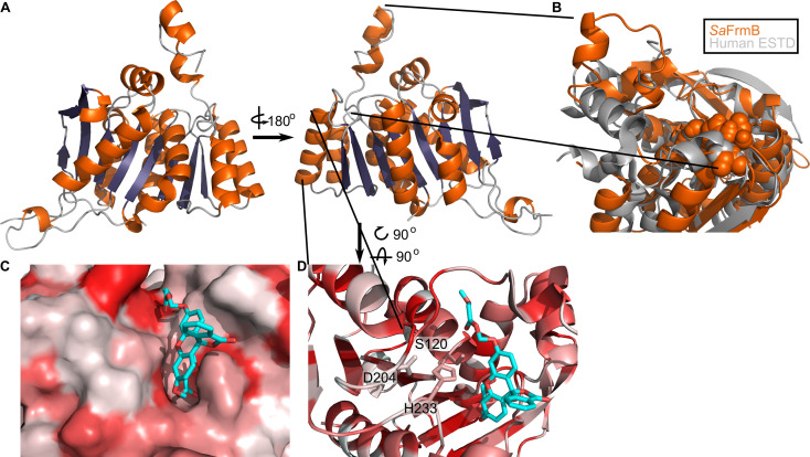
Figure 4—source data 1. Summary of crystallographic data collection and refinement statistics.

Figure 4.

Figure 4—figure supplement 1. Structural conservation of FrmB.

Figure 4—figure supplement 1.

(A) Overall structural alignment of FrmB (orange) with S. pneumonia EstA (PDB:2UZ0), B. intestinalis ferulic acid esterase (PDB:5VOL), and deep sea bacteria Est12 (PDB4RGY). (B) Conservation of the serine hydrolase catalytic triad in FrmB and related proteins.