

Abstract
Neurodegenerative diseases such as Alzheimer’s disease, Parkinson’s disease, Huntington’s disease, and amyotrophic lateral sclerosis are a heterogeneous group of debilitating disorders with multifactorial etiologies and pathogeneses that manifest distinct molecular mechanisms and clinical manifestations with abnormal protein dynamics and impaired bioenergetics. Mitochondrial dysfunction is emerging as an important feature in the etiopathogenesis of these age-related neurodegenerative diseases. The prevalence and incidence of these diseases is on the rise with the increasing global population and average lifespan. Although many therapeutic approaches have been tested, there are currently no effective treatment routes for the prevention or cure of these diseases. We present the current status of our knowledge and understanding of the involvement of mitochondrial dysfunction in these diseases and highlight recent advances in novel therapeutic strategies targeting neuronal bioenergetics as potential approach for treating these diseases.
Keywords: aging, Alzheimer's disease, amyotrophic lateral sclerosis, Huntington's disease, mitochondrial biogenesis, mitochondrial dysfunction, mtDNA mutations, neurodegeneration, oxidative stress, Parkinson's disease
Introduction
Neurodegenerative diseases (NDDs) such as Alzheimer’s disease (AD), Parkinson’s disease (PD), and Huntington’s disease (HD) and amyotrophic lateral sclerosis (ALS) are a heterogeneous group of progressively and irreversibly debilitating disorders with multifactorial etiologies and pathogeneses that manifest distinct molecular and clinical characteristics with impaired bioenergetics and abnormal protein dynamics (Yan et al., 2020). Mitochondrial dysfunction has emerged as an important feature in the etiopathogenesis of these age-related neurodegenerative diseases (Wang et al., 2019b).
As aging is the primary risk factor for most neurodegenerative disease (Mattson and Arumugam, 2018; Hou et al., 2019), the prevalence and incidence of these diseases continues to rise with the increasing global population and average lifespan (Erkkinen et al., 2018). Currently, no neurodegenerative disease is curable, and the treatments available only manage the symptoms or at best slow the progression of disease. With the global aging population expected to double by 2050 to 2 billion people (World Health Organisation, 2018), there is an urgent need for effective therapeutic approaches that are amenable of improving clinical course and outcome of these conditions to a significant extent.
Mitochondria are the energy powerhouses of the cell and generate ~90% of energy in the form of ATP by coupling the flux of electrons throughout the mitochondrial respiratory complexes I–IV with oxidative phosphorylation (OXPHOS).
The brain, at less than 3% of whole body weight (Attwell and Laughlin, 2001), utilizes approximately 25% of the total glucose required by the body (Rossi et al., 2001). This is mainly because neurons depend on OXPHOS to support their functions, and more than 50% of this energy consumption is devoted to maintaining synaptic homeostasis and plasticity. Dysfunctional mitochondrial bioenergetics therefore lead to altered neurometabolic coupling, neural processing and circuitry, and functional connectivity in mature neurons (Uittenbogaard and Chiaramello, 2014). This very high energy demand makes neurons especially susceptible to bioenergetic deficits that may arise from mitochondrial dysfunction. In addition to energy production, mitochondria also tightly regulate calcium homeostasis by modulating calcium uptake in an energy-dependent and pulsatile manner via the mitochondrial calcium uniporter and by providing ATP to stimulate the plasma membrane calcium- ATPase, thereby influencing synaptic transmission, cellular survival and metabolism (Zenisek and Matthews, 2000; De Stefani et al., 2011; Rizzuto et al., 2012). Under pathological conditions, mitochondrial calcium overload compromises the integrity of the mitochondrial inner membrane, thereby triggering mitochondrial permeability transition (MPT), a process when sustained results in cessation of ATP production, permeability of the outer mitochondrial membrane, cytochrome c leakage and subsequent neuronal cell death (Dong et al., 2006; Pivovarova and Andrews, 2010). Additionally, the release of mitochondrial DNA (mtDNA), and peptides from the mitochondrial matrix can activate an immune response that promotes a pro-inflammatory cascade (Chandel, 2015). The maintenance of an adequate and functional mitochondrial population during the lifetime of neurons is critical and involves a balance between mitochondrial biogenesis (Jornayvaz and Shulman, 2010; Golpich et al., 2017) , mitochondrial dynamics i.e. fusion and fission (Haun et al., 2013; Morciano et al., 2016), and mitophagy (de Castro et al., 2010; Ding and Yin, 2012; Stetler et al., 2013). These processes are often dysregulated in NDDs (Figure 1).
Figure 1.
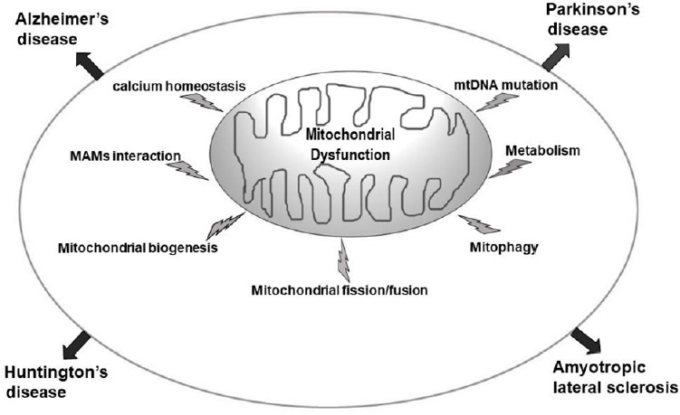
Mitochondrial dysfunction is implicated in sporadic age-related NDDs.
Of the multiple possible causes, mitochondrial impairments ranging from mitochondrial DNA (mtDNA) mutations, oxidative stress, altered gene expression, impaired mitobiogenesis, altered protein turnover and changed organelle dynamics (fission and fusion) are implicated. NDDs: Neurodegenerative diseases.
AD, the most common NDD, is marked by progressive loss of memory, characterized by the increased presence of extraneuronal amyloid plaques derived from the proteolytic processing of the amyloid precursor protein (APP) and intraneuronal neurofibrillary tangles made from hyperphosphorylated tau protein in the brain. Mitochondrial dysfunction is a prominent early feature of AD with lower energy metabolism being one of the primary abnormalities in this disease (Sheng et al., 2012). Mitochondrial OXPHOS impairment is observed in AD brains (Hauptmann et al., 2009) with the most consistent defect being a deficiency in cytochrome c oxidase (COX, complex IV) (Kish et al., 1992; Cardoso et al., 2004). Dysfunction of COX increases reactive oxygen species (ROS) production, reduces energy stores, and disturbs energy metabolism (Mutisya et al., 1994).
The second most common NDD is PD, a progressive neurological movement disorder linked to uncertain etiology having possible effects of genetic and environmental factors (Tomiyama et al., 2008; Korecka et al., 2013). While the cellular mechanisms that result in cell death in the nigrostriatal system in PD are still unclear (Korecka et al., 2013), mitochondrial dysfunction, oxidative stress, chronic inflammation, aberrant protein folding, and abnormal protein aggregation are accepted as the main cause (Greenamyre and Hastings, 2004; Ortega-Arellano et al., 2011; Golpich et al., 2015). Complex I deficit is the most common underlying mitochondrial dysfunction observed in PD (Keeney et al., 2006; Ng et al., 2012; Golpich et al., 2015).
HD is a genetic disorder caused by trinucleotide repeat (CAG) expansions in the huntingtin gene that causes early degeneration of medium spiny neurons in the striatum, resulting in continuous involuntary motor movements. Striatal metabolism is decreased well before atrophy, and the progression of the disease is more strongly correlated with glucose hypometabolism than with the number of CAG repeats (Antonini et al., 1996; Ciarmiello et al., 2006; Civitarese et al., 2010). Mitochondrial dysfunction is strongly associated with the pathogenesis of HD (Quintanilla et al., 2008; Chaturvedi et al., 2009; Chiang et al., 2011) and ATP levels and electron transport chain (ETC) activity are reduced in patients with HD (Cho et al., 2010). In late stage patients with HD, decreased activity of several OXPHOS components, including mitochondrial complexes II, III, and IV is observed in striatal neurons (Reddy et al., 2009).
ALS is a progressive adult-onset NDD that is accompanied by ongoing loss of motor neurons of central nervous system, causing muscle atrophy, respiratory failure and progressive weakness. ALS is characterized by accumulation of mutant proteins (like SOD1, FUS, TDP43, and Sqstm1/p62) (Vicencio et al., 2020). The etiology of ALS is still not fully understood but mitochondrial dysfunction has been identified as an initial phenomenon (Cozzolino and Carri, 2012). The loss of mitochondrial function precedes onset of diseases, which has been indicated in in vivo studies of ALS disease model by occurrence of defects in OXPHOS, calcium buffering, and mitochondrial transport (Orsini et al., 2015). ALS patients are hypercatabolic and have increased energy expenditure at rest (Funalot et al., 2009). ALS patients are also glucose intolerant (Pradat et al., 2010) and insulin resistant (Reyes et al., 1984) and the oxidative stress that contributes to the pathogenic pathway in ALS derives from defective OXPHOS (Lee, 2009).
At the subcellular level mechanistic similarities among these diseases include: mutations in mtDNA, impaired bioenergetics, increased ROS production, and abnormal protein dynamics, including the mitochondrial accumulation of disease specific proteins, impaired axonal transport, and programmed cell death (Armstrong, 2007; Camandola and Mattson, 2017). Bioenergetic deficits strongly influence the initiation and progression of disease. [18F]-fluorodeoxyglucose positron emission tomography imaging studies document glucose hypometabolism in brain regions affected in patients with AD, PD, ALS, and HD (Borghammer et al., 2010; Camandola and Mattson, 2017).
Given that bioenergetics deficits are associated with mitochondrial dysfunction including ETC deficiencies (Golpich et al., 2017): mitochondrial complex I, III, and COX deficiency in AD (Yamada et al., 2014), mitochondrial complex I and IV deficiency in PD (Hoglinger et al., 2003), mitochondrial complex II, III, and IV deficiency in HD (Fukui H and Moraes CT, 2007), and mitochondrial complex I, II, III, and IV deficiency in ALS (Menzies et al., 2002), developing novel therapeutic strategies targeting neuronal bioenergetics is a viable approach for treating these diseases.
Database Search Strategy
We used keyword search on both PubMed and Google Scholar for articles with keywords mitochondrial dysfunction in neurodegenerative diseases from 1999 to present.
Mitochondrial Genome
Unlike the nuclear genome which is only present in two copies in a post-mitotic cell, mtDNA is present in 10 s–1000 s of copies in each cell and undergoes lifelong replication in post-mitotic cells including neurons (Wei et al., 2017). The overall cellular content of mtDNA generally correlates with the underlying energy demand of the cell. The human mtDNA is circular and contains 16,569 base pairs and 37 genes. Of these genes, 22 encode transfer RNAs, two encode ribosomal RNAs (rRNAs; 12S and 16S), and the remaining 13 encode polypeptides. The 13 polypeptides encoded by mtDNA belong to the subunits of OXPHOS enzyme complexes I–V. Unlike nuclear DNA in which 98–99% is non-coding (Venter et al., 2001), most mtDNA genes are contiguous, generally separated by one or two non-coding base pairs, resulting in approximately 93% of mtDNA bases encoding proteins. Since germ cells have few mitochondria and are selectively degraded, the hereditary mode of most mitochondria is maternal inheritance, although biparental transmission of mtDNA in an autosomal dominant-like pattern has recently been demonstrated (Luo et al., 2018).
MtDNA Mutations
mtDNA do not have histone protection and are more likely to be exposed to ROS. Further, most of their entire coding regions lack repair mechanisms. As a result, mtDNA are highly susceptible to damage and mutations, with a 10- to 100-fold greater mutation rate than nuclear DNA (Stewart and Chinnery, 2015). The accumulation of mtDNA mutations impairs the electron transport chain (ETC), triggers oxidative damage, and generates ROS, resulting in bioenergetic deficits, dysregulated intracellular calcium levels (from calcium pump inactivation), activated and damaged membrane phospholipids and ultimately neurodegeneration (Reddy, 2009; Cha et al., 2015).
Different versions of the mitochondrial genome can coexist in each cell, where unlike the maternally inherited germ line variants (which are ‘homoplasmic’), the somatic mutations are usually present alongside the original wild-type molecules (heteroplasmy). Since each cell contains thousands of copies of mtDNA, the proportion of mutants in any one cell changes over time (Naue et al., 2015; Duan et al., 2018). Having several copies of mtDNA allows each mitochondrion to function normally, even when some copies are mutated, allowing cells to carry high loads of mutant mitochondrial DNA before their function is affected and disease symptoms emerge only when the mutant load exceeds a specific threshold (Stewart and Larsson, 2014). This threshold is lower for certain cell types, such as postmitotic neurons, which depend highly on mitochondria due to their high-energy metabolism (Carelli and Chan, 2014) and lack the capacity to proliferate, as a means of mitigating the damage that accumulates. Indeed, mtDNA deletions accumulate in the aging brain, reaching higher levels in regions vulnerable to neurodegeneration (Bender et al., 2006; Ross et al., 2013). Genetic evidence that mtDNA mutations can institute AD, PD HD and ALS like changes comes from cybrid studies (Swerdlow et al., 1997; Flint Beal, 2000; Trimmer and Bennett, 2009; Ferreira et al., 2010). Extensive analysis of such cybrid cell lines has revealed that their bioenergetic function declines with increasing time in cell culture (Trimmer et al., 2004). Indeed both inherited and acquired mtDNA mutations contribute to neural aging and NDDs (Cha et al., 2015; Keogh and Chinnery, 2015) and abnormal amounts of mtDNA have been found in the cerebrospinal fluid and brains of patients with neurodegenerative diseases (Hauptmann et al., 2009; Sheng et al., 2012).
Mitochondrial Oxidative Stress
Oxidative stress can occur when production of ROS, as well as reactive nitrogen species (RNS), outpaces the neuron’s ability to inactivate ROS/RNS and to repair damage (Jodeiri Farshbaf et al., 2016).
Mitochondria convert between 0.4–4% of the total consumed oxygen into superoxide radicals as a by-product of OXPHOS via electron leakage from the respiratory chain (Pieczenik and Neustadt, 2007). ROS comprise superoxide radicals, H2O2, and hydroxyl radicals (Naudi et al., 2012) while RNS include the nitric oxide radical (NO•), the nitrogen dioxide radical (•NO2), nitrite (NO2–) and peroxynitrite (ONOO–). Mitochondrial thioredoxin reductase/thioredoxin/peroxiredoxin-3,5 system, glutathione peroxidase, and glutathione anti-oxidant system effectively scavenge these ROS (Drose and Brandt, 2012). ROS stimulate mild uncoupling of mitochondria, and the resulting increase in proton conductance can have a negative feedback effect on ROS production (Naudi et al., 2012). Furthermore, the MPT pore (MPTP) is opened to reduce the electrochemical gradient and accelerate oxygen consumption, which decreases ROS production. Cytochrome c is also a powerful ROS scavenger. The presence of low ROS concentrations is physiologically normal, and ROS function as important intracellular second messengers (Murphy et al., 2011). However, excessive ROS generation is injurious to mtDNA and potentially leads to impaired ETC functions, reduced ATP synthesis, mitochondrial dysfunction, cell injury, and even apoptosis. ROS play key roles in the initiation and modulation of cell apoptosis (McCarroll et al., 2004). Similar to ROS, RNS are highly toxic (Bergendi, 1999). Microglia also play a role in generating RNS as nitric oxide synthases, inducible nitric oxide synthase and NADPH oxidase 2 (NOX2), and NADPH oxidase (NOX) are induced in these glial cells (Saha and Pahan, 2006). NOX2 activation can lead to a respiratory burst of superoxide flooding the mitochondria further contributing to neurodegeneration. NO• has been shown to play a role in neurodegenerative diseases by acting as a neurotoxin when excessively produced (Schulz et al., 1995).
Oxidative imbalance and resultant neuronal damage play a critical role in the initiation and progression of AD (Wang et al., 2014). The excessive accumulation of ROS in AD may induce mitochondrial dysfunction although the exact mechanisms underlying the disruption of redox balance still remain elusive (Zhao and Zhao, 2013). Aβ-induced mitochondrial dysfunction has been suggested to inhibit the efficient production of ATP and increase the generation of ROS in AD (Castellani et al., 2002; Zhao and Zhao, 2013). The activity of Complex I of the respiratory chain in SNc of patients with PD is reduced and may contribute to the generation of excessive ROS which in turn can induce apoptosis (Schapira, 2008; Blesa et al., 2015; Franco-Iborra et al., 2016). This mitochondrial Complex I deficiency is also present in the frontal cortex, fibroblasts, and blood platelets of PD patients (Blesa et al., 2015). Oxidative stress plays a crucial role in the neurodegenerative process of HD (Stack et al., 2008; Tasset et al., 2009). Indeed, susceptible neurons in the HD brain may not be able to handle well an increase in ROS/RNS production. Studies in transgenic mouse models of HD have shown an altered pattern of expression and activity of the enzyme nitric oxide synthase (NOS) (Deckel, 2001, 2002), as well as of the antioxidants SOD (Santamaria et al., 2001) and ascorbate (Rebec et al., 2002) in R6/1 and R6/2 HD transgenic mice (which express exon 1 of the human HD gene with 115 and 145 CAG repeats respectively (Mangiarini et al., 1996)). Furthermore, ROS production is also increased in the striatum of R6/1 mice (Perez-Severiano et al., 2004), while an increase in the levels of lipid peroxidation has recently been detected in several transgenic and knock-in HD mouse models (Lee et al., 2011). Free radical damage and abnormal free radical metabolism is evident in sporadic ALS and familial ALS patients (Pollari et al., 2014). The aberrant activity of mSOD1 leads to oxidative damage (Wiedau-Pazos et al., 1996; Crow et al., 1997) and other ALS-linked proteins, such as mutant TDP-43, promote oxidative stress in a motoneuron cell line (Duan et al., 2010). Excitotoxicity and oxidative stress caused by astrocytes arises from aberrant glutamate receptor function which leads to mis-regulated glutamate homeostasis (Rothstein et al., 1992).
Mitochondrial Dynamics
The movement, fusion and fission, remodeling of cristae, biogenesis and mitophagy constitute mitochondrial dynamics. These quality control processes are stringently regulated and adjusted to modify the overall mitochondrial morphology in response to changing energy demands and cellular stress (Eisner et al., 2018; Tilokani et al., 2018). Alteration in trafficking and fusion-fission dynamics in AD, PD, HD, and ALS has been shown (Kodavati et al., 2020).
Mitochondrial Fusion and Fission
Imbalances between mitochondrial fission and fusion have been proposed to cause neurodegenerative diseases; acute readjustment of such imbalances can have beneficial effects on mitochondrial structure and function and positively influence cell survival in various disease models (Chen and Chan, 2009; Itoh et al., 2013; Sebastián et al., 2017). Mitochondrial fusion is conserved in all eukaryotic cells and is essential for life (Chen et al., 2003). Fusion enables the transfer of mitochondrial components from viable mitochondria into those that are functioning at a sub-optimal level thereby mitigating damage and ensuring the maintenance of an efficient homogeneous mitochondrial population (Youle and van der Bliek, 2012). When cells are stressed, stress-induced mitochondrial hyperfusion, prevents mitophagy, maintains OXPHOS and preserves mitochondrial integrity (Wai and Langer, 2016). During starvation, stress-induced mitochondrial hyperfusion maintains ATP levels and protects cells from apoptosis (Gomes et al., 2011). Conversely, fusion-deficient cells have reduced growth, wide-spread differences in Δψm and reduced respiration (Chen et al., 2005). Thus, fusion of the mitochondrial network is cytoprotective, especially during stress (Neuspiel et al., 2005; Gomes et al., 2011), while the loss of fusion predisposes cells to apoptosis. Fusion occurs only between two partner mitochondria that have sufficient Δψm, whereas those that are depolarised are fusion-deficient (Twig et al., 2008) ensuring that only biochemically functional mitochondria can fuse back with the network, while dysfunctional mitochondria are isolated and degraded. Mitochondrial outer membrane fusion is mediated by the mitofusin proteins Mfn1 and Mfn2, which form homo and heterodimers between the two opposing membranes (Koshiba et al., 2004). Mitochondrial inner membrane fusion is regulated by optic atrophy protein 1 (OPA1). OPA1-depletion induces mitochondrial fragmentation (Cipolat et al., 2004; Wu et al., 2019). OPA1 also has roles in shaping the mitochondrial cristae junctions, which house the majority of ATP synthase, complex III and cytochrome c thus playing an important role in regulating OXPHOS and apoptosis (Olichon et al., 2003; Griparic et al., 2004; Cogliati et al., 2016).
The major mitochondrial fission protein is the mechanochemical GTPase dynamin-related protein 1 (Drp1), which constantly cycles between the cytoplasm and the mitochondrial outer membrane (Wasiak et al., 2007). Fission is required to maintain mitochondrial function and integrity and in postmitotic cells such as neurons it plays an important role in transport and distribution of mitochondria to distal neurites (Li et al., 2004; Verstreken et al., 2005; Lewis et al., 2018). It is also crucial for quality control as elimination of damaged mitochondria by mitophagy via the PINK1/parkin pathway (Narendra et al., 2008, 2010; Ashrafi et al., 2014; Pickrell and Youle, 2015) involves mitochondrial fission (Tanaka et al., 2010; Kageyama et al., 2014).
The balance between mitochondrial fusion and fission is impaired in AD neurons (Zhu et al., 2018). The balance of mitochondrial fusion and fission is disrupted in several models of PD (Santos and Cardoso, 2012). In PD cytoplasmic hybrid (cybrid) cells there is a significantly increased proportion of mitochondria with swollen vacuoles, pale matrices and few remaining cristae (Castellani et al., 2002). In HD brain there is a significant increase in Drp1 and decrease in Mfn1 (Reddy, 2014). This imbalance between the mitochondrial fusion and fission results in alterations of mitochondrial morphogenesis and negatively impacts important cellular mechanisms thus exacerbating neuronal death (Kim et al., 2010). Further, mutant HTT protein triggers mitochondrial fission prior to the emergence of neurological deficits and mutant HTT protein aggregates (Shirendeb et al., 2011, 2012; Reddy, 2014). ALS patients and animal models have altered mitochondrial dynamics with excessive fragmentation of mitochondria. Smaller mitochondria are observed in different models expressing ALS mutant SOD1 (Khalil and Liévens, 2017) and linked to a misexpression of mitochondrial dynamics genes. Mutant SOD1 mouse model spinal cord and skeletal muscles reveal downregulated Mfns (Liu et al., 2013; Russell et al., 2013) as well as OPA1, while levels of phosphorylated DRP1 and Fis1 are elevated (Liu et al., 2013). Inactivating DRP1 restores normal morphology of mitochondria and prevents death of mutant SOD1-expressing neurons (Song et al., 2013).
Mitochondrial Biogenesis
Increased mitochondrial biogenesis is a physiological response to stress conditions (e.g., cold, exercise, nutritional status), which is activated to meet the energetic requirements of tissues (Viscomi et al., 2015).
Mitochondrial biogenesis is essential for maintaining an adequate functional neuronal mitochondrial mass. It is a highly regulated process that requires coordination and crosstalk between the nuclear and mitochondrial genomes (Ryan and Hoogenraad, 2007) and occurs on a regular basis in healthy cells where mitochondria constantly divide and fuse with each other. Replication is regulated by the mtDNA polymerase γ (POLG) consisting of the catalytic subunit encoded by the POLG gene and auxiliary dimeric subunit encoded by the POLG2 gene (Graziewicz et al., 2006 ). mtDNA is transcribed by the mitochondrial RNA polymerase POLRMT (Tiranti et al., 1997). The key enhancer protein is TFAM (transcription factor A, mitochondrial), which ensures mtDNA unwinding and flexing required for the POLRMT binding to the mtDNA promoters. TFB2M (transcription factor B2, mitochondrial) acts as a specific dissociation factor that provides interaction between POLRMT and TFAM. Both TFB1M and TFB2M bind rRNA dimethyltransferases and, therefore, can function as rRNA modifiers (Rebelo et al., 2011). The major role of TFB1M is rRNA methylation and not its transcription factor function (Metodiev et al., 2009). Nuclear respiratory factors NRF1 and NRF2 regulate expression of the ETC subunits encoded by the nuclear genome (Evans and Scarpulla, 1990) and bind to the promoters of genes involved in mtDNA transcription. NRF1 binds to the specific promoter sites and regulates expression of TFAM (Virbasius and Scarpulla, 1994), TFB1M, and TFB2M (Gleyzer et al., 2005). Besides, nuclear respiratory factors, in particular NRF2, regulate expression of other mitochondrial enzymes, e.g., TOMM20 (translocase outer mitochondrial membrane), a key enzyme in the mitochondrial membrane transport (Blesa and Hernández-Yago, 2006). In turn, NRF1 and NRF2 are regulated by transcription coactivators, including PGC-1α (Scarpulla, 2008). PGC-1α is regulated on both the transcription and post-translation levels (Fernandez-Marcos and Auwerx, 2011). p38 MAPK is another factor that regulates the expression of PGC-1α by activating transcription factor 2 (Akimoto et al., 2005).
The impaired function of the master regulator for mitochondrial biogenesis PGC-1α has been recognized as a major contributor to the neuropathogenesis of several NDDs including AD, PD and HD (Tsunemi and La Spada, 2012; Uittenbogaard and Chiaramello, 2014). Reduced mitochondrial number and impaired mitochondrial gene expression contribute to mitochondrial dysfunction associated with AD (Zhu et al., 2013). Morphometric analyses performed on hippocampal neurons of autoptic brains from patients with AD have revealed a significant decrease in intact mitochondria (Hirai et al., 2001). The decreased levels of PGC-1α imply abnormal mitochondrial biogenesis as a key event for reduction of mitochondrial mass and bioenergetic functions in AD brain (Qin et al., 2009). The network of transcription factors regulating mitochondrial biogenesis, including NRF-1, NRF-2 and TFAM, are also altered suggesting a deficiency in the process of mitochondrial biogenesis (Sheng et al., 2012).
Abnormal PGC-1α-mediated mitochondrial biogenesis also interfaces with PD (Zheng et al., 2010; Shin et al., 2011). A meta-analysis of independent microarray analyses using microdissected human DA neurons from PD patients has documented a down-regulation of PGC-1α-regulated target genes, congruent with the concept of altered PGC-1α expression being the cause rather than the consequence of PD pathogenesis (Zheng et al., 2010).
In HD the severity of symptoms have been linked to sequence variants of PGC-1α (Chaturvedi et al., 2009; Weydt et al., 2009). PGC-1α dysfunction extends beyond the neuronal lineage to oligodendrocytes where PGC-1α regulates the expression of several genes required for proper myelination, such as myelin basic protein, in keeping with the observed deficient myelination in HD transgenic mice (Xiang et al., 2011). Finally, PGC-1α and its downstream effector NRF1 are downregulated in spinal cord and muscle tissues of patients and mutant SOD1 mice (Thau et al., 2012; Russell et al., 2013). Further, single-nucleotide polymorphisms reported in the brain-specific promoter region of PGC-1α modify age of onset and survival of ALS patients and mutant SOD1 mice (Eschbach et al., 2013).
Mitochondria Stress Response Signaling
Mitochondria produce most of the cellular ROS and the stress signaling that induces cellular senescence and apoptosis (Hamanaka and Chandel, 2010; Sena and Chandel, 2012). A major consequence of increased ROS and altered cellular redox state is the oxidation of thiol groups in cysteine residues in relevant proteins (Hamanaka and Chandel, 2010). FoxO transcription factors are activated in response to elevated ROS levels and induce anti-oxidant responses (increased expression of catalase and SOD2), cell cycle arrest and/or cell death (Kops et al., 2002; Fu and Tindall, 2008). Mitochondrial Akt, glycogen synthase kinase 3β (GSK3β), PKA, Abl, PKC, Src and Atm modulate the cellular stress response (Bera et al., 1995; Kumar et al., 2001; Majumder et al., 2001; Alto et al., 2002; Das et al., 2008; Robey and Hay, 2009; Mihaylova and Shaw, 2011). Akt phosphorylates and inactivates GSK-3β, which can localize to the mitochondria. Mitochondrial GSK-3β phosphorylates myeloid leukemia cell differentiation protein (MCL-1) and voltage-dependent anion channel (VDAC) (Juhaszova et al., 2004; Maurer et al., 2006) leading to MCL-1 degradation and induction of apoptosis (Maurer et al., 2006). The phosphorylation of VDAC by GSK-3β results in increased mitochondrial membrane permeability which also leads to apoptosis (Juhaszova et al., 2004; Martel et al., 2013). GSK-3β can also phosphorylate and promote the proteasomal degradation of c-Myc, cyclin D1, and β-catenin (Rubinfeld et al., 1996; Diehl et al., 1998). Hypoxia and other physiological stresses can induce the translocation of PKA to mitochondria (Carlucci et al., 2008; Kim et al., 2011) causing it to bind through Rab32 and other A-kinase AKAPs (163) resulting in the phosphorylation of VDAC (164), Drp1 (Kim et al., 2011), and other mitochondrial proteins.
Hypoxia, by inducing SIAH2, a mitochondrial ubiquitin ligase, destabilizes AKAP121 and limits oxidative capacity under conditions of low oxygen. Interestingly, AKAP121 also appears to promote mitochondrial localization of Src-tyrosine kinase (Livigni et al., 2006) where Src appears to regulate COX activity and respiratory activity (Miyazaki et al., 2003; Livigni et al., 2006), and other mitochondrial substrates for Src family kinases are likely (Tibaldi et al., 2008). Increased ROS induces protein kinase C-delta (PKCδ) associated with the mitochondria and this in turn recruits other signaling molecules, including the Abl tyrosine kinase that is associated with loss of membrane potential and non-apoptotic cell death (Kumar et al., 2001). Impaired oxidative metabolism and decreased ATP levels in neurons activate AMPK (Maurer et al., 2006). AMPK can also be activated by drugs such as metformin that inhibits complex I or resveratrol that inhibits the F0F1 ATPase (Mihaylova and Shaw, 2011). AMPK modulates mitochondrial metabolism and targets Acetyl CoA carboxylase-2 to the outer mitochondrial membrane (OMM) where it regulates lipid metabolism by controlling production of malonyl CoA (Mihaylova and Shaw, 2011). AMPK therefore plays a key role in mitochondrial homeostasis by ensuring that only functionally viable mitochondria are retained. Upon its activation it induces not only mitochondrial biogenesis through activation of PGC-1α (Zong et al., 2002; Jäger et al., 2007) but also initiates mitophagy through ULK1 activation and mTOR inhibition (Egan et al., 2011; Kim et al., 2011).
ATM kinase inhibition causes central nervous system neurodegeneration in animal models (Petersen et al., 2012). ATM kinase, is partly located at the mitochondria and is activated by mitochondrial uncoupling (Valentin-Vega et al., 2012). While the mitochondrial substrates of ATM are not known, loss of ATM in genetically engineered mouse models leads to mitochondrial dysfunction. ATM signaling is reduced in the neurons in vulnerable regions of the AD brain (Shen et al., 2016). A recent study of a mouse model of familial AD showed that the mutant amyloid precursor protein (APP) transgene caused aberrant persistence of gH2AX in the brain (Gleyzer et al., 2005). ATM is also involved in the pathogenesis of PD because ATM gene knockout (ATM KO) mice exhibit severe loss of tyrosine hydroxylase-positive DA nigro-striatal neurons, and midbrain DA neurons progressively degenerate with age (Eilam et al., 2003). PARK2 and ATM mutations sometimes occur synchronically at the same amino-acid residue, causing neuronal degeneration (Veeriah et al., 2010). ATM deficient neurons re-enter the cell cycle and die (Yang and Herrup, 2005; Rimkus et al., 2008), suggesting that ATM may protect neuron by stopping cells re-entering the cell cycle and lessening DNA damage. ATM impairment in glial cells may also trigger innate immune responses leading to cause neurodegeneration (Petersen et al., 2012). The histology of microglial cell in ATM KO mice is abnormal, and astrocytes from ATM KO mice show significant expressions of oxidative and endoplasmic reticulum stress and a senescence-like reaction (Kuljis et al., 1997; Liu et al., 2005). ATM deficiency may disturb DNA repair, trigger apoptosis, and accelerate aging and neuroinflammation. In HD, cells transfected with mutant HTT protein fragments showed elevated DNA damage and ATM activation (Giuliano et al., 2003; Illuzzi et al., 2009), and increased gH2AX was also found in fibro blasts from HD patients (Giuliano et al., 2003) and in HD mouse and patient striatal neurons (Enokido et al., 2010). Intriguingly, HD patient cells exhibit radiosensitivity, albeit less than A-T cells, suggesting subtle dysfunction in the DNA damage response (Ferlazzo et al., 2014).
Mitochondria and Inflammation
The local sterile inflammation has been shown to be finely linked with the development and the progression of different NDDs including AD, PD and ALS (Lin and Beal, 2006). Neuroinflammation is commonly driven through the abnormal activation of microglia and astrocytes, by damage-associated molecular patterns (DAMPs) molecules released from damaged and necrotic cells (Yin et al., 2016; Wilkins et al., 2017). Dysregulated activation of microglia and astrocytes results in persistent inflammasome activation, which, together with an increased level of DAMPs, leads to the establishment of low-grade chronic inflammation and thus to the development of age-related pathological processes (Kapetanovic et al., 2015). Noticeably, neuroinflammation drives the increased secretion of cytokines and chemokines not only within the brain, but also systemically (Licastro et al., 2000) and, in some cases, might lead to blood-brain barrier disruption with the consequent infiltration of peripheral immune cells (Wilkins et al., 2017). At the molecular level, neuroinflammation is mainly triggered by redox status (Innamorato et al., 2009): ROS are produced by microglia upon their activation by intrinsic or extrinsic factors [reviewed in Kierdorf and Prinz (2013)] and are released in the extracellular space. Uncontrolled ROS production might affect intracellular redox balance, thus inducing the expression of proinflammatory genes by acting as second messengers (Rojo et al., 2014). Consequently, abnormal activation of microglia leads to the release of reactive oxygen intermediates, proinflammatory cytokines, complement proteins and proteinases, driving a chronic inflammatory state responsible for triggering or maintaining neurodegenerative processes (Dheen et al., 2007). The main players involved in these processes are mitochondria, which not only generate ROS but also respond to ROS-induced cellular changes (Handy and Loscalzo, 2012).
Mitochondrial dysfunction can be both the leading cause of neuroinflammation and can be induced by it. Damaged mitochondria trigger the process of mitochondrial membrane permeabilization that initiates both apoptosis and necrosis. Adult neurons are resistant to apoptosis (Yuan et al., 2019). Necroptosis, a form of regulated necrotic cell death induced by tumor necrosis factor α (TNFα), is highly activated in NDDs (Ito et al., 2016; Yuan et al., 2019). Briefly, necroptosis is a form of programed cell death coordinated by receptor-interacting kinases 1 (RIPK1) and RIPK3 and mixed lineage kinase domain-like protein under caspase-8 deficient conditions. Necroptosis can be stimulated by TNF, other members of the TNF death ligand family, interferons, Toll-like receptor signaling and viral infection (Zhang et al., 2017). Ultimately, accumulation of damaged mitochondria can activate NLRP3 inflammasome-dependent inflammation in microglia. Moreover, damaged neurons are responsible for releasing DAMPs, such as mtDNA, in the extracellular environment, eliciting local inflammation (Nakahira et al., 2011) by increasing inflammasome activation, and thus IL-1β secretion, and also binding to microglial toll-like receptor-9, inducing TNFα and nitric oxide (NO) production (Iliev et al., 2004).
Mitochondrial Calcium Dyshomeostasis
Mitochondria actively regulate intracellular calcium levels; and when matrix calcium increases beyond physiological demands, the MPT pore (MPTP) opens, resulting in apoptotic or necrotic cell death (Rasola and Bernardi, 2011). The pathophysiological implications of altered calcium homeostasis has been described in AD (Yu et al., 2009), PD (Surmeier, 2007), HD (Bezprozvanny and Hayden, 2004) and ALS (von Lewinski and Keller, 2005). The common thread in these NDDs is that MPTP opening and altered calcium handling are involved (Lin and Beal, 2006; Wojda et al., 2008; Abramov et al., 2017) and precede neuronal death (Calì et al., 2012). In AD, the Aβ peptide is translocated to the mitochondria via the TOM (translocase of the outer membrane) machinery, where it localizes to the mitochondrial matrix and interacts with specific intra-mitochondrial proteins (Hansson Petersen et al., 2008) and impairs the activity of respiratory chain complexes III and IV (Caspersen et al., 2005). Aβ peptide can also induce calcium release from the endoplasmic reticulum (ER) resulting in mitochondrial calcium overload (Ferreiro et al., 2008; Sanz-Blasco et al., 2008) leading to MPTP opening and cell death (Du et al., 2008). In PD, α-synuclein plays a similar role in inducing mitochondrial dysfunction and cell death. α-synuclein localizes in the mitochondria (Nakamura et al., 2008; Di Maio et al., 2016) and inhibits complex I activity (Luth et al., 2014) leading to dose-dependent loss of mitochondrial membrane potential with an associated decrease in phosphorylation capacity (Banerjee et al., 2010).
Mutant, but not wild-type, huntingtin can interact with the outer mitochondrial membrane and significantly increase the susceptibility of mitochondria to calcium-induced MPTP opening. Mutant huntingtin induces mitochondrial swelling, MPTP opening, release of cytochrome c and subsequent activation of the apoptotic cascade. All these events were completely inhibited by CsA, indicating a role for calcium and MPTP opening in the pathogenesis of this disease (Choo et al., 2004).
In ALS, the mutant SOD1 protein impairs mitochondrial functions by aggregating in the mitochondria and binding to the Bcl-2 protein thereby reducing its anti-apoptotic properties (Pasinelli et al., 2004 ), and impairing the activity of complexes I, II and IV of the respiratory chain (Jung et al., 2002). Motoneurons expressing mutant SOD1 showed an early increase in mitochondrial calcium with loss of mitochondrial membrane potential, mitochondrial swelling and ER overload, followed by an increase in cytosolic calcium (Tradewell et al., 2011).
Mitophagy
Mitophagy is the selective degradation of mitochondria by autophagy. Mitophagy can effectively remove damaged or stressed mitochondria, which is essential for cellular health (Wang et al., 2019a).
An excess of ROS may function as an autophagy trigger (Kurz et al., 2007) and dysfunctional mitochondria that overproduce ROS, are indeed selectively targeted for mitophagy (Lemasters, 2005). Central to mitochondrial and cellular homeostasis, mitophagy is modulated by the PTEN-induced putative kinase 1 (PINK1)/Parkin pathway (Narendra and Youle, 2011) which primarily targets mitochondria devoid of membrane potential (ΔΨm). PINK1 accumulates on the outer membrane of dysfunctional mitochondria and recruit the E3 ubiquitin ligase Parkin (Valente et al., 2004; Jin et al., 2010; Narendra et al., 2010) that ubiquitinates several OMM proteins that are consequently targeted by P62/SQSTM1 (Geisler et al., 2010). F-box-containing proteins, sterol regulatory element binding transcription factor 1, and WD40 domain protein 7 (FBXW7) have been identified as the critical regulators in the PINK/Parkin pathway of mitophagy (Rasola and Bernardi, 2011). p62 recognizes ubiquitinated substrates and directly interacts with autophagosome-associated LC3 to recruit autophagosomal membranes to the mitochondria (Pankiv et al., 2007). Damaged mitochondria can also, independently of Parkin, increase FUNDC1 and Nix expression to recruit autophagosomes to mitochondria via direct interaction with LC3 (Novak et al., 2010; Liu et al., 2012). Ubiquitin ligases, like Smurf1, target depolarized mitochondria for mitophagy (Ding and Yin, 2012; Lokireddy et al., 2012; Fu et al., 2013). The transcription factor nuclear factor erythroid 2-related factor 2 (Nrf2) partly regulates p62 expression due to the presence of an antioxidant response element in its promoter region (Ishii et al., 2000; Jain et al., 2010). Electrophilic natural products such as isothiocyanate compound, sulforaphane which upregulate Nrf2 by interfering with its regulator protein, the redox sensitive ubiquitination facilitator Keap1 (Kelch-like ECH-associated protein 1) can potentially induce p62 expression (Kensler et al., 2007; Cheng et al., 2011). p62-mediated mitophagy inducer (PMI) (HB229), was recently developed to upregulate P62 via stabilization of Nrf2 and promote mitophagy. This compound bypasses the upstream steps of the mitophagic cascade and acts independently of the ΔΨm collapse, and does not mediate any apparent toxic effects on mouse embryonic fibroblast cells at the concentrations used in the assays (East et al., 2014). Parkin also modulates transport of mitochondria along microtubules to a perinuclear region where autophagosomes are concentrated (Narendra et al., 2010; Vives-Bauza et al., 2010). This is likely due to Parkin-mediated turnover of Miro, a protein required to tether kinesin motor protein complexes to the OMM (Wang et al., 2011). HDAC6, a ubiquitin-binding protein deacetylase is also recruited to mitochondria by Parkin (Cho et al., 2009) along microtubules (Lee et al., 2010a, b). Mitophagy is crucial for cellular homeostasis and its impairment is linked to several neurodegenerative diseases (de Castro et al., 2010; Karbowski and Neutzner, 2012). However, selective pharmacologic modulators of mitophagy that would facilitate dissection of the molecular steps involved in the removal of mitochondria from the network via this pathway are not presently available.
The development of AD is closely correlated with mitochondrial autophagy defects [219,220] and Parkin plays a key role in the removal of mitochondria. Parkin overexpression alleviates symptoms of AD (Ye et al., 2015). Supporting a role of mitophagy in AD, amyloid beta-derived diffusible ligands can induce the fragmentation of mitochondria and mitophagy (Wang et al., 2009; Nakamura et al., 2010). Sterol regulatory element binding transcription factor 1 is also one of the risk factors for sporadic PD (Rasola and Bernardi, 2011), and mitophagy thus may represent a common mechanism linking sporadic and familial PD (Bezprozvanny and Hayden, 2004; Surmeier, 2007; Yu et al., 2009). Mutation in the F-box domain is reported to be associated with the early-onset autosomal recessive PD. FBXW7 is involved in maintaining mitochondria and inducing mitophagy through direct interaction with Parkin, further confirming the significance of mitophagy in the pathogenesis of PD (von Lewinski and Keller, 2005).
Mitophagy is also altered in HD, and the mutant huntingtin induces selective autophagy (Khalil et al., 2015) and in cell culture models of HD, excessive mitochondrial fission partially mediates cytotoxicity (Guo et al., 2013). In addition, mutant Huntingtin is found to cause formation of spherical mitochondria in a nonapoptotic state, and it can also repress mitophagy, resulting in the impaired mitochondrial clearance (Rasola and Bernardi, 2011). In ALS, the reduced targeting of ubiquitinated mitochondria to autophagosomes may contribute to the disease pathogenesis (Benatar, 2007). In ALS models, the accumulation of damaged mitochondria can be detected by live cell imaging. Mitochondrial homeostasis can be reconstituted by inhibiting OPTN or TBK1 mutations, as well as pharmacological inhibition or genetic knockdown of PINK1 or Parkin. Altering TBK1/OPTN can significantly improve neuronal function and block disease progression. These data support the potential role of mitochondrial autophagy in ALS (Caspersen et al., 2005; Hansson Petersen et al., 2008; Calì et al., 2012).
Potential Interventions to Restore or Enhance Mitochondrial Function in NDDs
We conclude with potential mitochondria-targeted therapies that may be beneficial for these age-related NDDs.
Genetic therapies
Mitochondrial-targeted transcription activator-like effector nucleases (mitoTALENs) can correct mtDNA mutations in cultured human cells from patients with a mtDNA disease (Bacman et al., 2013; Yang et al., 2018) and correct induced mtDNA mutation in vivo in mouse models (Bacman et al., 2018). Mitochondrially targeted zinc-finger nucleases (mtZNFs)(Gammage et al., 2014) are another tool for specific removal of mtDNA mutations with a relatively low risk for interaction with the cell’s nuclear DNA. myZNFs are able to remove a pathogenic mtDNA mutation in vivo mouse experiment (Gammage et al., 2018). Almost all adult cells are heteroplasmic for mtDNA due to age-related acquired mtDNA mutations. The degree of mtDNA mutation load determines the onset of clinical symptoms, a phenomenon known as the threshold effect (Craven et al., 2017). Clinical symptomatology can be alleviated by shifting the existing equilibrium between healthy and mutated mtDNA in different ways (Rai et al., 2018). This is appealing for human clinical applications because it avoids the use of gene-editing techniques which might erroneously disrupt the function of normal genes of the treated cells (Gammage et al., 2014, 2018; Bacman et al., 2018; Peeva et al., 2018; Pereira et al., 2018).
Mitochondria-targeted antioxidants
Targeting detrimental neuronal ROS at the production stage without affecting ROS signaling would be ideal in preventing and treating AD. In this regard, it has been shown that mitochondria-targeted antioxidants potently sequester reactive oxygen intermediates and confer greater protection against oxidative damage in the mitochondria than untargeted cellular antioxidants. These mitochondria-targeted antioxidants such as (10-(6′-plastoquinonyl) decyltriphenyl-phosphonium) (SkQ1), MitoQ, MitoTEMPOL and MitoVitE prevent apoptosis by mitigating the oxidative damage more effectively than untargeted antioxidants such as 6-hydroxy-2,5,7,8-tetramethylchroman-2-carboxylic acid (Trolox) (Oyewole and Birch-Machin, 2015). Other such antioxidants include: 4,5-dihydroxybenzene-1,3-disulfonate (Tiron), which has been engineered to accumulate within the mitochondria by permeabilizing the mitochondrial membrane (Fang et al., 2012) and astaxanthin, a mitochondrion-permeable antioxidant, that can penetrate the blood-brain barrier and is effective in preventing and treating macular degeneration (Piermarocchi et al., 2012; Wu et al., 2014).
Antioxidants can also be targeted to mitochondria through the use of small, aromatic-cationic sequence motif called Szeto-Schiller (SS) tetrapeptides which enables them to be delivered and localized to the inner mitochondrial membrane with an approximate 1000–5000-fold accumulation (Smith and Murphy, 2011; Jin et al., 2014). Novel XJB peptides which consist of an electron and ROS scavenger (4-NH2-TEMPO) conjugated to the Leo-D-Phe-Pro-Val-Orn fragment of gramicidin S have been invented. This pentapeptide fragment can specifically target the XJB peptides to mitochondria. One of these peptides, XJB-5-131, improved mitochondrial function and enhanced the survival of neurons in a mouse model of HD (Xun et al., 2012). Another approach of targeting mitochondria with small bioactive molecules involves polymer based nano-carriers. These include biodegradable poly-lactide-co-gylcolide (PLGA) like PLGA CoQ10 nanoparticles (Nehilla et al., 2008) although their biological effects are yet to be fully elucidated.
N-Acetyl-5-methoxytryptamine (Melatonin), which is synthesized from tryptophan provides remarkable protection against oxidative stress in the brain and at physiological concentrations is more potent than vitamins C and E (Reiter et al., 1997, 1999, 2014; Galano et al., 2011). Melatonin and its metabolites function as broad-spectrum antioxidants (Tan et al., 2001, 2002; Ressmeyer et al., 2003). Melatonin also down regulates caspase-3 levels (Espino et al., 2010), which are linked directly to neuronal death (Louneva et al., 2008).
Enhancing mitochondrial biogenesis
The human mitochondrial genome can be manipulated from outside the cell to change expression and enhance mitochondrial energy production. TFAM has been engineered to rapidly pass through cell membranes and target mitochondria. Recombinant-human TFAM acts on cultured cells carrying a mtDNA disease (Iyer et al., 2012) as well as lab mice, energizing the DNA of the mice’s mitochondria, improving the memory of aged mice (Iyer et al., 2009; Thomas et al., 2012) and enabling them to run two times longer on their rotating rods than a control group cohort (Thomas et al., 2012). Expression of human TFAM significantly improved cognitive function, reducing accumulation of both 8-oxoguanine, an oxidized form of guanine, in mtDNA and intracellular Aβ in 3xTg-AD mice and increased expression of transthyretin, known to inhibit Aβ aggregation (Oka et al., 2016).
Calorie restriction to induce mitochondrial biogenesis
Calorie restriction (CR), i.e. the limitation of ingested calories without malnutrition, is known to enhance animal life span and prevent age-related diseases, including neurological deficits, brain atrophy, and cognitive decline (Colman et al., 2014). CR induces mitochondrial biogenesis (Cerqueira et al., 2012) in a NO•-mediated manner that culminates in increased mitophagy and the production of new, more efficient mitochondria that have reduced membrane potential, produce less ROS, consume increased levels of oxygen and exhibit an improved ATP/ROS ratio, leading to decreased energy expenditure (Onyango et al., 2010). In particular, the tissue-specific effects of CR include the prevention of the age-related loss of mtDNA in rat liver (Cassano et al., 2006) and the partial preservation of TFAM binding to mtDNA in rat brain with enhanced reserve respiratory capacity and improved survival in neurons (Picca et al., 2013).
Inducing mitophagy
Focal mitophagy eradicates degraded mitochondria and decreases ROS-induced neuronal death (Kubli and Gustafsson, 2012). Regulators of PINK1/parkin, metformin, and resveratrol have also been shown to increase mitophagy (Wang et al., 2019a). Sirtuin activating compounds or nicotinamide adenine dinucleotide (NAD) precursors such nicotinamide riboside (NR) or nicotinamide mononucleotide also induce mitophagy via sirtuins (Song et al., 2015). A novel potential inducer of mitophagy, PMI (P62-mediated mitophagy inducer), acts independently of the PINK1/parkin pathway, and therefore does not affect the mitochondrial network or cause loss of mitochondrial membrane potential (East et al., 2014).
Inactive glyceraldehyde-3-phosphate dehydrogenase (iGAPDH) can also induce mitophagy. It follows that iGAPDH serves as a molecular sensor for detecting and tagging damaged mitochondria as GAPDH is inactivated by mitochondrial ROS. Mitochondria-associated iGAPDH promotes direct uptake of damaged mitochondria into a lysosomal-like structure, a hybrid organelle of late endosome and lysosome (Yogalingam et al., 2013). Exogenously expressed, catalytically dead iGAPDH is sufficient to induce lysosomal-like structures to engulf damaged mitochondria for degradation (Yogalingam et al., 2013). Removing damaged or excessively ROS-producing mitochondria by modulating mitochondrial GAPDH may be key in protecting cells from damage.
Mild mitochondrial uncoupling
The pharmacological mitochondrial uncoupler 2,4-dinitrophenol (DNP) at low doses stimulates adaptive cellular stress-response signaling pathways in neurons including those involving brain-derived neurotrophic factor, the transcription factor cAMP response element-binding protein, and autophagy. Preclinical data show that low doses of DNP protects neurons and improves functional outcome in animal models of NDD (Geisler et al., 2017). The molecular reprogramming induced by DNP, which is similar to exercise and fasting is associated with improved learning and memory, suggesting potential therapeutic applications for DNP (Wei et al., 2015)
Enhancing proteasome function
Proteasome activation by small molecules is a promising strategy to treat or prevent NDDs characterized by the accumulation of toxic protein aggregates (Lee et al., 2010; Dantuma and Bott, 2014; Myeku et al., 2016).
Betulinic acid is a small molecule capable of increasing proteasome activity. It is a triterpene natural product that selectively enhances the chymotryptic-like site of proteasome activity. Synthetic modifications yielded inhibitory analogs, suggesting a complex structure activity relationship (Huang et al., 2007).
Another small molecule activator of the proteasome is Pyrazolone (Lee et al., 2010). It shows significant disease attenuation in vivo models of ALS (Trippier et al., 2014) and while affinity pull down experiments verify its association with the proteasome, the mechanism of its regulation is still unclear. PD169316 a small molecule p38 MAPK inhibitor very potently activates proteasome activity and enhances Proteolysis Targeting Chimeric (PROTAC)-mediated and ubiquitin-dependent protein degradation and decreasing the levels of both overexpressed and endogenous α-synuclein in a bimolecular fluorescence complementation assay (Outeiro et al., 2008). It does this without affecting the overall protein turnover while increasing the viability of cells overexpressing toxic α-synuclein assemblies (Leestemaker et al., 2017).
Synthetic peptides based upon the HbYX motif are the most common class of proteasome gate openers. Activity of these vary greatly depending on which protein activator protein they are modeled after, yet several have been reported to increase turnover of oxidized (Lau and Dunn, 2018; Jones and Tepe, 2019) proteins (Gillette and et al., 2008; Dal Vechio et al., 2014).
Finally, proteasome activity can be enhanced through activation of the transcription factor (Nuclear factor (erythroid-derived 2)-like 2 (Nrf2)). The antioxidant 3H-1,2-dithiole-3-thione (D3T) upregulates both 20S and 19S proteasome subunits, resulting in an increase in proteasome activity (Kwak et al., 2003). As a proof of concept, it has been shown that proteasome activation by genetic manipulation ameliorates the aging process and increases lifespan in different models including C. elegans, human fibroblasts and yeast cells (Chondrogianni et al., 2015).
Inhibiting excessive mitochondrial fission
Mitochondrial fission is mediated by dynamin-related protein 1 (Drp1), a large GTPase, that is recruited to the OMM from the cytosol by several mitochondrial outer membrane protein adaptors, including fission 1 (Fis1), mitochondrial fission factor (Mff), as well as mitochondrial dynamics protein (MiD) MiD49 and MiD51 (Palmer et al., 2011, 2013; Loson et al., 2013). Mitochondrial fragmentation (a pathological process) is caused by excessive mitochondrial fission and/or by impaired mitochondrial fusion (Knott et al., 2008; Chen and Chan, 2009).
Inhibiting excessive Drp1/Fis1-mediated mitochondrial fission may restore mitochondrial function and therefore benefit AD patients. Mdivi-1 is a Drp1 inhibitor (Cassidy-Stone et al., 2008) and its effects on Aβ-mediated mitochondrial dysfunction and AD-associated neuropathology in cultured neurons and APP/PS1 double-transgenic AD mice have been investigated (Joshi et al., 2017). The mdivi-1 treatment improves ATP production and cell viability. Its effects in vivo have been tested in TgCRND8 mice, an amyloid precursor line (Veeriah et al., 2010). Primary neuronal cultures from TgCRND8 mice treated with mdivi-1 showed significantly reduced amount of fractured mitochondria and increased mitochondrial membrane potential and ATP output and improves behavior in the spontaneous alteration task in a Y-maze apparatus (Veeriah et al., 2010).
P110, a seven-amino acid peptide, which specifically inhibits Drp1/Fis1 interaction without affecting the interaction of Drp1 with its other adaptors, attenuates Aβ42-induced mitochondrial recruitment of Drp1. The mitochondrial structure and function remains intact in cultured neurons of cells expressing mutant amyloid precursor protein (KM670/671NL), and in five different AD patient-derived fibroblasts that are treated with P110. The advantage of P110 is that it inhibits excessive mitochondrial fission in all tissues where excessive fission occurs without altering basal (physiological) fission even when used long term in vivo (Joshi et al., 2017).
Restoration of mitochondrial membrane potential
For more than three decades ursodeoxycholic acid (UDCA) has been used in the treatment of primary biliary sclerosis. It is a relatively safe drug with a limited side effect profile (Paumgartner and Beuers, 2002). UDCA and related compound tauroursodeoxycholic acid have also been tested in cell and animal AD models (Viana et al., 2009; Nũnes et al., 2012; Lo et al., 2013) and showed a putative protective effect (Ramalho et al., 2006; Viana et al., 2009; Lo et al., 2013; Dionísio et al., 2015). It has been shown that UDCA restores mitochondrial membrane potential in both sporadic AD and presenilin 1 (PSEN1) mutant fibroblasts via its actions on Drp1 while having no significant effect on mitochondrial morphology paving way for potential neuroprotective therapy for AD (Bell et al., 2018).
Ketogenic diet to shift heteroplasmy
Ketogenic treatment (i.e., low glucose, high ketone bodies) was shown to shift heteroplasmy in cybrid cell lines carrying deleted mtDNA. Although the mechanism of this shift is unclear, a selective stimulation of wild-type vs. mutant mtDNA replication has been proposed (Santra et al., 2004). While the neuroprotective mechanism has not been fully elucidated, several studies show that it increases the number and performance of mitochondria in neurons (Rho and Rogawski, 2007; Hughes et al., 2014). J147 is an experimental drug with reported effects against both AD and aging in mouse models of accelerated aging (Chen et al., 2011; Prior et al., 2013) and is in Phase 1 clinical trial as of January 2019. Enhanced neurogenic activity over J147 in human neural precursor cells has its derivative called CAD-31. CAD-31 enhances the use of free fatty acids for energy production by shifting of the metabolic profile of fatty acids toward the production of ketone bodies, a potent source of energy in the brain when glucose levels are low (Daugherty et al., 2017). The target molecule is a protein called ATP synthase, which is found in the mitochondria.
Endurance exercise to enhance mitochondria function
Endurance exercise is neuroprotective against NDDs. Exercise activates continuous oxidative stress that induces a series of counteractive mechanisms that enhance mitochondrial function and mitigate ROS-induced neurotoxicity, i.e. mitohormesis (Onyango et al., 2010; Radak et al., 2016), and this is especially important in the hippocampus, which is particularly sensitive to oxidative stress (Intlekofer and Cotman, 2013). In animal models of AD, physical exercise reduces the noxious effects of oxidative stress, the production of total cholesterol, and insulin resistance, while increasing vascularization and angiogenesis, improving glucose metabolism as well as neurotrophic functions, thereby facilitating neurogenesis and synaptogenesis, and as a consequence improving memory and cognitive functions (Koo et al., 2015; Paillard et al., 2015; Chen et al., 2016).
Targeting the inflammasome
The small molecule inhibitors of the NLRP3 inflammasome ameliorate AD pathology in animal models of AD (Dempsey et al., 2017; Yin et al., 2018). Further, CAD-31, a safe, orally active and brain-penetrant neurotrophic drug that targets inflammation has been shown to reduce synaptic loss, normalize cognitive skills and enhance brain bioenergetics in genetic mouse models of AD (Daugherty et al., 2017).
Cellular therapy
Cell-based therapies are a promising alternative currently being developed to enable the reversal of neurodegeneration either directly by replacing injured neurons or indirectly by stimulating neuronal repair via paracrine signaling at the injury site (Baraniak and McDevitt, 2010). Neurons and glial cells have successfully been generated from embryonic stem cells, neural stem cells, neural progenitor cells, mesenchymal stem cells, induced pluripotent stem cells, induced neuronal cells, and induced neuronal progenitor cells. Mesenchymal stem cells which are non-hematopoietic stem cells capable of differentiating into a multitude of cell lineages (Maijenburg et al., 2012), are the most commonly used cells in tissue engineering and regenerative medicine because they can promote host tissue repair through several different mechanisms including donor cell engraftment, release of cell signaling factors, and the transfer of healthy organelles to the host. Their transplantation into animal models of NDD have resulted in enhanced mitochondrial biogenesis, reversal of cognitive defects, clinical improvement and life extension of these animals (Kim et al., 2013; Zhang et al., 2015; Mendivil-Perez et al., 2017). It has been shown that human embryonic dopamine-neuron transplants survive in patients with severe PD and result in some clinical benefit in younger but not in older patients (Freed et al., 2001). While still in its formative phase, this new field shows great therapeutic promise for NDDs (Newell et al., 2018).
Intercellular mitochondrial transplantation
A novel approach for combating mitochondrial dysfunction is to supplement freshly isolated mitochondria (mitochondrial transplantation) to injury sites. This new paradigm of therapy delivers a supplement of healthy mitochondria into cells containing damaged mitochondria to enhance bioenergetics, reverse ROS production and restore mitochondrial function. A mechanism by which stroke induced activated astrocytes release mitochondria containing particles into damaged neurons through actin-dependent endocytosis to prevent neuronal death was recently discovered (Babenko et al., 2015; Hayakawa et al., 2016).
In stroke cases, astrocytes have been activated to release mitochondria-containing particles for intercellular transfer of mitochondria (to neurons) and it has been shown that supplement of freshly isolated mitochondria promotes neurite re-growth and restored the membrane potential of injured hippocampal neurons (Chien et al., 2018). In vivo PD models utilized a novel strategy of peptide-mediated allogeneic mitochondrial delivery (PMD) to improve functional incorporation of supplemented mitochondria into neurotoxin-induced PD rats. Direct microinjection of Pep-1-modified allogeneic mitochondria into medial forebrain bundle promoted cellular uptake of mitochondria compared to the injection of naïve mitochondria or xenogeneic PMD. The PMD successfully rescued impaired mitochondrial respiration, attenuated oxidative damage, sustained neuron survival, and restored locomotor activity of PD rats (Chang et al., 2016). The successful uptake of mitochondria by target tissues will depend upon the amount, quality of mitochondria and route of organelle delivery.
Using xenopeptides to bypass respiratory complexes defects
Single-peptide enzymes derived from yeast or low eukaryotes (xenopeptides) have been shown to bypass the block of the respiratory complexes (RC) due to defects in specific complexes in cellular and Drosophila models. The rationale for using these non-proton-pumping enzymes is that they re-establish the electron flow, thus reducing the accumulation of reduced intermediates and ROS production, while increasing ATP production by allowing proton pumping at the non-affected complexes. The NADH reductase (Ndi1), which in the yeast Saccharomyces cerevisiae transfers electrons from NADH to coenzyme Q, has been used to bypass complex I defects (Perales-Clemente et al., 2008; Sanz et al., 2010). Alternate oxidases, which in various organisms transfers electrons from coenzyme Q to molecular oxygen, have similarly been used to bypass complex III and complex IV defects. It is possible to safely use alternate oxidases to bypass OxPHOS defects in vivo in mammalian systems without affecting major physiological parameters (El-Khoury et al., 2013; Dhandapani et al., 2019 ).
Alternate energy sources
In NDDs the activity of several mitochondrial enzymes is reduced (Golpich et al., 2017). Acetyl-CoA is necessary for the TCA cycle and the production of NADH and FADH2, which donate electrons to the ETC. Introduction of alternate energy sources (“biofuels”) could potentially alleviate mitochondrial dysfunction in AD. Acetyl-L-carnitine (ALC) being an endogenous component of the inner mitochondrial membrane capable of readily crossing the blood-brain barrier and providing acetyl groups to facilitate the synthesis of acetyl-CoA would bypass the need for PDH (Pettegrew et al., 2000). Additionally, ALC increases the production of glutathione, giving it a bipartite effect and further increasing its therapeutic appeal (Pettegrew et al., 2000). ALC is shown to have beneficial effects for a number of NDDs, including AD (Pettegrew et al., 2000). Chronic ALC administration reduces neuronal degeneration after spinal cord injury (SCI) in rats (Karalija et al., 2012) and maintains mitochondrial function, improves functional recovery while protecting both white and gray matter within the spinal cord from further injury (Patel et al., 2010, 2012). ALC administration also reduces the number of damaged mitochondria, improves mitochondrial membrane potential, and decreases SCI-induced apoptosis in rats (Zhang et al., 2015).
Photobiomodulation
Transcranial photobiomodulation/low level laser therapy) uses red/near infra-red light to stimulate, preserve and regenerate cells and tissues. Light stimulation of mitochondrial cytochrome c oxidase and ion channels in neurons leads to the activation of signaling pathways, up-regulation of transcription factors, and increased expression of protective genes and has been shown to provides neuroprotection in animal models of various NDDs (Hennessy and Hamblin, 2017; Salehpour et al., 2018). “Remote photobiomodulation”, which involves targeting light at peripheral tissues, provides protection of the brain in an MPTP mouse model of PD, indicating that this may be a viable alternative strategy for overcoming penetration issues associated with transcranial photobiomodulation in humans where the scalp and skull may limit the utility of transcranial PBM in clinical contexts (Ganeshan et al., 2019).
Conclusion
NDDs comprise complex, mostly sporadic age-dependent diseases that are becoming increasingly prevalent, partly because the global population and average lifespan continue to increase. With only symptomatic treatments currently available, they represent a major threat to human health and are of great concern socioeconomically. Mitochondrial dysfunction is central in these diseases as impaired mitochondrial bioenergetics and dynamics likely are major etiological factors in their pathogeneses of and have many potential origins. Addressing these multiple mitochondrial deficiencies is a major challenge of mitochondrial systems biology. We have reviewed evidence for mitochondrial impairments ranging from mitochondrial DNA (mtDNA) mutations, to oxidative stress, altered gene expression, impaired mitobiogenesis, altered protein turnover and changed organelle dynamics (fission and fusion) and discussed potential approaches targeting each level of deficiency that might lead to the development of more effective, evidence based therapy (Figure 2).
Figure 2.
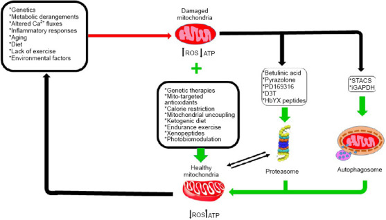
Schematic illustration of mitochondrial impairments observed in neurodegenerative diseases and potential therapeutic approaches for each level of deficiency.
Possible causes of mitochondrial impairment are shown in red and potential therapeutic approaches are depicted in green. Optimal proteasome function and mitochondrial health are tightly interdependent.
Footnotes
Conflicts of interest: The authors declare that they have no conflicts of interest.
Financial support: This work was supported by the European Regional Development Fund - Project MAGNET (No. CZ.02.1.01/0.0/0.0/15_003/0000492).
Copyright license agreement: The Copyright License Agreement has been signed by all authors before publication.
Plagiarism check: Checked twice by iThenticate.
Peer review: Externally peer reviewed.
C-Editors: Zhao M, Li CH; T-Editor: Jia Y
Funding: This work was supported by the European Regional Development Fund - Project MAGNET (No. CZ.02.1.01/0.0/0.0/15_003/0000492).
References
- 1.Abramov AY, Berezhnov AV, Fedotova EI, Zinchenko VP, Dolgacheva LP. Interaction of misfolded proteins and mitochondria in neurodegenerative disorders. Biochem Soc Trans. 2017;45:1025–1033. doi: 10.1042/BST20170024. [DOI] [PubMed] [Google Scholar]
- 2.Akimoto T, Pohnert SC, Li P, Zhang M, Gumbs C, Rosenberg PB, Williams RS, Yan Z. Exercise stimulates Pgc-1alpha transcription in skeletal muscle through activation of the p38 MAPK pathway. J Biol Chem. 2005;280:19587–19593. doi: 10.1074/jbc.M408862200. [DOI] [PubMed] [Google Scholar]
- 3.Alto NM, Soderling J, Scott JD. Rab32 is an A-kinase anchoring protein and participates in mitochondrial dynamics. J Cell Sci. 2002;158:659–668. doi: 10.1083/jcb.200204081. [DOI] [PMC free article] [PubMed] [Google Scholar]
- 4.Antonini A, Leenders KL, Spiegel R, Meier D, Vontobel P, Weigell-Weber M, Sanchez-Pernaute R, de Yébenez JG, Boesiger P, Weindl A, Maguire RP. Striatal glucose metabolism and dopamine D2 receptor binding in asymptomatic gene carriers and patients with Huntington’s disease. Brain. 1996;119(Pt 6):2085–2095. doi: 10.1093/brain/119.6.2085. [DOI] [PubMed] [Google Scholar]
- 5.Armstrong JS. Mitochondrial medicine: Pharmacological targeting of mitochondria in disease. Br J Pharmacol. 2007;151:1154–1165. doi: 10.1038/sj.bjp.0707288. [DOI] [PMC free article] [PubMed] [Google Scholar]
- 6.Ashrafi G, Schlehe JS, LaVoie MJ, Schwarz TL. Mitophagy of damaged mitochondria occurs locally in distal neuronal axons and requires PINK1 and Parkin. J Cell Biol. 2014;206:655–670. doi: 10.1083/jcb.201401070. [DOI] [PMC free article] [PubMed] [Google Scholar]
- 7.Attwell D, Laughlin SB. An energy budget for signaling in the grey matter of the brain. J Cereb Blood Flow Metab. 2001;21:1133–1145. doi: 10.1097/00004647-200110000-00001. [DOI] [PubMed] [Google Scholar]
- 8.Babenko VA, Silachev DN, Zorova LD, Pevzner IB, Khutornenko AA, Plotnikov EY, Sukhikh GT, Zorov DB. Improving the post-stroke therapeutic potency of mesenchymal multipotent stromal cells by Cocultivation with cortical neurons: the role of crosstalk between cells. Stem Cells Transl Med. 2015;4:1011–1020. doi: 10.5966/sctm.2015-0010. [DOI] [PMC free article] [PubMed] [Google Scholar]
- 9.Bacman SR, Williams SL, Pinto M, Peralta S, Moraes CT. Specific elimination of mutant mitochondrial genomes in patient-derived cells by mitoTALENs. Nat Med. 2013;19:1111–1113. doi: 10.1038/nm.3261. [DOI] [PMC free article] [PubMed] [Google Scholar]
- 10.Bacman SR, Kauppila JHK, Pereira CV, Nissanka N, Miranda M, Pinto M, Williams SL, Larsson NG, Stewart JB, Moraes CT. MitoTALEN reduces mutant mtDNA load and restores tRNAAla levels in a mouse model of heteroplasmic mtDNA mutation. Nat Med. 2018;24:1696–1700. doi: 10.1038/s41591-018-0166-8. [DOI] [PMC free article] [PubMed] [Google Scholar]
- 11.Banerjee K, Sinha M, Pham Cle L, Jana S, Chanda D, Cappai R, Chakrabarti S. Alpha-synuclein induced membrane depolarization and loss of phosphorylation capacity of isolated rat brain mitochondria: implications in Parkinson’s disease. FEBS Lett. 2010;584:1571–1576. doi: 10.1016/j.febslet.2010.03.012. [DOI] [PubMed] [Google Scholar]
- 12.Baraniak PR, McDevitt TC. Stem cell paracrine actions and tissue regeneration. Regen Med. 2010;5:121–143. doi: 10.2217/rme.09.74. [DOI] [PMC free article] [PubMed] [Google Scholar]
- 13.Bell SM, Barnes K, Clemmens H, Al-Rafiah AR, Al-Ofi EA, Leech V, Bandmann O, Shaw PJ, Blackburn DJ, Ferraiuolo L, Mortiboys H. Ursodeoxycholic acid improves mitochondrial function and redistributes Drp1 in fibroblasts from patients with either sporadic or familial Alzheimer’s disease. J Mol Biol. 2018;430:3942–3953. doi: 10.1016/j.jmb.2018.08.019. [DOI] [PMC free article] [PubMed] [Google Scholar]
- 14.Benatar M. Lost in translation: treatment trials in the SOD1 mouse and in human ALS. Neurobiol Dis. 2007;26:1–13. doi: 10.1016/j.nbd.2006.12.015. [DOI] [PubMed] [Google Scholar]
- 15.Bender A, Krishnan KJ, Morris CM, Taylor GA, Reeve AK, Perry RH, Jaros E, Hersheson JS, Betts J, Klopstock T, Taylor RW, Turnbull DM. High levels of mitochondrial DNA deletions in substantia nigra neurons in aging and Parkinson disease. Nat Genet. 2006;38:515–517. doi: 10.1038/ng1769. [DOI] [PubMed] [Google Scholar]
- 16.Bera AK, Ghosh S, Das S. Mitochondrial VDAC can be phosphorylated by cyclic AMP-dependent protein kinase. Biochem Biophys Res Commun. 1995;209:213–217. doi: 10.1006/bbrc.1995.1491. [DOI] [PubMed] [Google Scholar]
- 17.Bergendi L BL, Ďuracková Z, Ferenčik M. Chemistry, physiology and pathology of free radicals. Life Sci. 1999;65:1865–1874. doi: 10.1016/s0024-3205(99)00439-7. [DOI] [PubMed] [Google Scholar]
- 18.Bezprozvanny I, Hayden MR. Deranged neuronal calcium signaling and Huntington disease. Biochem Biophys Res Commun. 2004;322:1310–1317. doi: 10.1016/j.bbrc.2004.08.035. [DOI] [PubMed] [Google Scholar]
- 19.Blesa J, Trigo-Damas I, Quiroga-Varela A, Jackson-Lewis VR. Oxidative stress and Parkinson’s disease. Front Neuroanat. 2015;9:91. doi: 10.3389/fnana.2015.00091. [DOI] [PMC free article] [PubMed] [Google Scholar]
- 20.Blesa JR, Hernández-Yago J. Distinct functional contributions of 2 GABP-NRF-2 recognition sites within the context of the human TOMM70 promoter. Biochem Cell Biol. 2006;84:813–822. doi: 10.1139/o06-064. [DOI] [PubMed] [Google Scholar]
- 21.Borghammer P, Chakravarty M, Jonsdottir KY, Sato N, Matsuda H, Ito K, Arahata Y, Kato T, Gjedde A. Cortical hypometabolism and hypoperfusion in Parkinson’s disease is extensive: probably even at early disease stages. Brain Struct Funct. 2010;214:303–317. doi: 10.1007/s00429-010-0246-0. [DOI] [PubMed] [Google Scholar]
- 22.Calì T, Ottolini D, Brini M. Mitochondrial Ca2+ and neurodegeneration. Cell Calcium. 2012;52:73–85. doi: 10.1016/j.ceca.2012.04.015. [DOI] [PMC free article] [PubMed] [Google Scholar]
- 23.Camandola S, Mattson MP. Brain metabolism in health, aging, and neurodegeneration. EMBO J. 2017;36:1474–1492. doi: 10.15252/embj.201695810. [DOI] [PMC free article] [PubMed] [Google Scholar]
- 24.Cardoso SM, Proenca MT, Santos S, Santana I, Oliveira CR. Cytochrome c oxidase is decreased in Alzheimer’s disease platelets. Neurobiol Aging. 2004;25:105–110. doi: 10.1016/s0197-4580(03)00033-2. [DOI] [PubMed] [Google Scholar]
- 25.Carelli V, Chan DC. Mitochondrial DNA: impacting central and peripheral nervous systems. Neuron. 2014;84:1126–1142. doi: 10.1016/j.neuron.2014.11.022. [DOI] [PMC free article] [PubMed] [Google Scholar]
- 26.Carlucci A, Adornetto A, Scorziello A, Viggiano D, Foca M, Cuomo O. Proteolysis of AKAP121 regulates mitochondrial activity during cellular hypoxia and brain ischaemia. EMBO J. 2008;27:1073–8410. doi: 10.1038/emboj.2008.33. [DOI] [PMC free article] [PubMed] [Google Scholar]
- 27.Caspersen C, Wang N, Yao J, Sosunov A, Chen X, Lustbader JW, Xu HW, Stern D, McKhann G, Yan SD. Mitochondrial Abeta: a potential focal point for neuronal metabolic dysfunction in Alzheimer’s disease. FASEB J. 2005;19:2040–2041. doi: 10.1096/fj.05-3735fje. [DOI] [PubMed] [Google Scholar]
- 28.Cassano P, Sciancalepore AG, Lezza AM, Leeuwenburgh C, Cantatore P, Gadaleta MN. Tissue-specific effect of age and caloric restriction diet on mitochondrial DNA content. Rejuvenation Res. 2006;9:211–214. doi: 10.1089/rej.2006.9.211. [DOI] [PubMed] [Google Scholar]
- 29.Cassidy-Stone A, Chipuk JE, Ingerman E, Song C, Yoo C, Kuwana T, Kurth MJ, Shaw JT, Hinshaw JE, Green DR, Nunnari J. Chemical inhibition of the mitochondrial division dynamin reveals its role in Bax/Bak-dependent mitochondrial outer membrane permeabilization. Dev Cell. 2008;14:193–204. doi: 10.1016/j.devcel.2007.11.019. [DOI] [PMC free article] [PubMed] [Google Scholar]
- 30.Castellani R, Hirai K, Aliev G, Drew KL, Nunomura A, Takeda A, Cash AD, Obrenovich ME, Perry G, Smith MA. Role of mitochondrial dysfunction in Alzheimer’s disease. J Neurosci Res. 2002;70:357–360. doi: 10.1002/jnr.10389. [DOI] [PubMed] [Google Scholar]
- 31.Cerqueira FM, Cunha FM, Laurindo FR, Kowaltowski AJ. Calorie restriction increases cerebral mitochondrial respiratory capacity in a NO•-mediated mechanism: impact on neuronal survival. Free Radic Biol Med. 2012;52:123641. doi: 10.1016/j.freeradbiomed.2012.01.011. [DOI] [PubMed] [Google Scholar]
- 32.Cha MY, Kim DK, Mook-Jung I. The role of mitochondrial DNA mutation on neurodegenerative diseases. Exp Mol Med 47. 2015:e150. doi: 10.1038/emm.2014.122. [DOI] [PMC free article] [PubMed] [Google Scholar]
- 33.Chandel NS. Evolution of mitochondria as signaling organelles. Cell Metab. 2015;22:204–206. doi: 10.1016/j.cmet.2015.05.013. [DOI] [PubMed] [Google Scholar]
- 34.Chang JC, Wu SL, Liu KH, Chen YH, Chuang CS, Cheng FC, Su HL, Wei YH, Kuo SJ, Liu CS. Allogeneic/xenogeneic transplantation of peptide-labeled mitochondria in Parkinson’s disease: restoration of mitochondria functions and attenuation of 6-hydroxydopamine-induced neurotoxicity. Transl Res. 2016;170:40–56. doi: 10.1016/j.trsl.2015.12.003. [DOI] [PubMed] [Google Scholar]
- 35.Chaturvedi RK, Adhihetty P, Shukla S, Hennessy T, Calingasan N, Yang L, Starkov A, Kiaei M, Cannella M, Sassone J, Ciammola A, Squitieri F, Beal MF. Impaired PGC-1alpha function in muscle in Huntington’s disease. Hum Mol Genet. 2009;18:3048–3065. doi: 10.1093/hmg/ddp243. [DOI] [PMC free article] [PubMed] [Google Scholar]
- 36.Chen H, Chan DC. Mitochondrial dynamics-fusion, fission, movement, andmitophagy-in neurodegenerative diseases. Hum Mol Genet. 2009;18:R169–176. doi: 10.1093/hmg/ddp326. [DOI] [PMC free article] [PubMed] [Google Scholar]
- 37.Chen H, Chomyn A, Chan D. Disruption of fusion results in mitochondrial heterogeneity and dysfunction J Biol Chem 280:26185-26192. 2005 doi: 10.1074/jbc.M503062200. [DOI] [PubMed] [Google Scholar]
- 38.Chen H, Detmer SA, Ewald AJ, Griffin EE, Fraser SE, Chan DC. Mitofusins Mfn1 and Mfn2 coordinately regulate mitochondrial fusion and are essential for embryonic development. J Cell Biol. 2003;160:189–200. doi: 10.1083/jcb.200211046. [DOI] [PMC free article] [PubMed] [Google Scholar]
- 39.Chen Q, Prior M, Dargusch R, Roberts A, Riek R, Eichmann C, Chiruta C, Akaishi T, Abe K, Maher P, Schubert D. A novel neurotrophic drug for cognitive enhancement and Alzheimer’s disease. PLoS One 6. 2011:e27865. doi: 10.1371/journal.pone.0027865. [DOI] [PMC free article] [PubMed] [Google Scholar]
- 40.Chen W, Zhang X, Huang W. Role of physical exercise in Alzheimer’s disease (Review) Biomedical Reports. 2016;4:403–407. doi: 10.3892/br.2016.607. [DOI] [PMC free article] [PubMed] [Google Scholar]
- 41.Cheng X, Siow RC, Mann GE. Impaired redox signaling and antioxidant gene expression in endothelial cells in diabetes: a role for mitochondria and the nuclear factor-E2-related factor 2-Kelch-like ECH-associated protein 1 defense pathway. Antioxid Redox Signal. 2011;14:469–487. doi: 10.1089/ars.2010.3283. [DOI] [PubMed] [Google Scholar]
- 42.Chiang MC, Chern Y, Juo CG. The dysfunction of hepatic transcriptional factors in mice with Huntington’s disease. Biochim Biophys Acta. 2011;1812:1111–1120. doi: 10.1016/j.bbadis.2011.05.006. [DOI] [PubMed] [Google Scholar]
- 43.Chien L, Liang MZ, Chang CY, Wang C, Chen L. Mitochondrial therapy promotes regeneration of injured hippocampal neurons. Biochim Biophys Acta. 2018;1864:3001–3012. doi: 10.1016/j.bbadis.2018.06.012. [DOI] [PubMed] [Google Scholar]
- 44.Cho DH, Nakamura T, Lipton SA. Mitochondrial dynamics in cell death and neurodegeneration. Cell Mol Life Sci. 2010;67:3435–3447. doi: 10.1007/s00018-010-0435-2. [DOI] [PMC free article] [PubMed] [Google Scholar]
- 45.Cho DH, Nakamura T, Fang J, Cieplak P, Godzik A, Gu Z, Lipton SA. S-nitrosylation of Drp1 mediates beta-amyloid-related mitochondrial fission and neuronal injury. Science. 2009;324:102–105. doi: 10.1126/science.1171091. [DOI] [PMC free article] [PubMed] [Google Scholar]
- 46.Chondrogianni N, Voutetakis K, Kapetanou M, Delitsikou V, Papaevgeniou N, Sakellari M, Lefaki M, Filippopoulou K, Gonos ES. Proteasome activation: An innovative promising approach for delaying aging and retarding age-related diseases. Ageing Res Rev. 2015;23:37–55. doi: 10.1016/j.arr.2014.12.003. [DOI] [PubMed] [Google Scholar]
- 47.Choo YS, Johnson GVW, MacDonald M, Detloff PJ, Lesort M. Mutant huntingtin directly increases susceptibility of mitochondria to the calcium-induced permeability transition and cytochrome c release. Hum Mol Genet. 2004;13:1407–1420. doi: 10.1093/hmg/ddh162. [DOI] [PubMed] [Google Scholar]
- 48.Ciarmiello A, Cannella M, Lastoria S, Simonelli M, Frati L, Rubinsztein DC, Squitieri F. Brain white-matter volume loss and glucose hypometabolism precede the clinical symptoms of Huntington’s disease. J Nucl Med. 2006;47:215–222. [PubMed] [Google Scholar]
- 49.Cipolat S, Martins de Brito O, Dal Zilio B, Scorrano L. OPA1 requires mitofusin 1 to promote mitochondrial fusion. Proc Natl Acad Sci U S A. 2004;101:15927–15932. doi: 10.1073/pnas.0407043101. [DOI] [PMC free article] [PubMed] [Google Scholar]
- 50.Civitarese AE, MacLean PS, Carling S, Kerr-Bayles L, McMillan RP, Pierce A, Becker TC, Moro C, Finlayson J, Lefort N, Newgard CB, Mandarino L, Cefalu W, Walder K, Collier GR, Hulver MW, Smith SR, Ravussin E. Regulation of skeletal muscle oxidative capacity and insulin signaling by the mitochondrial rhomboid protease PARL. Cell Metab. 2010;11:412–426. doi: 10.1016/j.cmet.2010.04.004. [DOI] [PMC free article] [PubMed] [Google Scholar]
- 51.Cogliati S, Enriquez JA, Scorrano L. Mitochondrial cristae: where beauty meets functionality. Trends Biochem Sci. 2016;41:261–273. doi: 10.1016/j.tibs.2016.01.001. [DOI] [PubMed] [Google Scholar]
- 52.Colman RJ, Beasley TM, Kemnitz JW, Johnson SC, Weindruch R, Anderson RM. Caloric restriction reduces age-related and all-cause mortality in rhesus monkeys. Nat Commun. 2014;5:3557. doi: 10.1038/ncomms4557. [DOI] [PMC free article] [PubMed] [Google Scholar]
- 53.Cozzolino M, Carri MT. Mitochondrial dysfunction in ALS. Prog Neurobiol. 2012;97:54–66. doi: 10.1016/j.pneurobio.2011.06.003. [DOI] [PubMed] [Google Scholar]
- 54.Craven L, Alston CL, Taylor RW, Turnbull DM. Recent advances in mitochondrial Disease. Annu Rev Genomics Hum Genet. 2017;18:257–275. doi: 10.1146/annurev-genom-091416-035426. [DOI] [PubMed] [Google Scholar]
- 55.Crow JP, Ye YZ, Strong M, Kirk M, Barnes S, Beckman JS. Superoxide dismutase catalyzes nitration of tyrosines by peroxynitrite in the rod and head domains of neurofilament-L. J Neurochem. 1997;69:1945–1953. doi: 10.1046/j.1471-4159.1997.69051945.x. [DOI] [PubMed] [Google Scholar]
- 56.Dal Vechio FH, Cerqueira F, Augusto O, Lopes R, Demasi M. Peptides that activate the 20S proteasome by gate opening increased oxidized protein removal and reduced protein aggregation. Free Radic Biol Med. 2014;67:304–313. doi: 10.1016/j.freeradbiomed.2013.11.017. [DOI] [PubMed] [Google Scholar]
- 57.Dantuma NP, Bott LC. The ubiquitin-proteasome system in neurodegenerative diseases: precipitating factor, yet part of the solution. Front Mol Neurosci. 2014;7:70. doi: 10.3389/fnmol.2014.00070. [DOI] [PMC free article] [PubMed] [Google Scholar]
- 58.Das S, Wong R, Rajapakse N, Murphy E, Steenbergen C. Glycogen synthase kinase 3 inhibition slows mitochondrial adenine nucleotide transport and regulates voltage-dependent anion channel phosphorylation. Circ Res. 2008;103:983–991. doi: 10.1161/CIRCRESAHA.108.178970. [DOI] [PMC free article] [PubMed] [Google Scholar]
- 59.Daugherty D, Goldberg J, Fischer W, Dargusch R, Maher P, Schubert D. A novel Alzheimer’s disease drug candidate targeting inflammation and fatty acid metabolism. Alzheimers Res Ther. 2017;9:50. doi: 10.1186/s13195-017-0277-3. [DOI] [PMC free article] [PubMed] [Google Scholar]
- 60.de Castro IP, Martins LM, Tufi R. Mitochondrial quality control and neurological disease: an emerging connection. Expert Rev Mol Med 12. 2010:e12. doi: 10.1017/S1462399410001456. [DOI] [PMC free article] [PubMed] [Google Scholar]
- 61.De Stefani D, Raffaello A, Teardo E, Szabo I, Rizzuto R. A forty-kilodalton protein of the inner membrane is the mitochondrial calcium uniporter. Nature. 2011;476:336–340. doi: 10.1038/nature10230. [DOI] [PMC free article] [PubMed] [Google Scholar]
- 62.Deckel AW. Nitric oxide and nitric oxide synthase in Huntington’s disease. J Neurosci Res. 2001;64:99–107. doi: 10.1002/jnr.1057. [DOI] [PubMed] [Google Scholar]
- 63.Deckel AW, Tang V, Nuttal D, Gary K, Elder R. Altered neuronal nitric oxide synthase expression contributes to disease progression in Huntington’s disease transgenic mice. Brain Res. 2002;939:76–86. doi: 10.1016/s0006-8993(02)02550-7. [DOI] [PubMed] [Google Scholar]
- 64.Dempsey C, Rubio Araiz A, Bryson KJ, Finucane O, Larkin C, Mills EL, Robertson AAB, Cooper MA, O’Neill LAJ, Lynch MA. Inhibiting the NLRP3 inflammasome with MCC950 promotes non-phlogistic clearance of amyloid-β and cognitive function in APP/PS1 mice. Brain Behav Immun. 2017;61:306–316. doi: 10.1016/j.bbi.2016.12.014. [DOI] [PubMed] [Google Scholar]
- 65.Dhandapani PK, Lyyski AM, Paulin L, Khan NA, Suomalainen A, Auvinen P, Dufour E, Szibor M, Jacobs HT. Phenotypic effects of dietary stress in combination with a respiratory chain bypass in mice. Physiol Rep 7. 2019:e14159. doi: 10.14814/phy2.14159. [DOI] [PMC free article] [PubMed] [Google Scholar]
- 66.Dheen ST, Kaur C, Ling EA. Microglial activation and its implications in the brain diseases. Curr Med Chem. 2007;14:1189–1197. doi: 10.2174/092986707780597961. [DOI] [PubMed] [Google Scholar]
- 67.Di Maio R, Barrett PJ, Hoffman EK, Barrett CW, Zharikov A, Borah A, Hu X, McCoy J, Chu CT, Burton EA, Hastings TG, Greenamyre JT. α-Synuclein binds to TOM20 and inhibits mitochondrial protein import in Parkinson’s disease. Sci Transl Med. 2016;8:342ra78. doi: 10.1126/scitranslmed.aaf3634. [DOI] [PMC free article] [PubMed] [Google Scholar]
- 68.Diehl JA, Cheng M, Roussel MF, Sherr CJ. Glycogen synthase kinase-3 beta regulates cyclin D1 proteolysis and subcellular localisation. Genes Dev. 1998;12:3499–3511. doi: 10.1101/gad.12.22.3499. [DOI] [PMC free article] [PubMed] [Google Scholar]
- 69.Ding WX, Yin XM. Mitophagy, mechanisms, pathophysiological roles, and analysis. Biol Chem. 2012;393:547–564. doi: 10.1515/hsz-2012-0119. [DOI] [PMC free article] [PubMed] [Google Scholar]
- 70.Dionísio PA, Amaral JD, Ribeiro MF, Lo AC, D’Hooge R, Rodrigues CMP. Amyloid-β pathology is attenuated by tauroursodeoxycholic acid treatment in APP/PS1 mice after disease onset. Neurobiol Aging. 2015;36:228–240. doi: 10.1016/j.neurobiolaging.2014.08.034. [DOI] [PubMed] [Google Scholar]
- 71.Dong Z, Saikumar P, Weinberg JM, Venkatachalan MA. Calcium in cell injury and death. Annu Rev Pathol Mech Dis. 2006;1:405–434. doi: 10.1146/annurev.pathol.1.110304.100218. [DOI] [PubMed] [Google Scholar]
- 72.Drose S, Brandt U. Molecular mechanisms of superoxide production by the mitochondrial respiratory chain. Adv Exp Med Biol. 2012;748:145–169. doi: 10.1007/978-1-4614-3573-0_6. [DOI] [PubMed] [Google Scholar]
- 73.Du H, Guo L, Fang F, Chen D, Sosunov AA, McKhann GM, Yan Y, Wang C, Zhang H, Molkentin JD, Gunn-Moore FJ, Vonsattel JP, Arancio O, Chen JX, Yan SD. Cyclophilin D deficiency attenuates mitochondrial and neuronal perturbation and ameliorates learning and memory in Alzheimer’s disease. Nat Med. 2008;14:1097–1105. doi: 10.1038/nm.1868. [DOI] [PMC free article] [PubMed] [Google Scholar]
- 74.Duan M, Tu J, Lu Z. Recent advances in detecting mitochondrial DNA heteroplasmic variations. Molecules. 2018;23:2. doi: 10.3390/molecules23020323. [DOI] [PMC free article] [PubMed] [Google Scholar]
- 75.Duan W, Li X, Shi J, Guo Y, Li Z, Li C. Mutant TAR DNA-binding protein-43 induces oxidative injury in motor neuron-like cell. Neuroscience. 2010;169:1621–1629. doi: 10.1016/j.neuroscience.2010.06.018. [DOI] [PubMed] [Google Scholar]
- 76.East DA, Fagiani F, Crosby J, Georgakopoulos ND, Bertrand H, Schaap M, Campanella M. PMI: A ΔΨm independent pharmacological regulator of mitophagy. Chem Biol. 2014;21:1585–1596. doi: 10.1016/j.chembiol.2014.09.019. [DOI] [PMC free article] [PubMed] [Google Scholar]
- 77.Egan DF, Shackelford DB, Mihaylova MM, Gelino S, Kohnz RA, Mair W. Phosphorylation of ULK1 (hATG1) by AMP-activated protein kinase connects energy sensing to mitophagy. Science. 2011;331:456–461. doi: 10.1126/science.1196371. [DOI] [PMC free article] [PubMed] [Google Scholar]
- 78.Eilam R, Peter Y, Groner Y, Segal M. Late degeneration of nigro-striatal neurons in ATM−/− mice. Neuroscience. 2003;121:83–98. doi: 10.1016/s0306-4522(03)00322-1. [DOI] [PubMed] [Google Scholar]
- 79.Eisner V, Picard M, Hajnóczky G. Mitochondrial dynamics in adaptive and maladaptive cellular stress responses. Nat Cell Biol. 2018;20:755–765. doi: 10.1038/s41556-018-0133-0. [DOI] [PMC free article] [PubMed] [Google Scholar]
- 80.El-Khoury R, Dufour E, Rak M, Ramanantsoa N, Grandchamp N, Csaba Z, Duvillié B, Bénit P, Gallego J, Gressens P, Sarkis C, Jacobs HT, Rustin P. Alternative oxidase expression in the mouse enables bypassing cytochrome c oxidase blockade and limits mitochondrial ROS overproduction. PLoS Genet 9. 2013:e1003182. doi: 10.1371/journal.pgen.1003182. [DOI] [PMC free article] [PubMed] [Google Scholar]
- 81.Enokido Y, Tamura T, Ito H, Arumughan A, Komuro A, Shiwaku H, Sone M, Foulle R, Sawada H, Ishiguro H, Ono T, Murata M, Kanazawa I, Tomilin N, Tagawa K, Wanker EE, Okazawa H. Mutant huntingtin impairs Ku70-mediated DNA repair. J Cell Biol. 2010;189:425–443. doi: 10.1083/jcb.200905138. [DOI] [PMC free article] [PubMed] [Google Scholar]
- 82.Erkkinen MG, Kim MO, Geschwind MD. Clinical neurology and epidemiology of the major neurodegenerative diseases. Cold Spring Harb Perspect Biol 10. 2018:a033118. doi: 10.1101/cshperspect.a033118. [DOI] [PMC free article] [PubMed] [Google Scholar]
- 83.Eschbach J, Schwalenstöcker B, Soyal SM, Bayer H, Wiesner D, Akimoto C, Nilsson AC, Birve A, Meyer T, Dupuis L, Danzer KM, Andersen PM, Witting A, Ludolph AC, Patsch W, Weydt P. PGC-1α is a male-specific disease modifier of human and experimental amyotrophic lateral sclerosis. Hum Mol Genet. 2013;22:3477–3484. doi: 10.1093/hmg/ddt202. [DOI] [PubMed] [Google Scholar]
- 84.Espino J, Bejarano I, Redondo PC, Rosado JA, Barriga C, Reiter RJ, Pariente JA, Rodríguez AB. Melatonin reduces apoptosis induced by calcium signaling in human leukocytes: Evidence for the involvement of mitochondria and Bax activation. J Membr Biol. 2010;233(1-3):105–118. doi: 10.1007/s00232-010-9230-0. [DOI] [PubMed] [Google Scholar]
- 85.Evans MJ, Scarpulla RC. NRF-1: a trans-activator of nuclear-encoded respiratory genes in animal cells. Genes Dev. 1990;4:1023-1034. doi: 10.1101/gad.4.6.1023. [DOI] [PubMed] [Google Scholar]
- 86.Fang Y, Hu XH, Jia ZG, Xu MH, Guo ZY, Gao FH. Tiron protects against UVB-induced senescence-like characteristics in human dermal fibroblasts by the inhibition of superoxide anion production and glutathione depletion. Australas J Dermatol. 2012;53:172–180. doi: 10.1111/j.1440-0960.2012.00912.x. [DOI] [PubMed] [Google Scholar]
- 87.Ferlazzo ML, Sonzogni L, Granzotto A, Bodgi L, Lartin O, Devic C, Vogin G, Pereira S, Foray N. Mutations of the Huntington’s disease protein impact on the ATM-dependent signaling and repair pathways of the radiation-induced DNA double-strand breaks: corrective effect of statins and bisphosphonates. Mol Neurobiol. 2014;49:1200–1211. doi: 10.1007/s12035-013-8591-7. [DOI] [PubMed] [Google Scholar]
- 88.Fernandez-Marcos PJ, Auwerx J. Regulation of PGC-1α, a nodal regulator of mitochondrial biogenesis. Am J Clin Nutr. 2011;93:884S-890S. doi: 10.3945/ajcn.110.001917. [DOI] [PMC free article] [PubMed] [Google Scholar]
- 89.Ferreira IL, Nascimento MV, Ribeiro M, Almeida S, Cardoso SM, Grazina M, Pratas J, Santos MJ, Januário C, Oliveira CR, Rego AC. Mitochondrial-dependent apoptosis in Huntington’s disease human cybrids. Exp Neurol. 2010;222:243–255. doi: 10.1016/j.expneurol.2010.01.002. [DOI] [PubMed] [Google Scholar]
- 90.Ferreiro E, Oliveira CR, Pereira CMF. The release of calcium from the endoplasmic reticulum induced by amyloid-beta and prion peptides activates the mitochondrial apoptotic pathway. Neurobiol Dis. 2008;30:331–342. doi: 10.1016/j.nbd.2008.02.003. [DOI] [PubMed] [Google Scholar]
- 91.Flint Beal M. Mitochondria and the pathogenesis of ALS. Brain. 2000;123:1291–1292. doi: 10.1093/brain/123.7.1291. [DOI] [PubMed] [Google Scholar]
- 92.Franco-Iborra S, Vila M, Perier C. The Parkinson disease mitochondrial hypothesis: where are we at. Neuroscientist. 2016;22:266–277. doi: 10.1177/1073858415574600. [DOI] [PubMed] [Google Scholar]
- 93.Freed CR, Greene PE, Breeze RE, Tsai WY, DuMouchel W, Kao R, Dillon S, Winfield H, Culver S, Trojanowski JQ, Eidelberg D, Fahn S. Transplantation of embryonic dopamine neurons for severe Parkinson’s disease. N Engl J Med. 2001;344:710–719. doi: 10.1056/NEJM200103083441002. [DOI] [PubMed] [Google Scholar]
- 94.Fu M, St-Pierre P, Shankar J, Wang PT, Joshi B, Nabi IR. Regulation of mitophagy by the Gp78 E3 ubiquitin ligase. Mol Biol Cell. 2013;24:1153–1162. doi: 10.1091/mbc.E12-08-0607. [DOI] [PMC free article] [PubMed] [Google Scholar]
- 95.Fu Z, Tindall DJ. FOXOs, cancer and regulation of apoptosis. Oncogene. 2008;27:2312–2319. doi: 10.1038/onc.2008.24. [DOI] [PMC free article] [PubMed] [Google Scholar]
- 96.Fukui H, Moraes CT. Extended polyglutamine repeats trigger a feedback loop involving the mitochondrial complex III, the proteasome and huntingtin aggregates. Hum Mol Genet. 2007;16:783–797. doi: 10.1093/hmg/ddm023. [DOI] [PubMed] [Google Scholar]
- 97.Funalot B, Desport JC, Sturtz F, Camu W, Couratier P. High metabolic level in patients with familial amyotrophic lateral sclerosis. Amyotroph Lateral Scler. 2009;10:113–117. doi: 10.1080/17482960802295192. [DOI] [PubMed] [Google Scholar]
- 98.Galano A, Tan DX, Reiter RJ. Melatonin as a natural ally against oxidative stress: a physicochemical examination. J Pineal Res. 2011;51:1–16. doi: 10.1111/j.1600-079X.2011.00916.x. [DOI] [PubMed] [Google Scholar]
- 99.Gammage PA, Rorbach J, Vincent AI, Rebar EJ, Minczuk M. Mitochondrially targeted ZFNs for selective degradation of pathogenic mitochondrial genomes bearing large-scale deletions or point mutations. EMBO Mol Med. 2014;6:458–466. doi: 10.1002/emmm.201303672. [DOI] [PMC free article] [PubMed] [Google Scholar]
- 100.Gammage PA, Viscomi C, Simard ML, Costa ASH, Gaude E, Powell CA, Van Haute L, McCann BJ, Rebelo-Guiomar P, Cerutti R, Zhang L, Rebar EJ, Zeviani M, Frezza C, Stewart JB, Minczuk M. Genome editing in mitochondria corrects a pathogenic mtDNA mutation in vivo. Nat Med. 2018;24:1691–1695. doi: 10.1038/s41591-018-0165-9. [DOI] [PMC free article] [PubMed] [Google Scholar]
- 101.Ganeshan V, Skladnev NV, Kim JY, Mitrofanis J, Stone J, Johnstone DM. Pre-conditioning with remote photobiomodulation modulates the brain transcriptome and protects against MPTP insult in mice. Neuroscience. 2019;400:85–97. doi: 10.1016/j.neuroscience.2018.12.050. [DOI] [PubMed] [Google Scholar]
- 102.Geisler JG, Marosi K, Halpern J, Mattson MP. DNP, mitochondrial uncoupling, and neuroprotection: A little dab’ll do ya. Alzheimers Dement. 2017;13:582–591. doi: 10.1016/j.jalz.2016.08.001. [DOI] [PMC free article] [PubMed] [Google Scholar]
- 103.Geisler S, Holmstrom KM, Skujat D, Fiesel FC, Rothfuss OC, Kahle PJ, Springer W. PINK1/Parkin-mediated mitophagy is dependent on VDAC1 and p62/SQSTM1. Nat Cell Biol. 2010;12:119–131. doi: 10.1038/ncb2012. [DOI] [PubMed] [Google Scholar]
- 104.Gillette TG, Kumar B, Thompson D, Slaughter CA, DeMartino GN. Differential roles of the COOH termini of AAA subunits of PA700 (19 S regulator) in asymmetric assembly and activation of the 26 S proteasome. J Biol Chem. 2008;283:31813–31822. doi: 10.1074/jbc.M805935200. [DOI] [PMC free article] [PubMed] [Google Scholar]
- 105.Giuliano P, De Cristofaro T, Affaitati A, Pizzulo GM, Feliciello A, Criscuolo C, De Michele G, Filla A, Avvedimento EV, Varrone S. DNA damage induced by polyglutamine-expanded proteins. Hum Mol Genet. 2003;12:2301–2309. doi: 10.1093/hmg/ddg242. [DOI] [PubMed] [Google Scholar]
- 106.Gleyzer N, Vercauteren K, Scarpulla RC. Control of mitochondrial transcription specificity factors (TFB1M and TFB2M) by nuclear respiratory factors (NRF-1 and NRF-2) and PGC-1 family coactivators. Mol Cell Biol. 2005;25:1354–1366. doi: 10.1128/MCB.25.4.1354-1366.2005. [DOI] [PMC free article] [PubMed] [Google Scholar]
- 107.Golpich M, Rahmani B, Mohamed Ibrahim N, Dargahi L, Mohamed Z, Raymond AA, Ahmadiani A. Preconditioning as a potential strategy for the prevention of Parkinson’s disease. Mol Neurobiol. 2015;51:313–330. doi: 10.1007/s12035-014-8689-6. [DOI] [PubMed] [Google Scholar]
- 108.Golpich M, Amini E, Mohamed Z, Azman Ali R, Mohamed Ibrahim N, Ahmadiani A. Mitochondrial dysfunction and biogenesis in neurodegenerative diseases: pathogenesis and treatment. CNS Neurosci Ther. 2017;23:5–22. doi: 10.1111/cns.12655. [DOI] [PMC free article] [PubMed] [Google Scholar]
- 109.Gomes LC, Di Benedetto G, Scorrano L. During autophagy mitochondria elongate, are spared from degradation and sustain cell viability. Nat Cell Biol. 2011;13:589–598. doi: 10.1038/ncb2220. [DOI] [PMC free article] [PubMed] [Google Scholar]
- 110.Graziewicz MA, Longley MJ, Copeland WC. DNA polymerase gamma in mitochondrial DNA replication and repair. Chem Rev. 2006;106:383–405. doi: 10.1021/cr040463d. [DOI] [PubMed] [Google Scholar]
- 111.Greenamyre JT, Hastings TG. Parkinson’s: divergent causes, convergent mechanisms. Science. 2004;304:1120–1122. doi: 10.1126/science.1098966. [DOI] [PubMed] [Google Scholar]
- 112.Griparic L, Van Der Wel NN, Orozco IJ, Peters PJ, Van Der Bliek AM. Loss of the intermembrane space protein Mgm1/OPA1 induces swelling and localized constrictions along the lengths of mitochondria. J Biol Chem. 2004;279:18792–18798. doi: 10.1074/jbc.M400920200. [DOI] [PubMed] [Google Scholar]
- 113.Guo X, Disatnik MH, Monbureau M, Shamloo M, Mochly-Rosen D, Qi X. Inhibition of mitochondrial fragmentation diminishes Huntington’s disease-associated neurodegeneration. J Clin Invest. 2013;123:5371–5388. doi: 10.1172/JCI70911. [DOI] [PMC free article] [PubMed] [Google Scholar]
- 114.Hamanaka RB, Chandel NS. Mitochondrial reactive oxygen species regulate cellular signaling and dictate biological outcomes. Trends Biochem Sci. 2010;35:505–513. doi: 10.1016/j.tibs.2010.04.002. [DOI] [PMC free article] [PubMed] [Google Scholar]
- 115.Handy DE, Loscalzo J. Redox regulation of mitochondrial function. Antioxid Redox Signal. 2012;16:1323–1367. doi: 10.1089/ars.2011.4123. [DOI] [PMC free article] [PubMed] [Google Scholar]
- 116.Hansson Petersen CA, Alikhani N, Behbahani H, Wiehager B, Pavlov PF, Alafuzoff I, Leinonen V, Ito A, Winblad B, Glaser E, Ankarcrona M. The amyloid beta-peptide is imported into mitochondria via the TOM import machinery and localized to mitochondrial cristae. Proc Natl Acad Sci U S A. 2008;105:13145–13150. doi: 10.1073/pnas.0806192105. [DOI] [PMC free article] [PubMed] [Google Scholar]
- 117.Haun F, Nakamura T, Lipton SA. Dysfunctional mitochondrial dynamics in the pathophysiology of neurodegenerative diseases. J Cell Death. 2013;6:27–35. doi: 10.4137/JCD.S10847. [DOI] [PMC free article] [PubMed] [Google Scholar]
- 118.Hauptmann S, Scherping I, Dröse S, Brandt U, Schulz KL, Jendrach M, Leuner K, Eckert A, Müller WE. Mitochondrial dysfunction: an early event in Alzheimer pathology accumulates with age in AD transgenic mice. Neurobiol Aging. 2009;30:1574–1586. doi: 10.1016/j.neurobiolaging.2007.12.005. [DOI] [PubMed] [Google Scholar]
- 119.Hayakawa K, Esposito E, Wang X, Terasaki Y, Liu Y, Xing C, Ji X, Lo EH. Transfer of mitochondria from astrocytes to neurons after stroke. Nature. 2016;535:551–555. doi: 10.1038/nature18928. [DOI] [PMC free article] [PubMed] [Google Scholar]
- 120.Hennessy M, Hamblin MR. Photobiomodulation and the brain: a new paradigm. J Opt. 2017;19:013003. doi: 10.1088/2040-8986/19/1/013003. [DOI] [PMC free article] [PubMed] [Google Scholar]
- 121.Hirai K, Aliev G, Nunomura A, Fujioka H, Russell RL, Atwood CS, Johnson AB, Kress Y, Vinters HV, Tabaton M, Shimohama S, Cash AD, Siedlak SL, Harris PL, Jones PK, Petersen RB, Perry G, Smith MA. Mitochondrial abnormalities in Alzheimer’s disease. J Neurosci. 2001;21:3017–3023. doi: 10.1523/JNEUROSCI.21-09-03017.2001. [DOI] [PMC free article] [PubMed] [Google Scholar]
- 122.Höglinger GU, Carrard G, Michel PP, Medja F, Lombès A, Ruberg M, Friguet B, Hirsch EC. Dysfunction of mitochondrial complex I and the proteasome: interactions between two biochemical deficits in a cellular model of Parkinson’s disease. J Neurochem. 2003;86:1297–1307. doi: 10.1046/j.1471-4159.2003.01952.x. [DOI] [PubMed] [Google Scholar]
- 123.Hou Y, Dan X, Babbar M, Wei Y, Hasselbalch SG, Croteau DL, Bohr VA. Ageing as a risk factor for neurodegenerative disease. Nat Rev Neurol. 2019;15:565–581. doi: 10.1038/s41582-019-0244-7. [DOI] [PubMed] [Google Scholar]
- 124.Huang L, Ho P, Chen CH. Activation and inhibition of the proteasome by betulinic acid and its derivatives. FEBS Lett. 2007;581:4955–4959. doi: 10.1016/j.febslet.2007.09.031. [DOI] [PMC free article] [PubMed] [Google Scholar]
- 125.Hughes SD, Kanabus M, Anderson G, Hargreaves IP, Rutherford T, O’Donnell M, Cross JH, Rahman S, Eaton S, Heales SJ. The ketogenic diet component decanoic acid increases mitochondrial citrate synthase and complex I activity in neuronal cells. J Neurochem. 2014;129:426–433. doi: 10.1111/jnc.12646. [DOI] [PubMed] [Google Scholar]
- 126.Iliev AI, Stringaris AK, Nau R, Neumann H. Neuronal injury mediated via stimulation of microglial toll-like receptor-9 (TLR9) FASEB J 18:412-414. 2004 doi: 10.1096/fj.03-0670fje. [DOI] [PubMed] [Google Scholar]
- 127.Illuzzi J, Yerkes S, Parekh-Olmedo H, Kmiec EB. DNA breakage and induction of DNA damage response proteins precede the appearance of visible mutant huntingtin aggregates. J Neurosci Res. 2009;87:733–747. doi: 10.1002/jnr.21881. [DOI] [PubMed] [Google Scholar]
- 128.Innamorato NG, Lastres-Becker I, Cuadrado A. Role of microglial redox balance in modulation of neuroinflammation. Curr Opin Neurol. 2009;22:308–314. doi: 10.1097/WCO.0b013e32832a3225. [DOI] [PubMed] [Google Scholar]
- 129.Intlekofer KA, Cotman CW. Exercise counteracts declining hippocampal function in aging and Alzheimer’s disease. Neurobiol Dis. 2013;57:47–55. doi: 10.1016/j.nbd.2012.06.011. [DOI] [PubMed] [Google Scholar]
- 130.Ishii T, Itoh K, Takahashi S, Sato H, Yanagawa T, Katoh Y, Bannai S, Yamamoto M. Transcription factor Nrf2 coordinately regulates a group of oxidative stress-inducible genes in macrophages. J Biol Chem. 2000;275:16023–16029. doi: 10.1074/jbc.275.21.16023. [DOI] [PubMed] [Google Scholar]
- 131.Ito Y, Ofengeim D, Najafov A, Das S, Saberi S, Li Y, Hitomi J, Zhu H, Chen H, Mayo L, Geng J, Amin P, DeWitt JP, Mookhtiar AK, Florez M, Ouchida AT, Fan JB, Pasparakis M, Kelliher MA, Ravits J, et al. RIPK1 mediates axonal degeneration by promoting inflammation and necroptosis in ALS. Science. 2016;353:603–608. doi: 10.1126/science.aaf6803. [DOI] [PMC free article] [PubMed] [Google Scholar]
- 132.Itoh K, Nakamura K, Iijima M, Sesaki H. Mitochondrial dynamics in neurodegeneration. Trends Cell Biol. 2013;23:64–71. doi: 10.1016/j.tcb.2012.10.006. [DOI] [PMC free article] [PubMed] [Google Scholar]
- 133.Iyer S, Thomas R, Portell F, Dunham L, Quigley C, Bennett J., Jr Recombinant mitochondrial transcription factor A with N-terminal mitochondrial transduction domain increases respiration and mitochondrial gene expression. Mitochondrion. 2009;9:196–203. doi: 10.1016/j.mito.2009.01.012. [DOI] [PMC free article] [PubMed] [Google Scholar]
- 134.Iyer S, Bergquist K, Young K, Gnaiger E, Rao RR, Bennett JP., Jr Mitochondrial gene therapy improves respiration, biogenesis, and transcription in G11778A Leber’s hereditary optic neuropathy and T8993G Leigh’s syndrome cells. Hum Gene Ther. 2012;23:647–657. doi: 10.1089/hum.2011.177. [DOI] [PMC free article] [PubMed] [Google Scholar]
- 135.Jäger S, Handschin C, St-Pierre J, Spiegelman BM. AMP-activated protein kinase (AMPK) action in skeletal muscle via direct phosphorylation of PGC- 1alpha. Proc Natl Acad Sci U S A. 2007;104:12017–12022. doi: 10.1073/pnas.0705070104. [DOI] [PMC free article] [PubMed] [Google Scholar]
- 136.Jain A, Lamark T, Sjøttem E, Larsen KB, Awuh JA, Øvervatn A, McMahon M, Hayes JD, Johansen T. p62/SQSTM1 is a target gene for transcription factor NRF2 and creates a positive feedback loop by inducing antioxidant response element-driven gene transcription. J Biol Chem. 2010;285:22576–22591. doi: 10.1074/jbc.M110.118976. [DOI] [PMC free article] [PubMed] [Google Scholar]
- 137.Jin H, Kanthasamy A, Ghosh A, Anantharam V, Kalyanaraman B, Kanthasamy A. Mitochondria-targeted antioxidants for treatment of Parkinson’s disease: preclinical and clinical outcomes. Biochim Biophys Acta. 2014;1842:1282–1294. doi: 10.1016/j.bbadis.2013.09.007. [DOI] [PMC free article] [PubMed] [Google Scholar]
- 138.Jin SM, Lazarou M, Wang C, Kane LA, Narendra DP, Youle RJ. Mitochondrial membrane potential regulates PINK1 import and proteolytic destabilization by PARL. J Cell Biol. 2010;191:933–942. doi: 10.1083/jcb.201008084. [DOI] [PMC free article] [PubMed] [Google Scholar]
- 139.Jodeiri Farshbaf M, Ghaedi K, Megraw TL, Curtiss J, Shirani Faradonbeh M, Vaziri P, Nasr-Esfahani MH. Does PGC1α/FNDC5/BDNF elicit the beneficial effects of exercise on neurodegenerative disorders. Neuromolecular Med. 2016;18:1–15. doi: 10.1007/s12017-015-8370-x. [DOI] [PubMed] [Google Scholar]
- 140.Jones CL, Tepe JJ. Proteasome activation to combat proteotoxicity. Molecules. 2019;24:2841. doi: 10.3390/molecules24152841. [DOI] [PMC free article] [PubMed] [Google Scholar]
- 141.Jornayvaz FR, Shulman GI. Regulation of mitochondrial biogenesis. Essays Biochem. 2010;47:69–84. doi: 10.1042/bse0470069. [DOI] [PMC free article] [PubMed] [Google Scholar]
- 142.Joshi AU, Saw NL, Shamloo M, Mochly-Rosen D. Drp1/Fis1 interaction mediates mitochondrial dysfunction, bioenergetic failure and cognitive decline in Alzheimer’s disease. Oncotarget. 2017;9:6128–6143. doi: 10.18632/oncotarget.23640. [DOI] [PMC free article] [PubMed] [Google Scholar]
- 143.Juhaszova M, Zorov DB, Kim SH, Pepe S, Fu Q, Fishbein KW. Glycogen synthase kinase-3beta mediates convergence of protection signaling to inhibit the mitochondrial permeability transition pore. J Clin Invest. 2004;113:1535–4910. doi: 10.1172/JCI19906. [DOI] [PMC free article] [PubMed] [Google Scholar]
- 144.Jung C, Higgins CMJ, Xu Z. Mitochondrial electron transport chain complex dysfunction in a transgenic mouse model for amyotrophic lateral sclerosis. J Neurochem. 2002;83:535–545. doi: 10.1046/j.1471-4159.2002.01112.x. [DOI] [PubMed] [Google Scholar]
- 145.Kageyama Y, Hoshijima M, Seo K, Bedja D, Sysa-Shah P, Andrabi SA, Chen W, Höke A, Dawson VL, Dawson TM, Gabrielson K, Kass DA, Iijima M, Sesaki H. Parkin-independent mitophagy requires Drp1 and maintains the integrity of mammalian heart and brain. EMBO J. 2014;38:2798–2813. doi: 10.15252/embj.201488658. [DOI] [PMC free article] [PubMed] [Google Scholar]
- 146.Kapetanovic R, Bokil NJ, Sweet MJ. Innate immune perturbations, accumulating DAMPs and inflammasome dysregulation: A ticking time bomb in ageing. Ageing Res Rev. 2015;24:40–53. doi: 10.1016/j.arr.2015.02.005. [DOI] [PubMed] [Google Scholar]
- 147.Karalija A, Novikova LN, Kingham PJ, Wiberg M, Novikov LN. Neuroprotective effects of N-acetyl-cysteine and acetyl-L-carnitine after spinal cord injury in adult rats. PLoS One 7. 2012:e41086. doi: 10.1371/journal.pone.0041086. [DOI] [PMC free article] [PubMed] [Google Scholar]
- 148.Karbowski M, Neutzner A. Neurodegeneration as a consequence of failed mitochondrial maintenance. Acta Neuropathol. 2012;123:157–171. doi: 10.1007/s00401-011-0921-0. [DOI] [PubMed] [Google Scholar]
- 149.Keeney PM, Xie J, Capaldi RA, Bennett JP., Jr Parkinson’s disease brain mitochondrial complex I has oxidatively damaged subunits and is functionally impaired and misassembled. J Neurosci. 2006;26:5256–5264. doi: 10.1523/JNEUROSCI.0984-06.2006. [DOI] [PMC free article] [PubMed] [Google Scholar]
- 150.Kensler TW, Wakabayashi N, Biswal S. Cell survival responses to environmental stresses via the Keap1-Nrf2-ARE pathway. Annu Rev Pharmacol Toxicol. 2007;47:89–116. doi: 10.1146/annurev.pharmtox.46.120604.141046. [DOI] [PubMed] [Google Scholar]
- 151.Keogh MJ, Chinnery PF. Mitochondrial DNA mutations in neurodegeneration. Biochim Biophys Acta. 2015;1847:1401–1411. doi: 10.1016/j.bbabio.2015.05.015. [DOI] [PubMed] [Google Scholar]
- 152.Khalil B, Liévens JC. Mitochondrial quality control in amyotrophic lateral sclerosis: towards a common pathway. Neural Regen Res. 2017;12:1052–1061. doi: 10.4103/1673-5374.211179. [DOI] [PMC free article] [PubMed] [Google Scholar]
- 153.Khalil B, El Fissi N, Aouane A, Cabirol-Pol MJ, Rival T, Liévens JC. PINK1-induced mitophagy promotes neuroprotection in Huntington’s disease. Cell Death Dis 6. 2015:e1617. doi: 10.1038/cddis.2014.581. [DOI] [PMC free article] [PubMed] [Google Scholar]
- 154.Kierdorf K, Prinz M. Factors regulating microglia activation. Front Cell Neurosci. 2013;7:44. doi: 10.3389/fncel.2013.00044. [DOI] [PMC free article] [PubMed] [Google Scholar]
- 155.Kim J, Kundu M, Viollet B, Guan KL. AMPK and mTOR regulate autophagy through direct phosphorylation of Ulk1. Nat Cell Biol. 2011;13:132–141. doi: 10.1038/ncb2152. [DOI] [PMC free article] [PubMed] [Google Scholar]
- 156.Kim J, Moody JP, Edgerly CK, Bordiuk OL, Cormier K, Smith K, Beal MF, Ferrante RJ. Mitochondrial loss, dysfunction and altered dynamics in Huntington’s disease. Hum Mol Genet. 2010;19:3919–3935. doi: 10.1093/hmg/ddq306. [DOI] [PMC free article] [PubMed] [Google Scholar]
- 157.Kim SU, Lee HJ, Kim YB. Neural stem cell-based treatment for neurodegenerative diseases. Neuropathology. 2013;33:491–504. doi: 10.1111/neup.12020. [DOI] [PubMed] [Google Scholar]
- 158.Kish SJ, Bergeron C, Rajput A, Dozic S, Mastrogiacomo F, Chang LJ, Wilson JM, DiStefano LM, Nobrega JN. Brain cytochrome oxidase in Alzheimer’s disease. J Neurochem. 1992;59:776–779. doi: 10.1111/j.1471-4159.1992.tb09439.x. [DOI] [PubMed] [Google Scholar]
- 159.Knott AB, Perkins G, Schwarzenbacher R, Bossy-Wetzel E. Mitochondrial fragmentation in neurodegeneration. Nat Rev Neurosci. 2008;9:505–518. doi: 10.1038/nrn2417. [DOI] [PMC free article] [PubMed] [Google Scholar]
- 160.Kodavati M, Wang H, Hegde ML. Altered mitochondrial dynamics in motor neuron disease: an emerging perspective. Cells. 2020;9:1065. doi: 10.3390/cells9041065. [DOI] [PMC free article] [PubMed] [Google Scholar]
- 161.Koo JH, Kang EB, Kwon IS, Jang YC, Kim EJ, Lee Y, Cho IH, Cho JY. Endurance exercise confers neuroprotective mitochondrial phenotypes in the brain of Alzheimer’s disease mice. FASEB J. 2015;29:1055–1035. [Google Scholar]
- 162.Kops GJ, Dansen TB, Polderman E, Saarloos I, Wirtz KWA, Coffer PJ. Forkhead transcription factor FOXO3a protects quiescent cells from oxidative stress. Nature. 2002;419:316–321. doi: 10.1038/nature01036. [DOI] [PubMed] [Google Scholar]
- 163.Korecka JA, Eggers R, Swaab DF, Bossers K, Verhaagen J. Modeling early Parkinson’s disease pathology with chronic low dose MPTP treatment. Restor Neurol Neurosci. 2013;31:155–167. doi: 10.3233/RNN-110222. [DOI] [PubMed] [Google Scholar]
- 164.Koshiba T, Detmer SA, Kaiser JT, Chen H, McCaffery JM, Chan DC. Structural basis of mitochondrial tethering by mitofusin complexes. Science. 2004;305:858–862. doi: 10.1126/science.1099793. [DOI] [PubMed] [Google Scholar]
- 165.Kubli DA, Gustafsson ÅB. Mitochondria and mitophagy: The yin and yang of cell death control. Circ Res. 2012;111:1208–1221. doi: 10.1161/CIRCRESAHA.112.265819. [DOI] [PMC free article] [PubMed] [Google Scholar]
- 166.Kuljis RO, Xu Y, Aguila MC, Baltimore D. Degeneration of neurons, synapses, and neuropil and glial activation in a murine ATM knockout model of Ataxia-telangiectasia. Proc Natl Acad Sci U S A. 1997;94:12688–12693. doi: 10.1073/pnas.94.23.12688. [DOI] [PMC free article] [PubMed] [Google Scholar]
- 167.Kumar S, Bharti A, Mishra NC, Raina D, Kharbanda S, Saxena S. Targeting of the c-Abl tyrosine kinase to mitochondria in the necrotic cell death response to oxidative stress. J Biol Chem. 2001;276:17281–17510. doi: 10.1074/jbc.M101414200. [DOI] [PubMed] [Google Scholar]
- 168.Kurz T, Terman A, Brunk UT. Autophagy, ageing and apoptosis: the role of oxidative stress and lysosomal iron. Arch Biochem Biophys. 2007;462:220–230. doi: 10.1016/j.abb.2007.01.013. [DOI] [PubMed] [Google Scholar]
- 169.Kwak MK, Wakabayashi N, Greenlaw JL, Yamamoto M, Kensler TW. Antioxidants enhance mammalian proteasome expression through the Keap1-Nrf2 signaling pathway. Mol Cell Biol. 2003;23:8786–8794. doi: 10.1128/MCB.23.23.8786-8794.2003. [DOI] [PMC free article] [PubMed] [Google Scholar]
- 170.Lau JL, Dunn MK. Therapeutic peptides: Historical perspectives, current development trends, and future directions. Bioorg Med Chem. 2018;26:2700–2707. doi: 10.1016/j.bmc.2017.06.052. [DOI] [PubMed] [Google Scholar]
- 171.Lee BH, Lee MJ, Park S, Oh DC, Elsasser S, Chen PC, Gartner C, Dimova N, Hanna J, Gygi SP, Wilson SM, King RW, Finley D. Enhancement of proteasome activity by a small-molecule inhibitor of USP14. Nature. 2010;467:179–184. doi: 10.1038/nature09299. [DOI] [PMC free article] [PubMed] [Google Scholar]
- 172.Lee J, Kosaras B, Del Signore SJ, Cormier K, McKee A, Ratan RR, Kowall NW, Ryu H. Modulation of lipid peroxidation and mitochondrial function improves neuropathology in Huntington’s disease mice. Acta Neuropathol. 2011;121:487–498. doi: 10.1007/s00401-010-0788-5. [DOI] [PMC free article] [PubMed] [Google Scholar]
- 173.Lee J KM, Ferrante RJ, Kowall NW, Ryu H. Activation of Ets-2 by oxidative stress induces Bcl-xL expression and accounts for glial survival in amyotrophic lateral sclerosis. FASEB J. 2009;23:1739–1749. doi: 10.1096/fj.08-121046. [DOI] [PMC free article] [PubMed] [Google Scholar]
- 174.Lee JY, Nagano Y, Taylor JP, Lim KL, Yao TP. Disease-causing mutations in Parkin impair mitochondrial ubiquitination, aggregation, and HDAC6- dependent mitophagy. J Cell Biol. 2010a;189:671–679. doi: 10.1083/jcb.201001039. [DOI] [PMC free article] [PubMed] [Google Scholar]
- 175.Lee JY, Koga H, Kawaguchi Y, Tang W, Wong E, Gao YS. HDAC6 controls autophagosome maturation essential for ubiquitin-selective quality-control autophagy. EMBO J. 2010b;29:969–980. doi: 10.1038/emboj.2009.405. [DOI] [PMC free article] [PubMed] [Google Scholar]
- 176.Leestemaker Y, de Jong A, Witting KF, Penning R, Schuurman K, Rodenko B, Zaal EA, van de Kooij B, Laufer S, Heck AJR, Borst J, Scheper W, Berkers CR, Ovaa H. Proteasome Activation by Small Molecules. Cell Chem Biol. 2017;24:725–736. doi: 10.1016/j.chembiol.2017.05.010. [DOI] [PubMed] [Google Scholar]
- 177.Lemasters J. Selective mitochondrial autophagy, or mitophagy, as a targeted defense against oxidative stress, mitochondrial dysfunction, and aging. Rejuvenation Res. 2005;8:3–5. doi: 10.1089/rej.2005.8.3. [DOI] [PubMed] [Google Scholar]
- 178.Lewis TL, Kwon SK, Lee A, Shaw R, Polleux F. MFF-dependent mitochondrial fission regulates presynaptic release and axon branching by limiting axonal mitochondria size. Nat Commun. 2018;9:5008. doi: 10.1038/s41467-018-07416-2. [DOI] [PMC free article] [PubMed] [Google Scholar]
- 179.Li Z, Okamoto KI, Hayashi Y, Sheng M. The Importance of Dendritic Mitochondria in the Morphogenesis and Plasticity of Spines and Synapses. Cell. 2004;119:873–887. doi: 10.1016/j.cell.2004.11.003. [DOI] [PubMed] [Google Scholar]
- 180.Licastro F, Pedrini S, Caputo L, Annoni G, Davis LJ, Ferri C, Casadei V, Grimaldi LM. Increased plasma levels of interleukin-1, interleukin-6 and alpha-1-antichymotrypsin in patients with Alzheimer’s disease: Peripheral inflammation or signals from the brain. J Neuroimmunol. 2000;103:97–102. doi: 10.1016/s0165-5728(99)00226-x. [DOI] [PubMed] [Google Scholar]
- 181.Lin MT, Beal MF. Mitochondrial dysfunction and oxidative stress in neurodegenerative diseases. Nature. 2006;443:787–795. doi: 10.1038/nature05292. [DOI] [PubMed] [Google Scholar]
- 182.Liu L, Feng D, Chen G, Chen M, Zheng Q, Song P, Ma Q Q, Zhu C, Wang R, Qi W. Mitochondrial outer-membrane protein FUNDC1 mediates hypoxia-induced mitophagy in mammalian cells. Nat Cell Biol. 2012;14:177–185. doi: 10.1038/ncb2422. [DOI] [PubMed] [Google Scholar]
- 183.Liu N, Stoica G, Yan M, Scofield VL, Qiang W, Lynn WS, Wong PK. ATM deficiency induces oxidative stress and endoplasmic reticulum stress in astrocytes. Lab Investig. 2005;85:1471–1480. doi: 10.1038/labinvest.3700354. [DOI] [PubMed] [Google Scholar]
- 184.Liu W, Yamashita T, Tian F, Morimoto N, Ikeda Y, Deguchi K, Abe K. Mitochondrial fusion and fission proteins expression dynamically change in a murine model of amyotrophic lateral sclerosis. Curr Neurovasc Res. 2013;10:222–230. doi: 10.2174/15672026113109990060. [DOI] [PubMed] [Google Scholar]
- 185.Livigni A, Scorziello A, Agnese S, Adornetto A, Carlucci A, Garbi C. Mitochondrial AKAP121 links cAMP and src signaling to oxidative metabolism. Mol Biol Cell. 2006;17:263–271. doi: 10.1091/mbc.E05-09-0827. [DOI] [PMC free article] [PubMed] [Google Scholar]
- 186.Lo AC, Callaerts-Vegh Z, Nũnes AF, Rodrigues CM, D’Hooge R. Tauroursodeoxycholic acid (TUDCA) supplementation prevents cognitive impairment and amyloid deposition in APP/PS1 mice. Neurobiol Dis. 2013;50:21–29. doi: 10.1016/j.nbd.2012.09.003. [DOI] [PubMed] [Google Scholar]
- 187.Lokireddy S, Wijesoma IW, Teng S, Bonala S, Gluckman PD, McFarlane C, Sharma M, Kambadur R. The ubiquitin ligase Mul1 induces mitophagy in skeletal muscle in response to muscle-wasting stimuli. Cell Metab. 2012;16:613–624. doi: 10.1016/j.cmet.2012.10.005. [DOI] [PubMed] [Google Scholar]
- 188.Loson OC, Song Z, Chen H, Chan DC. Fis1, Mff, MiD49, and MiD51 mediate Drp1 recruitment in mitochondrial fission. Mol Biol Cell. 2013;24:659–667. doi: 10.1091/mbc.E12-10-0721. [DOI] [PMC free article] [PubMed] [Google Scholar]
- 189.Louneva N, Cohen JW, Han LY, Talbot K, Wilson RS, Bennett DA, Trojanowski JQ, SE A. Caspase-3 is enriched in postsynaptic densities and increased in Alzheimer’s disease. Am J Pathol. 2008;173:1488–1495. doi: 10.2353/ajpath.2008.080434. [DOI] [PMC free article] [PubMed] [Google Scholar]
- 190.Luo S, Valencia CA, Zhang J, Lee NC, Slone J, Gui B, Wang X, Li Z, Dell S, Brown J, Chen SM, Chien YH, Hwu WL, Fan PC, Wong LJ, Atwal PS, Huang T. Biparental Inheritance of Mitochondrial DNA in Humans. Proc Natl Acad Sci U S A. 2018;115:13039–13044. doi: 10.1073/pnas.1810946115. [DOI] [PMC free article] [PubMed] [Google Scholar]
- 191.Luth ES, Stavrovskaya IG, Bartels T, Kristal BS, Selkoe DJ. Soluble, prefibrillar α-synuclein oligomers promote complex I-dependent, Ca2+-induced mitochondrial dysfunction. J Biol Chem. 2014;289:21490–21507. doi: 10.1074/jbc.M113.545749. [DOI] [PMC free article] [PubMed] [Google Scholar]
- 192.Maijenburg MW, van der Schoot CE, Voermans C. Mesenchymal stromal cell migration: possibilities to improve cellular therapy. Stem Cells Dev. 2012;21:19–29. doi: 10.1089/scd.2011.0270. [DOI] [PubMed] [Google Scholar]
- 193.Majumder PK, Mishra NC, Sun X, Bharti A, Kharbanda S, Saxena S, Kufe D. Targeting of protein kinase C delta to mitochondria in the oxidative stress response. Cell Growth Differ. 2001;12:465–470. [PubMed] [Google Scholar]
- 194.Mangiarini L, Sathasivam K, Seller M, Cozens B, Harper A, Hetherington C, Lawton M, Trottier Y, Lehrach H, Davies SW, Bates GP. Exon 1 of the HD gene with an expanded CAG repeat is sufficient to cause a progressive neurological phenotype in transgenic mice. Cell. 1996;87:493–506. doi: 10.1016/s0092-8674(00)81369-0. [DOI] [PubMed] [Google Scholar]
- 195.Martel C, Allouche M, Esposti DD, Fanelli E, Boursier C, Henry C. Glycogen synthase kinase 3-mediated voltage-dependent anion channel phosphorylation controls outer mitochondrial membrane permeability during lipid accumulation. Hepatology. 2013;57:93–102. doi: 10.1002/hep.25967. [DOI] [PubMed] [Google Scholar]
- 196.Mattson MP, Arumugam TV. Hallmarks of brain aging: adaptive and pathological modification by metabolic states. Cell Metabolism. 2018;27:1176–1199. doi: 10.1016/j.cmet.2018.05.011. [DOI] [PMC free article] [PubMed] [Google Scholar]
- 197.Maurer U, Chavret C, Wagman AS, Dejardin E, Green DR. Glycogen synthase kinase-3 regulates mitochondrial outer membrane permeabilization and apoptosis by destabilization of MCL-1. Mol Cell. 2006;21:749–760. doi: 10.1016/j.molcel.2006.02.009. [DOI] [PubMed] [Google Scholar]
- 198.McCarroll SA, Murphy CT, Zou S, Pletcher SD, Chin CS, Jan YN, Kenyon C, Bargmann CI, Li H. Comparing genomic expression patterns across species identifies shared transcriptional profile in aging. Nat Genet. 2004;36:197–204. doi: 10.1038/ng1291. [DOI] [PubMed] [Google Scholar]
- 199.Mendivil-Perez M, Soto-Mercado V, Guerra-Librero A, Fernandez-Gil BI, Florido J, Shen YQ, Tejada MA, Capilla-Gonzalez V, Rusanova I, Garcia-Verdugo JM, Acuña-Castroviejo D, López LC, Velez-Pardo C, Jimenez-Del-Rio M, Ferrer JM, Escames G. Melatonin enhances neural stem cell differentiation and engraftment by increasing mitochondrial function. J Pineal Res. 2017 doi: 10.1111/jpi.12415. doi: 101111/jpi12415. [DOI] [PubMed] [Google Scholar]
- 200.Menzies FM, Cookson MR, Taylor RW, Turnbull DM, Chrzanowska-Lightowlers ZM, Dong L, Figlewicz DA, Shaw PJ. Mitochondrial dysfunction in a cell culture model of familial amyotrophic lateral sclerosis. Brain. 2002;125(Pt 7):1522–1533. doi: 10.1093/brain/awf167. [DOI] [PubMed] [Google Scholar]
- 201.Metodiev MD, Lesko N, Park CB, Cámara Y, Shi Y, Wibom R, Hultenby K, Gustafsson CM, Larsson NG. Methylation of 12S rRNA is necessary for in vivo stability of the small subunit of the mammalian mitochondrial ribosome. Cell Metab. 2009;9:386–397. doi: 10.1016/j.cmet.2009.03.001. [DOI] [PubMed] [Google Scholar]
- 202.Mihaylova MM, Shaw RJ. The AMPK signaling pathway coordinates cell growth, autophagy and metabolism. Nat Rev Cell Biol. 2011;13:1016–1023. doi: 10.1038/ncb2329. [DOI] [PMC free article] [PubMed] [Google Scholar]
- 203.Miyazaki T, Neff L, Tanaka S, Horne WC, Baron R. Regulation of cytochrome c oxidase activity by c-Src in osteoclasts. J Cell Biol. 2003;160:709–718. doi: 10.1083/jcb.200209098. [DOI] [PMC free article] [PubMed] [Google Scholar]
- 204.Morciano G, Pedriali G, Sbano L, Iannitti T, Giorgi C, Pinton P. Intersection of mitochondrial fission and fusion machinery with apoptotic pathways: Role of Mcl-1. Biol Cell. 2016;108:279–293. doi: 10.1111/boc.201600019. [DOI] [PubMed] [Google Scholar]
- 205.Murphy MP, Holmgren A, Larsson NG, Halliwell B, Chang CJ, Kalyanaraman B, Rhee SG, Thornalley PJ, Partridge L, Gems D, Nystrom T, Belousov V, Schumacker PT, Winterbourn CC. Unraveling the biological roles of reactive oxygen species. Cell Metab. 2011;13:361–366. doi: 10.1016/j.cmet.2011.03.010. [DOI] [PMC free article] [PubMed] [Google Scholar]
- 206.Mutisya EM, Bowling AC, Beal MF. Cortical cytochrome oxidase activity is reduced in Alzheimer’s disease. J Neurochem. 1994;63:2179–2184. doi: 10.1046/j.1471-4159.1994.63062179.x. [DOI] [PubMed] [Google Scholar]
- 207.Myeku N, Clelland CL, Emrani S, Kukushkin NV, Yu WH, Goldberg AL, Duff KE. Tau-driven 26S proteasome impairment and cognitive dysfunction can be prevented early in disease by activating cAMP-PKA signaling. Nat Med. 2016;22:46–53. doi: 10.1038/nm.4011. [DOI] [PMC free article] [PubMed] [Google Scholar]
- 208.Nakahira K, Haspel JA, Rathinam VA, Lee SJ, Dolinay T, Lam HC, Englert JA, Rabinovitch M, Cernadas M, Kim HP, Fitzgerald KA, Ryter SW, Choi AM. Autophagy proteins regulate innate immune responses by inhibiting the release of mitochondrial DNA mediated by the NALP3 inflammasome. Nat Immunol. 2011;12:222–230. doi: 10.1038/ni.1980. [DOI] [PMC free article] [PubMed] [Google Scholar]
- 209.Nakamura K, Nemani VM, Wallender EK, Kaehlcke K, Ott M, Edwards RH. Optical reporters for the conformation of alpha-synuclein reveal a specific interaction with mitochondria. J Neurosci. 2008;28:12305–12317. doi: 10.1523/JNEUROSCI.3088-08.2008. [DOI] [PMC free article] [PubMed] [Google Scholar]
- 210.Nakamura T, Cieplak P, Cho DH, Godzik A, Lipton SA. S-nitrosylation of Drp1 links excessive mitochondrial fission to neuronal injury in neurodegeneration. Mitochondrion. 2010;10:573–578. doi: 10.1016/j.mito.2010.04.007. [DOI] [PMC free article] [PubMed] [Google Scholar]
- 211.Narendra D, Tanaka A, Suen D-F, Youle RJ. Parkin is recruited selectively to impaired mitochondria and promotes their autophagy. J Cell Biol. 2008;183:795–803. doi: 10.1083/jcb.200809125. [DOI] [PMC free article] [PubMed] [Google Scholar]
- 212.Narendra D, Kane LA, Hauser DN, Fearnley IM, Youle RJ. p62/SQSTM1 is required for Parkin-induced mitochondrial clustering but not mitophagy; VDAC1 is dispensable for both. Autophagy. 2010;6:1090–1106. doi: 10.4161/auto.6.8.13426. [DOI] [PMC free article] [PubMed] [Google Scholar]
- 213.Narendra DP, Youle RJ. Targeting mitochondrial dysfunction: role for PINK1 and Parkin in mitochondrial quality control. Antioxid Redox Signal. 2011;14:1929–1938. doi: 10.1089/ars.2010.3799. [DOI] [PMC free article] [PubMed] [Google Scholar]
- 214.Narendra DP, Jin SM, Tanaka A, Suen DF, Gautier CA, Shen J, Cookson MR, Youle RJ. PINK1 is selectively stabilized on impaired mitochondria to activate Parkin. PLoS Biol 8. 2010:e1000298. doi: 10.1371/journal.pbio.1000298. [DOI] [PMC free article] [PubMed] [Google Scholar]
- 215.Naudi A, Jove M, Ayala V, Cassanye A, Serrano J, Gonzalo H, Boada J, Prat J, Portero-Otin M, Pamplona R. Cellular dysfunction in diabetes as maladaptive response to mitochondrial oxidative stress. Exp Diabetes Res. 2012;2012:696215. doi: 10.1155/2012/696215. [DOI] [PMC free article] [PubMed] [Google Scholar]
- 216.Naue J, Horer S, Sanger T. Evidence for frequent and tissue-specific sequence heteroplasmy in human mitochondrial DNA. Mitochondrion. 2015;20:82–94. doi: 10.1016/j.mito.2014.12.002. [DOI] [PubMed] [Google Scholar]
- 217.Nehilla BJ, Bergkvist M, Popat KC, Desai TA. Purified and surfactant-free coenzyme Q10-loaded biodegradable nanoparticles. Int J Pharm. 2008;348(1-2):107–114. doi: 10.1016/j.ijpharm.2007.07.001. [DOI] [PubMed] [Google Scholar]
- 218.Neuspiel M, Zunino R, Gangaraju S, Rippstein P, McBride H. Activated mitofusin 2 signals mitochondrial fusion, interferes with Bax activation, and reduces susceptibility to radical induced depolarization. J Biol Chem. 2005;280:25060–25070. doi: 10.1074/jbc.M501599200. [DOI] [PubMed] [Google Scholar]
- 219.Newell C, Sabouny R, Hittel DS, Shutt TE, Khan A, Klein MS, Shearer J. Mesenchymal stem cells shift mitochondrial dynamics and enhance oxidative phosphorylation in recipient cells. Front Physiol. 2018;13:1572. doi: 10.3389/fphys.2018.01572. [DOI] [PMC free article] [PubMed] [Google Scholar]
- 220.Ng CH, Guan MS, Koh C, Ouyang X, Yu F, Tan EK, O’Neill SP, Zhang X, Chung J, Lim KL. AMP kinase activation mitigates dopaminergic dysfunction and mitochondrial abnormalities in Drosophila models of Parkinson’s disease. J Neurosci. 2012;32:14311–14317. doi: 10.1523/JNEUROSCI.0499-12.2012. [DOI] [PMC free article] [PubMed] [Google Scholar]
- 221.Novak I, Kirkin V, McEwan DG, Zhang J, Wild P, Rozenknop A, Rogov V, Löhr F, Popovic D, Occhipinti A, Reichert AS, Terzic J, Dötsch V, Ney PA, Dikic I. Nix is a selective autophagy receptor for mitochondrial clearance. EMBO Rep. 2010;11:45–51. doi: 10.1038/embor.2009.256. [DOI] [PMC free article] [PubMed] [Google Scholar]
- 222.Nunes AF, Amaral JD, Lo AC, Fonseca MB, Viana RJ, Callaerts-Vegh Z, D’Hooge R, Rodrigues CM. TUDCA, a bile acid, attenuates amyloid precursor protein processing and amyloid-β deposition in APP/PS1 mice. Mol Neurobiol. 2012;45:440–454. doi: 10.1007/s12035-012-8256-y. [DOI] [PubMed] [Google Scholar]
- 223.Olichon A, Baricault L, Gas N, Guillou E, Valette A, Belenguer P, Lenaers G. Loss of OPA1 perturbates the mitochondrial inner membrane structure and integrity, leading to cytochrome c release and apoptosis. J Biol Chem. 2003;278:7743–7746. doi: 10.1074/jbc.C200677200. [DOI] [PubMed] [Google Scholar]
- 224.Onyango IG, Lu J, Rodova M, Lezi E, Crafter AB, Swerdlow RH. Regulation of neuron mitochondrial biogenesis and relevance to brain health. Biochim Biophys Acta. 2010;1802:228–234. doi: 10.1016/j.bbadis.2009.07.014. [DOI] [PubMed] [Google Scholar]
- 225.Orsini M, Oliveira AB, Nascimento OJ, Reis CH, Leite MA, de Souza JA, Pupe C, de Souza OG, Bastos VH, de Freitas MR, Teixeira S, Bruno C, Davidovich E, Smidt B. Amyotrophic lateral sclerosis: new perpectives and update. Neurol Int. 2015;7:5885. doi: 10.4081/ni.2015.5885. [DOI] [PMC free article] [PubMed] [Google Scholar]
- 226.Ortega-Arellano HF, Jimenez-Del-Rio M, Velez-Pardo C. Life span and locomotor activity modification by glucose and polyphenols in Drosophila melanogaster chronically exposed to oxidative stress-stimuli: implications in Parkinson’s disease. Neurochem Res. 2011;36:1073–1086. doi: 10.1007/s11064-011-0451-0. [DOI] [PubMed] [Google Scholar]
- 227.Outeiro TF, Putcha P, Tetzlaff JE, Spoelgen R, Koker M, Carvalho F, Hyman BT, McLean PJ. Formation of toxic oligomeric alpha- synuclein species in living cells. PLoS One 3. 2008:e1867. doi: 10.1371/journal.pone.0001867. [DOI] [PMC free article] [PubMed] [Google Scholar]
- 228.Oyewole AO, Birch-Machin MA. Mitochondria-targeted antioxidants. FASEB J. 2015;29:4766–4771. doi: 10.1096/fj.15-275404. [DOI] [PubMed] [Google Scholar]
- 229.Paillard T, Rolland Y, de Souto Barreto P. Protective effects of physical exercise in alzheimer’s disease and parkinson’s disease: a narrative review. J Clin Neurol. 2015;11:212–219. doi: 10.3988/jcn.2015.11.3.212. [DOI] [PMC free article] [PubMed] [Google Scholar]
- 230.Palmer CS, Osellame LD, Laine D, Koutsopoulos OS, Frazier AE, Ryan MT. MiD49 and MiD51, new components of the mitochondrial fission machinery. EMBO Rep. 2011;12:565–573. doi: 10.1038/embor.2011.54. [DOI] [PMC free article] [PubMed] [Google Scholar]
- 231.Palmer CS, Elgass KD, Parton RG, Osellame LD, Stojanovski D, Ryan MT. Adaptor proteins MiD49 and MiD51 can act independently of Mff and Fis1 in Drp1 recruitment and are specific for mitochondrial fission. J Biol Chem. 2013;288:27584–27593. doi: 10.1074/jbc.M113.479873. [DOI] [PMC free article] [PubMed] [Google Scholar]
- 232.Pankiv S, Clausen TH, Lamark T, Brech A, Bruun JA, Outzen H, Øvervatn A, Bjørkøy G, Johansen T. p62/SQSTM1 binds directly to Atg8/LC3 to facilitate degradation of ubiquitinated protein aggregates by autophagy. J Biol Chem. 2007;282:24131–24145. doi: 10.1074/jbc.M702824200. [DOI] [PubMed] [Google Scholar]
- 233.Pasinelli P, Belford ME, Lennon N, Bacskai BJ, Hyman BT, Trotti D, Brown RH., Jr Amyotrophic lateral sclerosis-associated SOD1 mutant proteins bind and aggregate with Bcl-2 in spinal cord mitochondria. Neuron. 2004;43:19–30. doi: 10.1016/j.neuron.2004.06.021. [DOI] [PubMed] [Google Scholar]
- 234.Patel SP, Sullivan PG, Lyttle TS, Rabchevsky AG. Acetyl-L-carnitine ameliorates mitochondrial dysfunction following contusion spinal cord injury. J Neurochem. 2010;114:291–301. doi: 10.1111/j.1471-4159.2010.06764.x. [DOI] [PMC free article] [PubMed] [Google Scholar]
- 235.Patel SP, Sullivan PG, Lyttle TS, Magnuson DSK, Rabchevsky AG. Acetyl-L-carnitine treatment following spinal cord injury improves mitochondrial function correlated with remarkable tissue sparing and functional recovery. Neuroscience. 2012;210:296–307. doi: 10.1016/j.neuroscience.2012.03.006. [DOI] [PMC free article] [PubMed] [Google Scholar]
- 236.Paumgartner G, Beuers U. Ursodeoxycholic acid in cholestatic liver disease: mechanisms of action and therapeutic use revisited. Hepatology. 2002;36:525–531. doi: 10.1053/jhep.2002.36088. [DOI] [PubMed] [Google Scholar]
- 237.Peeva V, Blei D, Trombly G, Corsi S, Szukszto MJ, Rebelo-Guiomar P, Gammage PA, Kudin AP, Becker C, Altmüller J, Minczuk M, Zsurka G, Kunz WS. Linear mitochondrial DNA is rapidly degraded by components of the replication machinery. Nat Commun. 2018;9:1727. doi: 10.1038/s41467-018-04131-w. [DOI] [PMC free article] [PubMed] [Google Scholar]
- 238.Perales-Clemente E, Bayona-Bafaluy MP, Perez-Martos A, Barrientos A, Fernandez-Silva P, Enriquez JA. Restoration of electron transport without proton pumping in mammalian mitochondria. Proc Natl Acad Sci U S A. 2008;105:18735–18739. doi: 10.1073/pnas.0810518105. [DOI] [PMC free article] [PubMed] [Google Scholar]
- 239.Pereira CV, Bacman SR, Arguello T, Zekonyte U, Williams SL, Edgell DR, Moraes CT. mitoTev-TALE: a monomeric DNA editing enzyme to reduce mutant mitochondrial DNA levels. EMBO Mol Med 10. 2018:e8084. doi: 10.15252/emmm.201708084. [DOI] [PMC free article] [PubMed] [Google Scholar]
- 240.Perez-Severiano F, Santamaria A, Pedraza-Chaverri J, MedinaCampos ON, Rios C, Segovia J. Increased formation of reactive oxygen species, but no changes in glutathione peroxidase activity, in striata of mice transgenic for the Huntington’s disease mutation. Neurochem Res. 2004;29:729–733. doi: 10.1023/b:nere.0000018843.83770.4b. [DOI] [PubMed] [Google Scholar]
- 241.Petersen AJ, Rimkus SA, Wassarman DA. ATM kinase inhibition in glial cells activates the innate immune response and causes neurodegeneration in Drosophila. Proc Natl Acad Sci U S A. 2012;109:E656–664. doi: 10.1073/pnas.1110470109. [DOI] [PMC free article] [PubMed] [Google Scholar]
- 242.Pettegrew JW, Levine J, McClure RJ. Acetyl-L-carnitine physical-chemical, metabolic, and therapeutic properties: relevance for its mode of action in Alzheimer’s disease and geriatric depression. Mol Psychiatry. 2000;5:616–632. doi: 10.1038/sj.mp.4000805. [DOI] [PubMed] [Google Scholar]
- 243.Picca A, Fracasso F, Pesce V, Cantatore P, Joseph AM, Leeuwenburgh C, Gadaleta MN, Lezza AM. Age- and calorie restriction-related changes in rat brain mitochondrial DNA and TFAM binding. Age (Dordr) 2013;35:1607–1620. doi: 10.1007/s11357-012-9465-z. [DOI] [PMC free article] [PubMed] [Google Scholar]
- 244.Pickrell AM, Youle RJ. The roles of PINK1, parkin, and mitochondrial fidelity in Parkinson’s disease. Neuron. 2015;85:257–273. doi: 10.1016/j.neuron.2014.12.007. [DOI] [PMC free article] [PubMed] [Google Scholar]
- 245.Pieczenik SR, Neustadt J. Mitochondrial dysfunction and molecular pathways of disease. Exp Mol Pathol. 2007;83:84–92. doi: 10.1016/j.yexmp.2006.09.008. [DOI] [PubMed] [Google Scholar]
- 246.Piermarocchi S, Saviano S, Parisi V, Tedeschi M, Panozzo G, Scarpa G, Boschi G, Lo Giudice G Carmis Study Group. Carotenoids in Age-related Maculopathy Italian Study (CARMIS): two-year results of a randomized study. Eur J Ophthalmol. 2012;22:216–225. doi: 10.5301/ejo.5000069. [DOI] [PubMed] [Google Scholar]
- 247.Pivovarova NB, Andrews SB. Calcium-dependent mitochondrial function and dysfunction in neurons. FEBS J. 2010;277:3622–3636. doi: 10.1111/j.1742-4658.2010.07754.x. [DOI] [PMC free article] [PubMed] [Google Scholar]
- 248.Pollari E, Goldsteins G, Bart G, Koistinaho J, Giniatullin R. The role of oxidative stress in degeneration of the neuromuscular junction in amyotrophic lateral sclerosis. Front Cell Neurosci. 2014;8:131. doi: 10.3389/fncel.2014.00131. [DOI] [PMC free article] [PubMed] [Google Scholar]
- 249.Pradat PF, Bruneteau G, Gordon PH, Dupuis L, Bonnefont-Rousselot D, Simon D, Salachas F, Corcia P, Frochot V, Lacorte JM, Jardel C, Coussieu C, Le Forestier N, Lacomblez L, Loeffler JP, Meininger V. Impaired glucose tolerance in patients with amyotrophic lateral sclerosis. Amyotroph Lateral Scler. 2010;11:166–171. doi: 10.3109/17482960902822960. [DOI] [PubMed] [Google Scholar]
- 250.Prior M, Dargusch R, Ehren JL, Chiruta C, Schubert D. The neurotrophic compound J147 reverses cognitive impairment in aged Alzheimer’s disease mice. Alzheimers Res Ther. 2013;5:25. doi: 10.1186/alzrt179. [DOI] [PMC free article] [PubMed] [Google Scholar]
- 251.Qin W, Haroutunian V, Katsel P, Cardozo CP, Ho L, Buxbaum JD, Pasinetti GM. PGC-1α expression decreases in the Alzheimer disease brain as a function of dementia. Arch Neurol. 2009;66:352–361. doi: 10.1001/archneurol.2008.588. [DOI] [PMC free article] [PubMed] [Google Scholar]
- 252.Quintanilla RA, Jin YN, Fuenzalida K, Bronfman M, Johnson GV. Rosiglitazone treatment prevents mitochondrial dysfunction in mutant Huntingtin-expressing cells possible role of peroxisome proliferator-activated receptor-γ (PPARγ ) in the pathogenesis of Huntington disease. J Biol Chem. 2008;283:25628–25637. doi: 10.1074/jbc.M804291200. [DOI] [PMC free article] [PubMed] [Google Scholar]
- 253.Radak Z Z, Suzuki K, Higuchi M, Balogh L, Boldogh I, Koltai E. Physical exercise, reactive oxygen species and neuroprotection. Free Radic Biol Med. 2016;98:187–196. doi: 10.1016/j.freeradbiomed.2016.01.024. [DOI] [PubMed] [Google Scholar]
- 254.Rai PK, Craven L, Hoogewijs K, Russell OM, Lightowlers RN. Advances in methods for reducing mitochondrial DNA disease by replacing or manipulating the mitochondrial genome. Essays Biochem. 2018;62:455–465. doi: 10.1042/EBC20170113. [DOI] [PMC free article] [PubMed] [Google Scholar]
- 255.Ramalho RM, Borralho PM, Castro RE, Solá S, Steer CJ, Rodrigues CM. Tauroursodeoxycholic acid modulates p53-mediated apoptosis in Alzheimer’s disease mutant neuroblastoma cells. J Neurochem. 2006;98:1610–1618. doi: 10.1111/j.1471-4159.2006.04007.x. [DOI] [PubMed] [Google Scholar]
- 256.Rasola A, Bernardi P. Mitochondrial permeability transition in Ca2+-dependent apoptosis and necrosis. Cell Calcium. 2011;50:222–233. doi: 10.1016/j.ceca.2011.04.007. [DOI] [PubMed] [Google Scholar]
- 257.Rebec GV, Barton SJ, Ennis MD. Dysregulation of ascorbate release in the striatum of behaving mice expressing the Huntington’s disease gene. J Neurosci. 2002;22:RC202. doi: 10.1523/JNEUROSCI.22-02-j0006.2002. [DOI] [PMC free article] [PubMed] [Google Scholar]
- 258.Rebelo AP, Dillon LM, Moraes CT. Mitochondrial DNA transcription regulation and nucleoid organization. J Inherit Metab Dis. 2011;34:941–951. doi: 10.1007/s10545-011-9330-8. [DOI] [PubMed] [Google Scholar]
- 259.Reddy PH. Increased mitochondrial fission and neuronal dysfunction in Huntington’s disease: implications for molecular inhibitors of excessive mitochondrial fission. Drug Discov Today. 2014;19:951–955. doi: 10.1016/j.drudis.2014.03.020. [DOI] [PMC free article] [PubMed] [Google Scholar]
- 260.Reddy PH. Role of mitochondria in neurodegenerative diseases: mitochondria as a therapeutic target in Alzheimer’s disease. CNS Spectr. 2009;14:8–13. doi: 10.1017/s1092852900024901. [DOI] [PMC free article] [PubMed] [Google Scholar]
- 261.Reddy PH, Mao P, Manczak M. Mitochondrial structural and functional dynamics in Huntington’s disease. Brain Res Rev. 2009;60:33–48. doi: 10.1016/j.brainresrev.2009.04.001. [DOI] [PMC free article] [PubMed] [Google Scholar]
- 262.Reiter R, Tang L, Garcia JJ, Muñoz-Hoyos A. Pharmacological actions of melatonin in oxygen radical pathophysiology. Life Sci. 1997;60:2255–2271. doi: 10.1016/s0024-3205(97)00030-1. [DOI] [PubMed] [Google Scholar]
- 263.Reiter RJ, Tan DX, Galano A. Melatonin: exceeding expectations. Physiology (Bethesda) 2014;29:325–333. doi: 10.1152/physiol.00011.2014. [DOI] [PubMed] [Google Scholar]
- 264.Reiter RJ, Tan D, Kim SJ, Manchester LC, Qi W, Garcia JJ, Cabrera JC, El-Sokkary G, Rouvier-Garay V. Augmentation of indices of oxidative damage in life-long melatonin-deficient rats. Mech Ageing Dev. 1999;110:157–173. doi: 10.1016/s0047-6374(99)00058-5. [DOI] [PubMed] [Google Scholar]
- 265.Ressmeyer AR, Mayo JC, Zelosko V, Sáinz RM, Tan DX, Poeggeler B, Antolín I, Zsizsik BK, Reiter RJ, Hardeland R. Antioxidant properties of the melatonin metabolite N1-acetyl-5-methoxykynuramine (AMK): scavenging of free radicals and prevention of protein destruction. Redox Rep. 2003;8:205–213. doi: 10.1179/135100003225002709. [DOI] [PubMed] [Google Scholar]
- 266.Reyes ET, Perurena OH, Festoff BW, Jorgensen R, Moore WV. Insulin resistance in amyotrophic lateral sclerosis. J Neurol Sci. 1984;63:317–324. doi: 10.1016/0022-510x(84)90154-0. [DOI] [PubMed] [Google Scholar]
- 267.Rho JM, Rogawski MA. The ketogenic diet: stoking the powerhouse of the cell. Epilepsy Curr. 2007;7:58–60. doi: 10.1111/j.1535-7511.2007.00170.x. [DOI] [PMC free article] [PubMed] [Google Scholar]
- 268.Rimkus SA, Katzenberger RJ, Trinh AT, Dodson GE, Tibbetts RS, Wassarman DA. Mutations in String/CDC25 inhibit cell cycle re-entry and neurodegeneration in a Drosophila model of Ataxia telangiectasia. Genes Dev. 2008;22:1205–1220. doi: 10.1101/gad.1639608. [DOI] [PMC free article] [PubMed] [Google Scholar]
- 269.Rizzuto R, De Stefani D, Raffaello A, Mammucar C. Mitochondria as sensors and regulators of calcium signalling. Nat Rev. 2012;2012:566–578. doi: 10.1038/nrm3412. [DOI] [PubMed] [Google Scholar]
- 270.Robey RB, Hay N. Is Akt the “Warburg kinase”– Akt energy metabolism interactions and oncogenesis. Semin Cancer Biol. 2009;19:25–31. doi: 10.1016/j.semcancer.2008.11.010. [DOI] [PMC free article] [PubMed] [Google Scholar]
- 271.Rojo AI, McBean G, Cindric M, Egea J, Lopez MG, Rada P, Zarkovic N, Cuadrado A. Redox control of microglial function: Molecular mechanisms and functional significance. Antioxid Redox Signal. 2014;21:1766–1801. doi: 10.1089/ars.2013.5745. [DOI] [PMC free article] [PubMed] [Google Scholar]
- 272.Ross JM, Stewart JB, Hagström E, Brené S, Mourier A, Coppotelli G, Freyer C, Lagouge M, Hoffer BJ, Olson L, Larsson NG. Germline mitochondrial DNA mutations aggravate ageing and can impair brain development. Nature. 2013;501:412–415. doi: 10.1038/nature12474. [DOI] [PMC free article] [PubMed] [Google Scholar]
- 273.Rossi S, Zanier ER, Mauri I, Columbo A, Stocchetti N. Brain temperature, body core temperature, and intracranial pressure in acute cerebral damage. J Neurol Neurosurg Psychiatry. 2001;71:448–454. doi: 10.1136/jnnp.71.4.448. [DOI] [PMC free article] [PubMed] [Google Scholar]
- 274.Rothstein JD, Martin LJ, Kuncl RW. Decreased glutamate transport by the brain and spinal-cord in amyotrophic lateral sclerosis. N Engl J Med. 1992;326:1464–1468. doi: 10.1056/NEJM199205283262204. [DOI] [PubMed] [Google Scholar]
- 275.Rubinfeld B, Albert I, Porfiri E E, Fiol C, Munemitsu S, Polakis P. Binding of GSK3B to the APC-b-catenin complex and regulation of complex assembly. Science. 1996;272:1023–1610. doi: 10.1126/science.272.5264.1023. [DOI] [PubMed] [Google Scholar]
- 276.Russell AP, Wada S, Vergani L, Hock MB, Lamon S, Léger B, Ushida T, Cartoni R, Wadley GD, Hespel P, Kralli A, Soraru G, Angelini C, Akimoto T. Disruption of skeletal muscle mitochondrial network genes and miRNAs in amyotrophic lateral sclerosis. Neurobiol Dis. 2013;49:107–117. doi: 10.1016/j.nbd.2012.08.015. [DOI] [PubMed] [Google Scholar]
- 277.Ryan MT, Hoogenraad NJ. Mitochondrial-nuclear communications. Annu Rev Biochem. 2007;76:701–722. doi: 10.1146/annurev.biochem.76.052305.091720. [DOI] [PubMed] [Google Scholar]
- 278.Saha RN, Pahan K. Regulation of inducible nitric oxide synthase gene in glial cells. Antioxid Redox Signal. 2006;8(5-6):929–947. doi: 10.1089/ars.2006.8.929. [DOI] [PMC free article] [PubMed] [Google Scholar]
- 279.Salehpour F, Mahmoudi J, Kamari F, Sadigh-Eteghad S, Rasta SH, Hamblin MR. Brain photobiomodulation therapy: a narrative review. Mol Neurobiol. 2018;55:6601–6636. doi: 10.1007/s12035-017-0852-4. [DOI] [PMC free article] [PubMed] [Google Scholar]
- 280.Santamaría A, Pérez-Severiano F, Rodríguez-Martínez E, Maldonado PD, Pedraza-Chaverri J, Ríos C, Segovia J. Comparative analysis of superoxide dismutase activity between acute pharmacological models and a transgenic mouse model of Huntington’s disease. Neurochem Res. 2001;26:419–424. doi: 10.1023/a:1010911417383. [DOI] [PubMed] [Google Scholar]
- 281.Santos D, Cardoso SM. Mitochondrial dynamics and neuronal fate in Parkinson’s disease. Mitochondrion. 2012;12:428–437. doi: 10.1016/j.mito.2012.05.002. [DOI] [PubMed] [Google Scholar]
- 282.Santra S, Gilkerson RW, M D, Schon EA. Ketogenic treatment reduces deleted mitochondrial DNAs in cultured human cells. Ann Neurol. 2004;56:662–669. doi: 10.1002/ana.20240. [DOI] [PubMed] [Google Scholar]
- 283.Sanz-Blasco S, Valero RA, Rodríguez-Crespo I, Villalobos C, Núñez L. Mitochondrial Ca2+ overload underlies Aβ oligomers neurotoxicity providing an unexpected mechanism of neuroprotection by NSAIDs. PLoS One 3. 2008:e2718. doi: 10.1371/journal.pone.0002718. [DOI] [PMC free article] [PubMed] [Google Scholar]
- 284.Sanz A, Soikkeli M, Portero-Otín M, Wilson A, Kemppainen E, McIlroy G, Ellilä S, Kemppainen KK, Tuomela T, Lakanmaa M, Kiviranta E, Stefanatos R, Dufour E, Hutz B, Naudí A, Jové M, Zeb A, Vartiainen S, Matsuno-Yagi A, Yagi T, et al. Expression of the yeast NADH dehydrogenase Ndi1 in Drosophila confers increased lifespan independently of dietary restriction. Proc Natl Acad Sci U S A. 2010;107:9105–9110. doi: 10.1073/pnas.0911539107. [DOI] [PMC free article] [PubMed] [Google Scholar]
- 285.Scarpulla RC. Transcriptional paradigms in mammalian mitochondrial biogenesis and function. Physiol Rev. 2008;88:611–638. doi: 10.1152/physrev.00025.2007. [DOI] [PubMed] [Google Scholar]
- 286.Schapira AH. Mitochondria in the aetiology and pathogenesis of Parkinson’s disease. Lancet Neurol. 2008;7:97–109. doi: 10.1016/S1474-4422(07)70327-7. [DOI] [PubMed] [Google Scholar]
- 287.Schulz JB, Matthews RT, Beal MF. Role of nitric oxide in neurodegenerative diseases. Curr Opin Neurol. 1995;8:480–486. doi: 10.1097/00019052-199512000-00016. [DOI] [PubMed] [Google Scholar]
- 288.Sebastián D, Palacín M, Zorzano A. Mitochondrial dynamics: coupling mitochondrial fitness with healthy aging. Trends Mol. 2017;23:201–215. doi: 10.1016/j.molmed.2017.01.003. [DOI] [PubMed] [Google Scholar]
- 289.Sena LA, Chandel NS. Physiological roles of mitochondrial reactive oxygen species. Mol Cell. 2012;48:158–167. doi: 10.1016/j.molcel.2012.09.025. [DOI] [PMC free article] [PubMed] [Google Scholar]
- 290.Shen X, Chen J, Li J, Kofler J, Herrup K. Neurons in vulnerable regions of the Alzheimer’s disease brain display reduced ATM signaling. eNeuro. 2016;3:ENEURO0124–152016. doi: 10.1523/ENEURO.0124-15.2016. [DOI] [PMC free article] [PubMed] [Google Scholar]
- 291.Sheng B, Wang X, Su B, Lee HG, Casadesus G, Perry G, Zhu X. Impaired mitochondrial biogenesis contributes to mitochondrial dysfunction in Alzheimer’s disease. J Neurochem. 2012;120:419–429. doi: 10.1111/j.1471-4159.2011.07581.x. [DOI] [PMC free article] [PubMed] [Google Scholar]
- 292.Shin JH, Ko HS, Kang H, Lee Y LY, Pletinkova O, Troconso JC, Dawson VL, Dawson TM. PARIS (ZNF746) repression of PGC-1α contributes to neurodegeneration in Parkinson’s disease. Cell. 2011;144:689–702. doi: 10.1016/j.cell.2011.02.010. [DOI] [PMC free article] [PubMed] [Google Scholar]
- 293.Shirendeb U, Reddy AP, Manczak M, Calkins MJ, Mao P, Tagle DA, Reddy PH. Abnormal mitochondrial dynamics, mitochondrial loss and mutant huntingtin oligomers in Huntington’s disease: implications for selective neuronal damage. Hum Mol Genet. 2011;20:1438–1455. doi: 10.1093/hmg/ddr024. [DOI] [PMC free article] [PubMed] [Google Scholar]
- 294.Shirendeb UP, Calkins MJ, Manczak M, Anekonda V, Dufour B, McBride JL, Mao P, Reddy PH. Mutant huntingtin’s interaction with mitochondrial protein Drp1 impairs mitochondrial biogenesis and causes defective axonal transport and synaptic degeneration in Huntington’s disease. Hum Mol Genet. 2012;21:406–420. doi: 10.1093/hmg/ddr475. [DOI] [PMC free article] [PubMed] [Google Scholar]
- 295.Smith RA, Murphy MP. Mitochondria-targeted antioxidants as therapies. Discov Med. 2011;11:106–114. [PubMed] [Google Scholar]
- 296.Song W, Song Y, Kincaid B BB, Bossy-Wetzel E. Mutant SOD1G93A triggers mitochondrial fragmentation in spinal cord motor neurons: Neuroprotection by SIRT3 and PGC-1α. Neurobiol Dis. 2013;51:72–81. doi: 10.1016/j.nbd.2012.07.004. [DOI] [PMC free article] [PubMed] [Google Scholar]
- 297.Song YM, Lee YH, Kim JW, Ham DS, Kang ES, Cha BS, Lee HC, Lee BW. Metformin alleviates hepatosteatosis by restoring SIRT1-mediated autophagy induction via an AMP-activated protein kinase-independent pathway. Autophagy. 2015;11:46–59. doi: 10.4161/15548627.2014.984271. [DOI] [PMC free article] [PubMed] [Google Scholar]
- 298.Stack EC, Matson WR, Ferrante RJ. Evidence of oxidant damage in Huntington’s disease: translational strategies using antioxidants. Ann N Y Acad Sci. 2008;1147:79–92. doi: 10.1196/annals.1427.008. [DOI] [PubMed] [Google Scholar]
- 299.Stetler RA, Leak R, Gao Y, Chen J. The dynamics of the mitochondrial organelle as a potential therapeutic organelle. J Cereb Blood Flow Metab. 2013;33:22–32. doi: 10.1038/jcbfm.2012.158. [DOI] [PMC free article] [PubMed] [Google Scholar]
- 300.Stewart JB, Larsson NG. Keeping mtDNA in shape between generations. PLoS Genet. 2014;10:10. doi: 10.1371/journal.pgen.1004670. [DOI] [PMC free article] [PubMed] [Google Scholar]
- 301.Stewart JB, Chinnery PF. The dynamics of mitochondrial DNA heteroplasmy: implications for human health and disease. Nat Rev Genet. 2015;16:530–542. doi: 10.1038/nrg3966. [DOI] [PubMed] [Google Scholar]
- 302.Surmeier DJ. Calcium, ageing, and neuronal vulnerability in Parkinson’s disease. Lancet. 2007;6:933–938. doi: 10.1016/S1474-4422(07)70246-6. [DOI] [PubMed] [Google Scholar]
- 303.Swerdlow RH, Parks JK, Cassarino DS, Maguire DJ, Maguire RS, Bennett JP, Jr, Davis RE, Parker WD., Jr Cybrids in Alzheimer’s disease: a cellular model of the disease. Neurology. 1997;49:918–925. doi: 10.1212/wnl.49.4.918. [DOI] [PubMed] [Google Scholar]
- 304.Tan DX, Manchester LC, Burkhardt S, Sainz RM, Mayo JC, Kohen R, Shohami E, Huo YS, Hardeland R, RJ R. N1-acetyl-N2-formyl-5-methoxykynuramine, a biogenic amine and melatonin metabolite, functions as a potent antioxidant. FASEB J. 2001;15:2294–2296. doi: 10.1096/fj.01-0309fje. [DOI] [PubMed] [Google Scholar]
- 305.Tan DX, Reiter RJ, Manchester LC, Yan MT, El-Sawi M, Sainz RM, Mayo JC, Kohen R, Allegra M, R. H Chemical and physical properties and potential mechanisms: melatonin as a broad spectrum antioxidant and free radical scavenger. Curr Top Med Chem. 2002;2:181–197. doi: 10.2174/1568026023394443. [DOI] [PubMed] [Google Scholar]
- 306.Tanaka A, Cleland MM, Xu S, Narendra DP, Suen DF, Karbowski M, Youle RJ. Proteasome and p97 mediate mitophagy and degradation of mitofusins induced by Parkin. J Cell Biol. 2010;191:1367–1380. doi: 10.1083/jcb.201007013. [DOI] [PMC free article] [PubMed] [Google Scholar]
- 307.Tasset I, Sanchez F, Tunez I. The molecular bases of Huntington’s disease: the role played by oxidative stress. Rev Neurol. 2009;49:424–429. [PubMed] [Google Scholar]
- 308.Thau N, Knippenberg S, Körner S, Rath KJ, Dengler R, Petri S. Decreased mRNA expression of PGC-1α and PGC-1α-regulated factors in the SOD1G93A ALS mouse model and in human sporadic ALS. J Neuropathol Exp Neurol. 2012;71:1064–1074. doi: 10.1097/NEN.0b013e318275df4b. [DOI] [PubMed] [Google Scholar]
- 309.Thomas RR, Khan SM, Smigrodzki RM, Onyango IG, Dennis J, Khan OM, Portell FR, Bennett JP. RhTFAM treatment stimulates mitochondrial oxidative metabolism and improves memory in aged mice. Aging (Albany NY) 2012;4:620–635. doi: 10.18632/aging.100488. [DOI] [PMC free article] [PubMed] [Google Scholar]
- 310.Tibaldi E, Brunati AM, Massimino ML, Stringaro A, Colone M, Agostinelli E, Arancia G, Toninello A. Src-Tyrosine kinases are major agents in mitochondrial tyrosine phosphorylation. J Cell Biochem. 2008;104:840–910. doi: 10.1002/jcb.21670. [DOI] [PubMed] [Google Scholar]
- 311.Tilokani L, Nagashima S, Paupe V, Prudent J. Mitochondrial dynamics: overview of molecular mechanisms. Essays Biochem. 2018;62:341–360. doi: 10.1042/EBC20170104. [DOI] [PMC free article] [PubMed] [Google Scholar]
- 312.Tiranti V, Savoia A, Forti F, D’Apolito MF, Centra M, Rocchi M, Zeviani M. Identification of the gene encoding the human mitochondrial RNA polymerase (h-mtRPOL) by cyberscreening of the Expressed Sequence Tags database. Hum Mol Genet. 1997;6:615–625. doi: 10.1093/hmg/6.4.615. [DOI] [PubMed] [Google Scholar]
- 313.Tomiyama H, Mizuta I, Li Y, Funayama M, Yoshino H, Li L, Murata M, Yamamoto M, Kubo SI, Mizuno Y, Toda T, Hattori N. LRRK2 P755L variant in sporadic Parkinson’s disease. J Hum Genet. 2008;53(11-12):1012–1015. doi: 10.1007/s10038-008-0336-5. [DOI] [PubMed] [Google Scholar]
- 314.Tradewell ML, Cooper LA, Minotti S, Durham HD. Calcium dysregulation, mitochondrial pathology and protein aggregation in a culture model of amyotrophic lateral sclerosis: Mechanistic relationship and differential sensitivity to intervention. Neurobiol Dis. 2011;42:265–275. doi: 10.1016/j.nbd.2011.01.016. [DOI] [PubMed] [Google Scholar]
- 315.Trimmer PA, Bennett JP., Jr The cybrid model of sporadic Parkinson’s disease. Exp Neurol. 2009;218:320–325. doi: 10.1016/j.expneurol.2009.03.016. [DOI] [PMC free article] [PubMed] [Google Scholar]
- 316.Trimmer PA, Keeney PM, Borland MK, Simon FA, Almeida J, Swerdlow RH, Parks JP, Parker WD, Jr, Bennett JP., Jr Mitochondrial abnormalities in cybrid cell models of sporadic Alzheimer’s disease worsen with passage in culture. Neurobiol Dis. 2004;15:29–39. doi: 10.1016/j.nbd.2003.09.011. [DOI] [PubMed] [Google Scholar]
- 317.Trippier PC, Zhao KT, Fox SG, Schiefer IT, Benmohamed R, Moran J, Kirsch DR, Morimoto RI, Silverman RB. Proteasome activation is a mechanism for pyrazolone small molecules displaying therapeutic potential in amyotrophic lateral sclerosis ACS Chem Neurosci 5:823-829. 2014 doi: 10.1021/cn500147v. [DOI] [PMC free article] [PubMed] [Google Scholar]
- 318.Tsunemi T, La Spada AR. PGC-1α at the intersection of bioenergetics regulation and neuronal function: From Huntington’s disease to Parkinson’s disease and beyond. Prog Neurobiol. 2012;97:142–151. doi: 10.1016/j.pneurobio.2011.10.004. [DOI] [PMC free article] [PubMed] [Google Scholar]
- 319.Twig G, Elorza A, Molina AJ, Mohamed H, Wikstrom JD, Walzer G, Stiles L, Haigh SE, Katz S, Las G, Alroy J, Wu M, Py BF, Yuan J, Deeney JT, Corkey BE, Shirihai OS. Fission and selective fusion govern mitochondrial segregation and elimination by autophagy. Embo J. 2008;27:433–446. doi: 10.1038/sj.emboj.7601963. [DOI] [PMC free article] [PubMed] [Google Scholar]
- 320.Uittenbogaard M, Chiaramello A. Mitochondrial biogenesis: A therapeutic target for neurodevelopmental disorders and neurodegenerative diseases. Curr Pharm Des. 2014;20:5574–5559. doi: 10.2174/1381612820666140305224906. [DOI] [PMC free article] [PubMed] [Google Scholar]
- 321.Valente EM, Abou-Sleiman PM, Caputo V, Muqit MM, Harvey K, Gispert S, Ali Z, Del Turco D, Bentivoglio AR, Healy DG, Albanese A, Nussbaum R, González-Maldonado R, Deller T, Salvi S, Cortelli P, Gilks WP, Latchman DS, Harvey RJ, Dallapiccola B, et al. Hereditary early-onset Parkinson’s disease caused by mutations in PINK1. Science. 2004;304:1158–1160. doi: 10.1126/science.1096284. [DOI] [PubMed] [Google Scholar]
- 322.Valentin-Vega YA, MacLean KH, Tait-Mulder J, Milasta S, Steeves M, Dorsey FC, Cleveland JL, Green DR, Kastan MB. Mitochondrial dysfunction in ataxia-telangiectasia. Blood. 2012;119:1490–5001. doi: 10.1182/blood-2011-08-373639. [DOI] [PMC free article] [PubMed] [Google Scholar]
- 323.Veeriah S, Taylor BS, Meng S, Fang F, Yilmaz E, Vivanco I, Janakiraman M, Schultz N, Hanrahan AJ, Pao W, Ladanyi M, Sander C, Heguy A, Holland EC, Paty PB, Mischel PS, Liau L, Cloughesy TF, Mellinghoff IK, Solit DB, et al. Somatic mutations of the Parkinson’s disease-associated gene PARK2 in glioblastoma and other human malignancies. Nat Genet. 2010;42:77–82. doi: 10.1038/ng.491. [DOI] [PMC free article] [PubMed] [Google Scholar]
- 324.Venter JC, Adams MD, Myers EW, Li PW, Mural RJ, Sutton GG, Smith HO, Yandell M, Evans CA, Holt RA, Gocayne JD, Amanatides P, Ballew RM, Huson DH, Wortman JR, Zhang Q, Kodira CD, Zheng XH, Chen L, Skupski M, et al. The sequence of the human genome. Science. 2001;291:1304–1351. doi: 10.1126/science.1058040. [DOI] [PubMed] [Google Scholar]
- 325.Verstreken P, Ly CV, Venken KJT, Koh TW, Zhou Y, Bellen HJ. Synaptic mitochondria are critical for mobilization of reserve pool vesicles at drosophila neuromuscular junctions. Neuron. 2005;47:365–378. doi: 10.1016/j.neuron.2005.06.018. [DOI] [PubMed] [Google Scholar]
- 326.Viana RJ, Nunes AF, Castro RE, Ramalho RM, Meyerson J, Fossati S, Ghiso J, Rostagno A, Rodrigues CM. Tauroursodeoxycholic acid prevents E22Q Alzheimer’s Abeta toxicity in human cerebral endothelial cells. Cell Mol Life Sci. 2009;66:1094–1104. doi: 10.1007/s00018-009-8746-x. [DOI] [PMC free article] [PubMed] [Google Scholar]
- 327.Viana RJ, Nunes AF, Castro RE, Ramalho RM, Meyerson J, Fossati S, Ghiso J, Rostagno A, Rodrigues CM. Tauroursodeoxycholic acid prevents E22Q Alzheimer’s Abeta toxicity in human cerebral endothelial cells. Cell Mol Life Sci. 2009;66:1094–1104. doi: 10.1007/s00018-009-8746-x. [DOI] [PMC free article] [PubMed] [Google Scholar]
- 328.Vicencio E, Beltrán S, Labrador L, Manque P, Nassif M, Woehlbier U. Implications of selective autophagy dysfunction for ALS pathology. Cells. 2020;9:381. doi: 10.3390/cells9020381. [DOI] [PMC free article] [PubMed] [Google Scholar]
- 329.Virbasius JV, Scarpulla RC. Activation of the human mitochondrial transcription factor A gene by nuclear respiratory factors: a potential regulatory link between nuclear and mitochondrial gene expression in organelle biogenesis. Proc Natl Acad Sci U S A. 1994;91:1309–1313. doi: 10.1073/pnas.91.4.1309. [DOI] [PMC free article] [PubMed] [Google Scholar]
- 330.Viscomi C, Bottani E, Zeviani M. Emerging concepts in the therapy of mitochondrial disease. Biochim Biophys Acta. 2015;1847(6-7):544–557. doi: 10.1016/j.bbabio.2015.03.001. [DOI] [PubMed] [Google Scholar]
- 331.Vives-Bauza C, Zhou C, Huang Y, Cui M, de Vries RL, Kim J. PINK1- dependent recruitment of Parkin to mitochondria in mitophagy. Proc Natl Acad Sci U S A. 2010;107:378–383. doi: 10.1073/pnas.0911187107. [DOI] [PMC free article] [PubMed] [Google Scholar]
- 332.von Lewinski F, Keller BU. Ca2+, mitochondria and selective motoneuron vulnerability: implications for ALS. Trends Neurosci. 2005;28:494–500. doi: 10.1016/j.tins.2005.07.001. [DOI] [PubMed] [Google Scholar]
- 333.Wai T, Langer T. Mitochondrial dynamics and metabolic regulation. Trends Endocrinol Metab. 2016;27:105–117. doi: 10.1016/j.tem.2015.12.001. [DOI] [PubMed] [Google Scholar]
- 334.Wang X, Winter D, Ashrafi G, Schlehe J, Wong YL, Selkoe D. PINK1 and Parkin target Miro for phosphorylation and degradation to arrest mitochondrial motility. Cell. 2011;147:893–906. doi: 10.1016/j.cell.2011.10.018. [DOI] [PMC free article] [PubMed] [Google Scholar]
- 335.Wang X, Wang W, Li L, Perry G, Lee HG, Zhu X. Oxidative stress and mitochondrial dysfunction in Alzheimer’s disease. Biochim Biophys Acta. 2014;1842:1240–1247. doi: 10.1016/j.bbadis.2013.10.015. [DOI] [PMC free article] [PubMed] [Google Scholar]
- 336.Wang X, Su B, Lee HG, Li X, Perry G, Smith MA, Zhu X. Impaired balance of mitochondrial fission and fusion in Alzheimer’s disease. J Neurosci. 2009;29:9090–9103. doi: 10.1523/JNEUROSCI.1357-09.2009. [DOI] [PMC free article] [PubMed] [Google Scholar]
- 337.Wang Y, Liu N, Lu B. Mechanisms and roles of mitophagy in neurodegenerative diseases. CNS Neurosci Ther. 2019a;25:859–875. doi: 10.1111/cns.13140. [DOI] [PMC free article] [PubMed] [Google Scholar]
- 338.Wang Y, Xu E, Musich PR, Lin F. Mitochondrial dysfunction in neurodegenerative diseases and the potential countermeasure. CNS Neurosci Ther. 2019b;25:816–824. doi: 10.1111/cns.13116. [DOI] [PMC free article] [PubMed] [Google Scholar]
- 339.Wasiak S, Zunino R, McBride HM. Bax/Bak promote sumoylation of DRP1 and its stable association with mitochondria during apoptotic cell death. J Cell Biol. 2007;177:439–450. doi: 10.1083/jcb.200610042. [DOI] [PMC free article] [PubMed] [Google Scholar]
- 340.Wei H, Liu L, Chen Q. Selective removal of mitochondria via mitophagy: distinct pathways for different mitochondrial stresses. Biochim Biophys Acta. 2015;1853:2784–2790. doi: 10.1016/j.bbamcr.2015.03.013. [DOI] [PubMed] [Google Scholar]
- 341.Wei W, Keogh MJ, Wilson I, Coxhead J, Ryan S, Rollinson S, Griffin H, Kurzawa-Akanbi M, Santibanez-Koref M, Talbot K, Turner MR, McKenzie CA, Troakes C, Attems J, Smith C, Al Sarraj S, Morris CM, Ansorge O, Pickering-Brown S, Ironside JW, et al. Mitochondrial DNA point mutations and relative copy number in 1363 disease and control human brains. Acta Neuropathol Commun. 2017;5:13. doi: 10.1186/s40478-016-0404-6. [DOI] [PMC free article] [PubMed] [Google Scholar]
- 342.Weydt P, Soyal SM, Gellera C, Didonato S, Weidinger C, Oberkofler H, Landwehrmeyer GB, Patsch W. The gene coding for PGC-1alpha modifies age at onset in Huntington’s Disease. Mol Neurodegener. 2009;4:3. doi: 10.1186/1750-1326-4-3. [DOI] [PMC free article] [PubMed] [Google Scholar]
- 343.World Health Organisation. Ageing and health. 2018. https://www.who.int/news-room/fact-sheets/detail/ageing-and-health .
- 344.Wiedau-Pazos M, Goto JJ, Rabizadeh S, Gralla EB, Roe JA, Lee MK, Valentine JS, Bredesen DE. Altered reactivity of superoxide dismutase in familial amyotrophic lateral sclerosis. Science. 1996;271:515–518. doi: 10.1126/science.271.5248.515. [DOI] [PubMed] [Google Scholar]
- 345.Wilkins HM, Weidling IW, Ji Y, Swerdlow RH. Mitochondria-derived damage-associated molecular patterns in neurodegeneration. Front Immunol. 2017;8:508. doi: 10.3389/fimmu.2017.00508. [DOI] [PMC free article] [PubMed] [Google Scholar]
- 346.Wojda U, Salinska E, Kuznicki J. Calcium ions in neuronal degeneration. IUBMB Life. 2008;60:575–590. doi: 10.1002/iub.91. [DOI] [PubMed] [Google Scholar]
- 347.Wu W, Zhao D, Shah SZA, Zhang X, Lai M, Yang D, Wu X, Guan Z, Li J, Zhao H, Li W, Gao H, Zhou X, Qiao J, Yang L. OPA1 overexpression ameliorates mitochondrial cristae remodeling, mitochondrial dysfunction, and neuronal apoptosis in prion diseases. Cell Death Dis. 2019;10:710. doi: 10.1038/s41419-019-1953-y. [DOI] [PMC free article] [PubMed] [Google Scholar]
- 348.Wu W, Wang X, Xiang Q, Meng X, Peng Y, Du N, Liu Z, Sun Q, Wang C, Liu X. Astaxanthin alleviates brain aging in rats by attenuating oxidative stress and increasing BDNF levels. Food Funct. 2014;5:158–166. doi: 10.1039/c3fo60400d. [DOI] [PubMed] [Google Scholar]
- 349.Xiang Z, Valenza M, Cui L, Leoni V, Jeong HK, Brilli E, Zhang J, Peng Q, Duan W, Reeves SA, Cattaneo E, Krainc D. Peroxisome-proliferator-activated receptor gamma coactivator 1 α contributes to dysmyelination in experimental models of Huntington’s disease. J Neurosci. 2011;31:9544–9553. doi: 10.1523/JNEUROSCI.1291-11.2011. [DOI] [PMC free article] [PubMed] [Google Scholar]
- 350.Xun Z, Rivera-Sánchez S, Ayala-Peña S, Lim J, Budworth H, Skoda EM, Robbins PD, Niedernhofer LJ, Wipf P, McMurray CT. Targeting of XJB-5-131 to mitochondria suppresses oxidative DNA damage and motor decline in a mouse model of Huntington’s disease. Cell Rep. 2012;2:1137–1142. doi: 10.1016/j.celrep.2012.10.001. [DOI] [PMC free article] [PubMed] [Google Scholar]
- 351.Yamada ES, Respondek G, Müssner S, de Andrade A, Höllerhage M, Depienne C, Rastetter A, Tarze A, Friguet B, Salama M, Champy P, Oertel WH, Höglinger GU. Annonacin, a natural lipophilic mitochondrial complex I inhibitor, increases phosphorylation of tau in the brain of FTDP-17 transgenic mice. Exp Neurol. 2014;253:113–125. doi: 10.1016/j.expneurol.2013.12.017. [DOI] [PubMed] [Google Scholar]
- 352.Yan X, Wang B, Hu Y, Wang S, Zhang X. Abnormal mitochondrial quality control in neurodegenerative diseases. Front Cell Neurosci. 2020;14:138. doi: 10.3389/fncel.2020.00138. [DOI] [PMC free article] [PubMed] [Google Scholar]
- 353.Yang Y, Herrup K. Loss of neuronal cell cycle control in Ataxia-telangiectasia: A unified disease mechanism. J Neurosci. 2005;25:2522–2529. doi: 10.1523/JNEUROSCI.4946-04.2005. [DOI] [PMC free article] [PubMed] [Google Scholar]
- 354.Yang Y, Wu H, Kang X, Liang Y, Lan T, Li T, Tan T, Peng J, Zhang Q, An G, Liu Y, Yu Q, Ma Z, Lian Y, Soh BS, Chen Q, Liu P, Chen Y, Sun X, et al. Targeted elimination of mutant mitochondrial DNA in MELAS-iPSCs by mitoTALENs. Protein Cell. 2018;9:283–297. doi: 10.1007/s13238-017-0499-y. [DOI] [PMC free article] [PubMed] [Google Scholar]
- 355.Ye X, Sun X, Starovoytov V, Cai Q. Parkin-mediated mitophagy in mutant hAPP neurons and Alzheimer’s disease patient brains. Hum Mol Genet. 2015;24:2938–2951. doi: 10.1093/hmg/ddv056. [DOI] [PMC free article] [PubMed] [Google Scholar]
- 356.Yin F, Sancheti H, Patil I, Cadenas E. Energy metabolism and inflammation in brain aging and Alzheimer’s disease. Free Radic Biol Med. 2016;100:108–122. doi: 10.1016/j.freeradbiomed.2016.04.200. [DOI] [PMC free article] [PubMed] [Google Scholar]
- 357.Yin J, Zhao F, Chojnacki JE, Fulp J, Klein WL, Zhang S, Zhu X. NLRP3 Inflammasome inhibitor ameliorates amyloid pathology in a mouse model of Alzheimer’s disease. Mol Neurobiol. 2018;55:1977–1987. doi: 10.1007/s12035-017-0467-9. [DOI] [PMC free article] [PubMed] [Google Scholar]
- 358.Yogalingam G, Hwang S, Ferreira JC, Mochly-Rosen D. Glyceraldehyde3-phosphate dehydrogenase (GAPDH) phosphorylation by protein kinase Cδ (PKCδ) inhibits mitochondria elimination by lysosomal-like structures following ischemia and reoxygenation-induced injury. J Biol Chem. 2013;288:18947–18960. doi: 10.1074/jbc.M113.466870. [DOI] [PMC free article] [PubMed] [Google Scholar]
- 359.Youle RJ, van der Bliek AM. Mitochondrial fission, fusion and stress. Science. 2012;337:1062–1065. doi: 10.1126/science.1219855. [DOI] [PMC free article] [PubMed] [Google Scholar]
- 360.Yu JT, Chang RC, Tan L. Calcium dysregulation in Alzheimer’s disease: from mechanisms to therapeutic opportunities. Prog Neurobiol. 2009;89:240–255. doi: 10.1016/j.pneurobio.2009.07.009. [DOI] [PubMed] [Google Scholar]
- 361.Yuan J, Amin P, Ofengeim D. Necroptosis and RIPK1-mediated neuroinflammation in CNS diseases. Nat Rev Neurosci. 2019;20:19–33. doi: 10.1038/s41583-018-0093-1. [DOI] [PMC free article] [PubMed] [Google Scholar]
- 362.Zenisek D, Matthews G. The role of mitochondria in presynaptic calcium handling at a ribbon synapse. Neuron. 2000;25:229–237. doi: 10.1016/s0896-6273(00)80885-5. [DOI] [PubMed] [Google Scholar]
- 363.Zhang S, Tang MB, Luo HY, Shi CH, Xu YM. Necroptosis in neurodegenerative diseases: A potential therapeutic target. Cell Death Dis 8. 2017:e2905. doi: 10.1038/cddis.2017.286. [DOI] [PMC free article] [PubMed] [Google Scholar]
- 364.Zhang W, Gu GJ, Shen X, Zhang Q, Wang GM, Wang PJ. Neural stem cell transplantation enhances mitochondrial biogenesis in a transgenic mouse model of Alzheimer’s disease-like pathology. Neurobiol Aging. 2015;36:1282–1292. doi: 10.1016/j.neurobiolaging.2014.10.040. [DOI] [PubMed] [Google Scholar]
- 365.Zhang ZY, Fan ZK, Cao Y, Jia ZQ, Li G, Zhi XD, Yu DS, Lv G. Acetyl-L-carnitine ameliorates mitochondrial damage and apoptosis following spinal cord injury in rats. Neurosci Lett. 2015;604:18–23. doi: 10.1016/j.neulet.2015.06.021. [DOI] [PubMed] [Google Scholar]
- 366.Zhao Y, Zhao B. Oxidative stress and the pathogenesis of Alzheimer’s disease. Oxid Med Cell Longev. 2013;2013:316523. doi: 10.1155/2013/316523. [DOI] [PMC free article] [PubMed] [Google Scholar]
- 367.Zheng B, Liao Z, Locascio JJ, Lesniak KA, Roderick SS, Watt ML, Eklund AC, Zhang-James Y, Kim PD, Hauser MA, Grünblatt E, Moran LB, Mandel SA, Riederer P, Miller RM, Federoff HJ, Wüllner U, Papapetropoulos S, Youdim MB, Cantuti-Castelvetri I, et al. PGC-1α, a potential therapeutic target for early intervention in Parkinson’s disease. Sci Transl Med. 2010;2:52ra73. doi: 10.1126/scitranslmed.3001059. [DOI] [PMC free article] [PubMed] [Google Scholar]
- 368.Zhu T, Chen JL, Wang Q, Shao W, Qi B. Modulation of mitochondrial dynamics in neurodegenerative diseases: an insight into prion diseases. Front Aging Neurosci. 2018;10:336. doi: 10.3389/fnagi.2018.00336. [DOI] [PMC free article] [PubMed] [Google Scholar]
- 369.Zhu X, Perry G, Smith MA, Wang X. Abnormal mitochondrial dynamics in the pathogenesis of Alzheimer’s disease. J Alzheimers Dis. 2013;33:S253–262. doi: 10.3233/JAD-2012-129005. [DOI] [PMC free article] [PubMed] [Google Scholar]
- 370.Zong H, Ren JM, Young LH, Pypaert M, Mu J, Birnbaum MJ, Shulman GI. AMP kinase is required for mitochondrial biogenesis in skeletal muscle in response to chronic energy deprivation. Proc Natl Acad Sci U S A. 2002;99:15983–15987. doi: 10.1073/pnas.252625599. [DOI] [PMC free article] [PubMed] [Google Scholar]