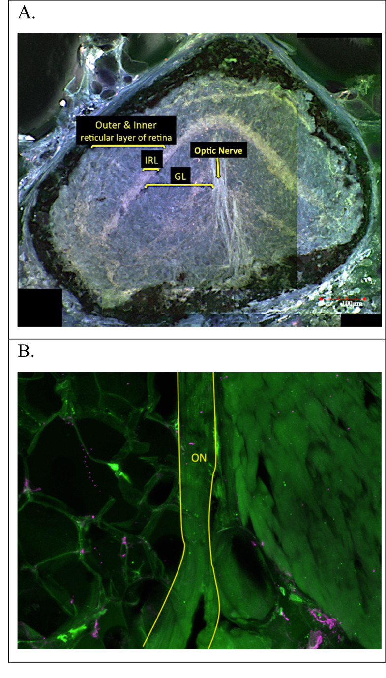
Figure 4. Adult E. rathbuni ocular sections.
Sections showing undifferentiated tissue layers surrounded by pigment epithelium (A). The optic nerve is attached to the posterior region of the vestigial eye (A) and can also be seen at higher magnification and outlined in yellow (B). Sections are labeled after Eigenmann (1900) as follows: optic nerve (ON), ganglion layer (GL), inner reticular layer (IRL). outer and inner reticular layer of the retina.

