Figure 2:

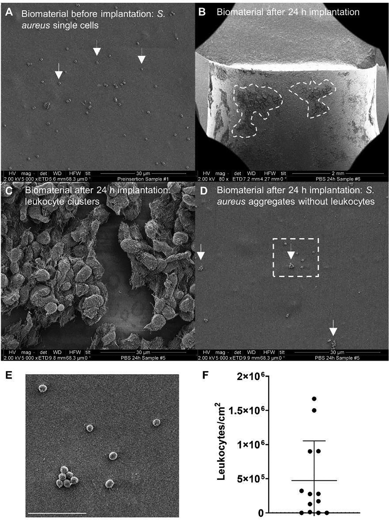
Immune cell recruitment to an inoculated peritoneal implant is heterogeneous. (A) SEM micrographs showing adherent S. aureus cells on the surface of a silicone tube prior to implantation into the mouse peritoneum. (B-D) Representative SEM micrographs from silicone tubes removed from the mouse peritoneal cavity after 24 h. (B) At low (80x) magnification, distinct areas of significant immune cell recruitment can be observed as dark patches on the implant surface (highlighted by dashed lines). (C) Higher magnification (5000x) reveals the dark patches outlined in (B) to be leukocyte clusters. (D) Higher magnification (5000x) reveals sections of the implant devoid of leukocytes. (E) Inset of the region denoted by the white box in (D). Scale bar = 5 μm. (F) Estimated leukocyte numbers on the implant surface from SEM images, normalized to FOV area (n=13 FOVs).
