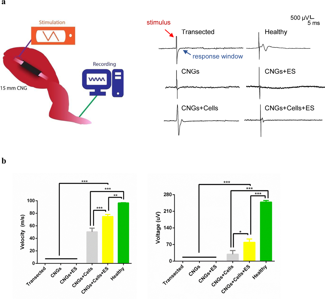
Fig. 5.
The electrophysiological analysis demonstrates functional recovery and reinnervation of regenerated sciatic nerves with their affected muscles. (a) A schematic showing the proximal stimulation of the sciatic nerve and recording of the compound action potential from the paw 12 weeks after CNG implantation (left panel). Following proximal stimulation, compound action potential was observed in the following groups: healthy control (healthy), unstimulated hNPC-containing CNGs (CNGs+Cells), and stimulated hNPC-containing CNGs (CNGs+Cells+ES). The transected control (Transected), unstimulated (CNGs) and stimulated CNGs (CNGs+ES) did not exhibit any signals following proximal stimulation (right panel). (b) The conduction velocity measured from the electrophysiology showed stimulated hNPC-containing CNGs (CNGs+Cells+ES) with the fastest nerve speed of the experimental groups in response to stimulation (left panel). The stimulated hNPC-containing CNGs (CNGs+Cells+ES) also showed increased amplitude from compound action potential following proximal stimulation (right panel) (n=3, *p<0.05, **p<0.01, ***p<0.001). Data were analyzed by one-way ANOVA followed by Tukey’s test.

