Fig. 8.
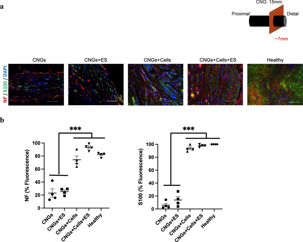
Both unstimulated and stimulated hNPC-containing CNGs demonstrates a high level of biomarkers: neurofilament (NF) for axons and S100 for Schwann cells. (a) Unstimulated (CNGs+Cells) and stimulated hNPC-containing CNGs (CNGs+Cells+ES) displayed more aligned axons (red) with Schwann cells (green) wrapping around the nuclei (DAPI: blue). The unstimulated (CNGs) and stimulated CNGs (CNGs+ES) presented fewer aligned axons with the scattered Schwann cell marker (scale bar: 50 μm). (b) Normalized fluorescent intensity showed a higher expression level of neurofilament and Schwann cells for unstimulated (CNGs+Cells) and stimulated hNPC-containing CNGs (CNGs+Cells+ES). (n=4, ***p<0.001) Data were analyzed by one-way ANOVA followed by Tukey’s test.

