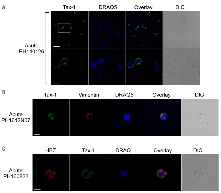
Figure 2.
Tax-1 subcellular localization in peripheral blood mononuclear cells of patients with acute adult T-cell leukemia-lymphoma. (A) Peripheral blood mononuclear cells (PBMC) of representative acute adult T-cell leukemia-lymphoma patients PH40126 (A) and PH1612N07 (B) were stained with the A51-2 anti-Tax-1 monoclonal antibody (mAb) followed by Alexa Fluor 488-conjugated goat-anti-mouse IgG2a antibody (green) and analyzed by confocal microscopy. Counterstaining of the nuclear or cytoplasmic compartments was performed by using DRAQ5 fluorescence probe to detect the nucleus (blue) and anti-vimentin rabbit polyclonal antibody followed by goat anti-rabbit IgG conjugated to Alexa Fluor 546 (B, red) to stain the cytoplasmic compartment. (C) PBMC of representative acute ATL patient PH160822 were costained with the 4D4-F3 anti-HBZ mAb followed by Alexa Fluor 546-conjugated goat anti-mouse IgG1 antibody (red) and with the A51-2 anti-Tax- 1 mAb followed by Alexa Fluor 488-conjugated goat-anti-mouse IgG2a antibody (green) and analyzed by confocal microscopy. DRAQ5 fluorescence probe was used to detect the nucleus (blue). DIC represents the differential interference contrast image. At least 300 cells were analyzed. All scale bars are 5mm.

