Severe acute respiratory syndrome coronavirus 2 (SARS-CoV-2) is a novel coronavirus that from December 2019 is spreading throughout the world causing a pandemic of Coronavirus Disease 2019 (COVID-19).1 COVID-19 is characterized by severe activation of inflammatory response that is thought to be a major cause of disease severity and death. Immune response to COVID-19 infection is constituted by two steps: during the phase of incubation, an adaptive immune response is able to control virus proliferation preventing disease progression. In the later phase, an uncontrolled inflammatory response could determine major clinical complications. This response, known as cytokine storm, is induced by the activation of T cells, natural killer (NK) cells, monocytes/ macrophage, neutrophils and any cells that run into the virus itself or degrade viral products. During the cytokine storm TNF-α, IFN-γ, IL-1b, IL-6, IL-12 and IL-17 are of crucial importance.2 Given the aberrant response of immune cells to viral infection, acute respiratory distress syndrome appears to be the most acute and fatal event of this disease. It’s evident that COVID-19 poses several challenges to the management of patients with hematologic malignancies since patients appear to be vulnerable to COVID-19.3 Chronic lymphocytic leukemia (CLL) is the most common leukemia in Western countries that mainly affects older people. CLL is characterized by several clinical complications related to alterations in the immune system. Predisposition to infections in CLL patients includes both immunodeficiency related to the leukemia itself and the results of cumulative immunosuppression caused by treatments. Given these evidences CLL patients might represent a high-risk group for SARSCoV- 2 infection and the outcome of patients needed admission is poor with high mortality rate.4 As supposed by different studies, BTK inhibitors (BTKi), in particular ibrutinib, may have a possible protective effect against lung injury in patients with COVID-19, tempering the hyper-inflammatory state.5,6 Some different retrospective studies, in particular an observational study conducted in Europe identified treatment with ibrutinib or BTKi as a factor associated with better outcome during COVID-19, with a shorter hospitalization of CLL patients. On the contrary, a similar study conducted in US did not confirm this observation. Of importance, BTKi treatment was interrupted at the onset of COVID-19 in the majority of cases.4,7 On this line, even though off-label administration of acalabrutinib in patients with severe COVID-19 had demonstrated reduction of the host inflammatory immune response and improved clinical outcomes, the CALAVI phase II trial for acalabrutinib in patients hospitalized with respiratory symptoms did not meet the primary efficacy endpoint of increasing the proportion of patients who remained alive and free of respiratory failure (https://www.astrazeneca.com/media-centre/press-releases/ 2020/update-on-calavi-phase-ii-trials-for-calquence-inpatients- hospitalised-with respiratory-symptoms-of-covid- 19.html).8 Ibrutinib exerts an immunomodulatory action, both on innate and adaptive immunity, sustaining a Th1 polarization and, at the same time, an anti-inflammatory polarization of macrophages.9,10
In the present study, we examined the modulation of ibrutinib treatment on the cytokine release by T cells and monocytes in CLL patients during infection with SARSCoV- 2.
Blood samples from patients that matched standard diagnostic criteria for CLL were obtained from the Hematology Unit of Modena Hospital, Italy with a protocol approved by the local Institutional Review Board and adhered to the tenets of the Declaration of Helsinki. Peripheral blood mononuclear cells (PBMC) were isolated and used fresh or cryopreserved until use. This study was performed on samples isolated from CLL patients who have not experienced SARS-CoV-2 infection. PBMC isolated from CLL patients were treated with ibrutinib or vehicle then stimulated with SARS-CoV-2 Peptide Pools Protein S, S1, S+, N and M (Miltenyi Biotech) and analyzed using cytokine secretion assay (CSA) for TNF-α and IFN-γ (Miltenyi Biotech). In order to identify the monocytic and T-cell population, PBMC were stained with CD14 and CD3 antibody respectively. For the ex vivo analysis, PBMC from CLL patients before and after 3 months of ibrutinib therapy were collected and stored in liquid nitrogen. PBMC were stimulated with SARS-CoV-2 Peptide Pools Protein and monocytes or T cells were identified by staining with CD14 or CD3. Secretion of TNF-α and IFN-γ was analyzed by CSA. Conditioned media were collected by centrifugation at 1,600 rpm for 10 minutes and stored at -20°C before being assayed. The levels of IL-6 were determined by human enzyme linked immunosorbent assay kit high sensitivity (Abcam). All data are presented as mean ± standard error of the mean. The t-test or Wilcoxon matched-pair signed rank test were used to determine statistical significance (GraphPad v6, GraphPad Software Inc).
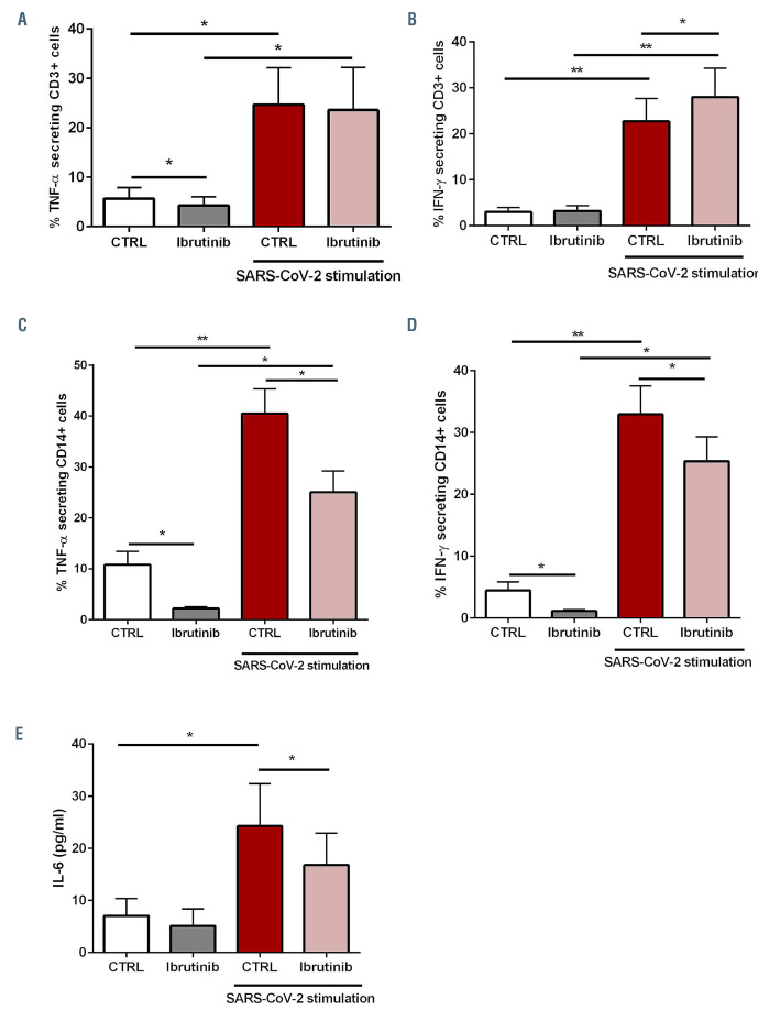
CLL is typically characterized by perturbations of the immune system, involving both innate and adaptive immune responses. Firstly, we examined the impact of SARS-CoV-2 infection in vitro on the adaptive and innate immunity in CLL collected from treatment-naϊve patients. As shown in Figure 1, following stimulation with SARS-CoV-2 peptides, we measured a strong and significant release of pro-inflammatory cytokines both by CD3+ T cells and CD14+ monocytes characterized by significant increase of TNF-α and IFN-γ secretion (*P<0.05; **P<0.01). Then, we analyzed if treatment with ibrutinib can modify the immunological response during infection. Ibrutinib targets T cells by irreversibly binding Tec family kinase, ITK, skewing toward Th1 phenotype and affecting Th2 immunity.9 ITK is critically involved in T-cellmediated immune response and depletion of its expression impairs influenza virus replication.11 As shown in Figure 1A and B and in the Online Supplementary Figure S1 ibrutinib did not determine a significant alteration in TNF-α secretion either in presence or absence of stimulation with SARS-CoV-2 peptides (P=not significant [ns]), in addition secretion of IFN-γ was not altered by ibrutinib and after stimulation we detected a slight increase (*P<0.05). Ibrutinib targets BTK in CLL-derived macrophages (nurse-like cells [NLC]) potentiating its immunosuppressive M2-skewed features.10 BTK inhibition hampered the inflammatory response of NLC during A. fumigatus infection.12 According to this evidence, we aimed to characterize the response of CLL monocytes to SARS-CoV-2 in the presence of ibrutinib. As reported in Figure 1C and D and in the Online Supplementary Figure S1, ibrutinib strongly impaired the release of TNF-α and IFN-γ by CLL monocytes induced by SARS-CoV-2 (*P<0.05). IL-6 is one of the key mediators of inflammation and cytokine storm in COVID-19 patients. Therefore, we measured the amount of secreted IL-6 in conditioned media collected after stimulation with sSARS-CoV-2 peptides either in the presence or absence of ibrutinib treatment. As shown in Figure 1E, SARSCoV- 2 caused an increased release of IL-6 in the conditioned media (*P<0.05) that was significantly mitigated by BTK inhibition (*P<0.05).
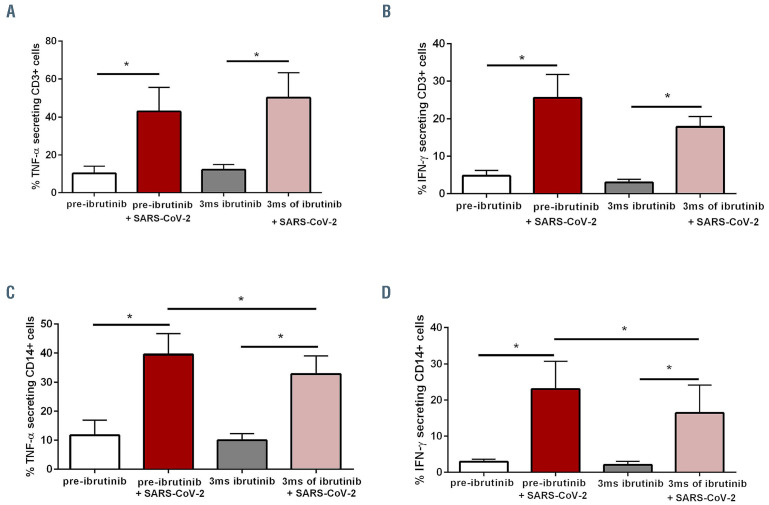
Furthermore, we planned to analyze ex vivo matched samples isolated from CLL patients before treatment and after 3 months of treatment with ibrutinib comparing the response of CD3+ and CD14+ cells to SARS-CoV-2. Our data did not show any significant modification in proinflammatory cytokines release by CD3 during treatment with ibrutinib (Figure 2A and B; Online Supplementary Figure S2; P=ns). On the contrary, the secretion of TNF-α**********and IFN-γ by monocytes measured during the first 3 months of therapy was significantly reduced as compared to pre-treatment condition (Figure 2C and D; Online Supplementary Figure S2; *P<0.05).
Ibrutinib treatment continuation in CLL patients affected by COVID-19 is feeding an important clinical and scientific debate about the opportunity to maintain or discontinue treatment in the presence of SARS-CoV-2 infection. In this scenario, some concerns are related to the evidence that an immunosuppressive activity of ibrutinib has been related to the occurrence of early-onset invasive fungal infections in treated-patients. For this reason, the choice of ibrutinib discontinuation may be related to a potentially increased risk of secondary infections and impaired humoral immunity.13 Our results demonstrate how ibrutinib may have protective effects on COVID-19 in CLL patients by limiting the cytokine storm preventing lung injury. On one hand, ibrutinib may influence T-cell function, by skewing T cells from a Th2-dominant to a Th1 and CD8+ cytotoxic population promoting Th1 immunity.9 On the other hand, BTK is involved in the regulation of macrophage lineage commitment towards M1 polarization and ibrutinib is able to skew towards an immunosuppressive phenotype also in response to proinflammatory stimuli as fungal infection.12 Inhibition of BTK, expressed by myeloid immune cells, affects the induction of inflammatory cytokines through the NFkB pathway and in mice intratracheal injection of BTK small interfering RNA confers potent protection against sepsisinduced acute lung injury with significant reduction of pulmonary edema, inflammatory cytokines and neutrophil infiltration in the lung tissues.14 In the setting of influenza A infection, ibrutinib mitigates inflammation and improves resolution of infection.15 Our results seem to strongly support a protective action of ibrutinib mitigating the feared cytokine storm sustaining the suggestion that there are no evident reasons to prematurely interrupt ibrutinib in these patients as recently suggested.13 In the light of these observations, since ibrutinib seems to not worsen COVID-19 specific immune response, our study provides a biological rationale for continuation of BTKi in CLL patients who develop COVID-19. Noteworthy, a better overview on the potential use of BTKi against COVID-19 is under evaluation in clinical trials (clinicaltrials.gov Identifier: NCT04439006, NCT04375397) conducted in patients not affected by B-cell malignancies.
Figure 1.
Immunomodulatory modifications induced by ibrutinib in chronic lymphocytic leukemia T cells and monocytes during infection by SARS-CoV-2. Chronic lymphocytic leukemia (CLL) peripheral blood mononuclear cells (PBMC) were isolated, treated with ibrutinib 1 mM for 90 minutes and then stimulated with severe acute respiratory syndrome coronavirus 2 (SARS-CoV-2) peptides (1 mg/mL) for additional 6 hours. TNF-α and IFN-γ secretion levels have been determined by cytokine secretion assay kit gating CD3+ and CD14+ populations by flow cytometry. (A and B) Bar diagrams show the percentage of TNF-α and IFN-γ**********secretion by T cells either in the presence or absence of stimulation by SARS-CoV-2 peptides (n=7; *P<0.05,**P<0.01). (C and D) Bar diagrams show the percentage of TNF-α and IFN-γ secretion by monocytes either in the presence or absence of stimulation by SARS-CoV-2 (n=7; *P<0.05; **P<0.01). (E) Conditioned media were collected for human enzyme linked immunosorbent assay determination of IL-6. Bar diagrams represent the mean protein concentration measured in 11 separated experiments. Values presented are in pg/mL (n=11, *P<0.05).
Figure 2.
Immunological alterations in chronic lymphocytic leukemia patients under ibrutinib therapy during infection by SARS-CoV-2. Chronic lymphocytic leukemia (CLL) peripheral blood mononuclear cells (PBMC) were isolated from CLL patients before and after 3 months of treatment with ibrutinib and stimulated with severe acute respiratory syndrome coronavirus 2 (SARS-CoV-2) peptides (1 mg/ml) for 6 hours. TNF-α and IFN-γ secretion levels have been determined by cytokine secretion assay kit gating CD3+ and CD14+ populations by flow cytometry. (A and B) Bar diagrams show the percentage of TNF-α and IFN-γ secretion by T cells either in the presence or absence of stimulation by SARS-CoV-2 (n=7; *P<0.05). (C and D) Bar diagrams show the percentage of TNF-α and IFN-γ**********secretion by monocytes either in the presence or absence of stimulation by SARS-CoV-2 (n=7; *P<0.05).
Supplementary Material
Funding Statement
Funding: this work was supported by Associazione Italiana per la Ricerca sul Cancro (IG21436 RM), Progetto Dipartimenti di Eccellenza 2018-2022. SF is supported by Fondazione Umberto Veronesi, Milan, Italy. The funding bodies had no role in study design, data collection and analysis, decision to publish or preparation of the manuscript.
References
- 1.Zhou P, Yang X-L, Wang X-G, et al. A pneumonia outbreak associated with a new coronavirus of probable bat origin. Nature. 2020; 579(7798):270-273. [DOI] [PMC free article] [PubMed] [Google Scholar]
- 2.Riva G, Nasillo V, Tagliafico E, Trenti T, Comoli P, Luppi M. COVID- 19: more than a cytokine storm. Crit Care. 2020;24(1):549. [DOI] [PMC free article] [PubMed] [Google Scholar]
- 3.Passamonti F, Cattaneo C, Arcaini L, et al. Clinical characteristics and risk factors associated with COVID-19 severity in patients with haematological malignancies in Italy: a retrospective, multicentre, cohort study. Lancet Haematol. 2020;7(10):e737-e745. [DOI] [PMC free article] [PubMed] [Google Scholar]
- 4.Mato AR, Roeker LE, Lamanna N, et al. Outcomes of COVID-19 in patients with CLL: a multicenter international experience. Blood. 2020;136(10):1134-1143. [DOI] [PMC free article] [PubMed] [Google Scholar]
- 5.Treon SP, Castillo JJ, Skarbnik AP, et al. The BTK inhibitor ibrutinib may protect against pulmonary injury in COVID-19-infected patients. Blood. 2020;135(21):1912-1915. [DOI] [PMC free article] [PubMed] [Google Scholar]
- 6.Thibaud S, Tremblay D, Bhalla S, Zimmerman B, Sigel K, Gabrilove J. Protective role of Bruton tyrosine kinase inhibitors in patients with chronic lymphocytic leukaemia and COVID-19. Br J Haematol. 2020;190(2):e73-e76. [DOI] [PMC free article] [PubMed] [Google Scholar]
- 7.Scarfò L, Chatzikonstantinou T, Rigolin GM, et al. COVID-19 severity and mortality in patients with chronic lymphocytic leukemia: a joint study by ERIC, the European Research Initiative on CLL, and CLL Campus. Leukemia. 2020;34(9):2354-2363. [DOI] [PMC free article] [PubMed] [Google Scholar]
- 8.Roschewski M, Lionakis MS, Sharman JP, et al. Inhibition of Bruton tyrosine kinase in patients with severe COVID-19. Science Immunol. 2020;5(48):eabd0110. [DOI] [PMC free article] [PubMed] [Google Scholar]
- 9.Dubovsky JA, Beckwith KA, Natarajan G, et al. Ibrutinib is an irreversible molecular inhibitor of ITK driving a Th1-selective pressure in T lymphocytes. Blood. 2013;122(15):2539-2549. [DOI] [PMC free article] [PubMed] [Google Scholar]
- 10.Fiorcari S, Maffei R, Audrito V, et al. Ibrutinib modifies the function of monocyte/macrophage population in chronic lymphocytic leukemia. Oncotarget. 2016;7(40):65968–65981. [DOI] [PMC free article] [PubMed] [Google Scholar]
- 11.Fan K, Jia Y, Wang S, et al. Role of Itk signalling in the interaction between influenza A virus and T-cells. J Gen Virol. 2012;93(Pt 5):987-997. [DOI] [PubMed] [Google Scholar]
- 12.Fiorcari S, Maffei R, Vallerini D, et al. BTK inhibition impairs the innate response against fungal infection in patients with chronic lymphocytic leukemia. Front Immunol. 2020;11:2158. [DOI] [PMC free article] [PubMed] [Google Scholar]
- 13.Chong EA, Roeker LE, Shadman M, Davids MS, Schuster SJ, Mato AR. BTK inhibitors in cancer patients with COVID-19: “The winner will be the one who controls that chaos” (Napoleon Bonaparte). Clin Cancer Res. 2020;26(14):3514-3516. [DOI] [PMC free article] [PubMed] [Google Scholar]
- 14.Zhou P, Ma B, Xu S, et al. Knockdown of Burton’s tyrosine kinase confers potent protection against sepsis-induced acute lung injury. Cell Biochem Biophys. 2014;70(2):1265-1275. [DOI] [PubMed] [Google Scholar]
- 15.Florence JM, Krupa A, Booshehri LM, Davis SA, Matthay MA, Kurdowska AK. Inhibiting Bruton’s tyrosine kinase rescues mice from lethal influenza-induced acute lung injury. Am J Physiol Lung Cell Mol Physiol. 2018;315(1):L52-L58. [DOI] [PMC free article] [PubMed] [Google Scholar]
Associated Data
This section collects any data citations, data availability statements, or supplementary materials included in this article.


