. 2021 Aug 3;10:e67604. doi: 10.7554/eLife.67604

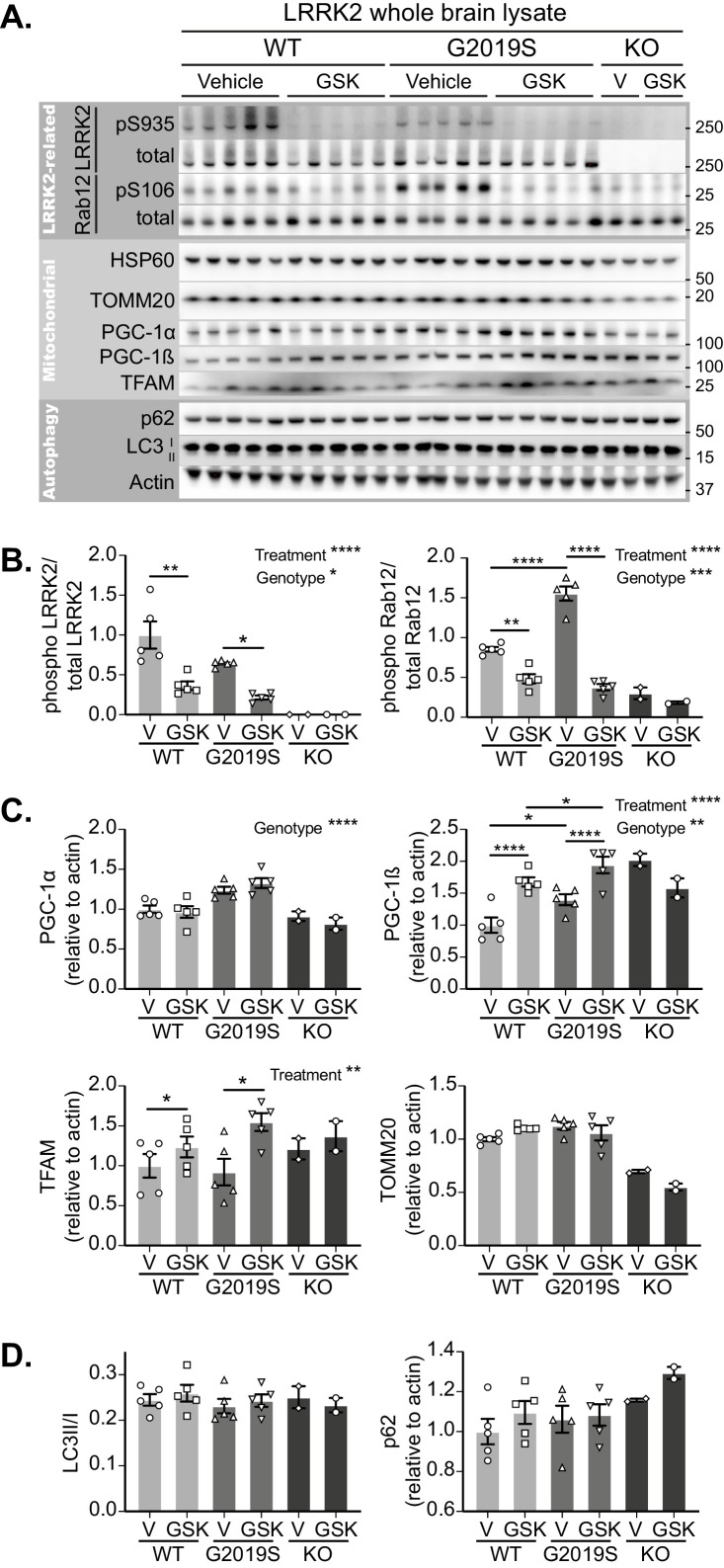
Figure 5. GSK3357679A treatment inhibits LRRK2 in the brain.

(A) Immunoblots of the indicated LRRK2-related, mitochondrial and autophagy proteins from brain lysates of LRRK2 WT, LRRK2 G2019S, and LRRK2 KO mito-QC mice treated with vehicle or GSK3357679A. (B) Quantitation of phosphorylation data from the LRRK2-related proteins displayed in A. (C) Quantitation of mitochondria- and mitochondrial biogenesis-related proteins displayed in A. (D) Quantitation of autophagy-related proteins displayed in A. Overall data is represented as mean +/- SEM. Statistical significance is displayed as *p<0.05, **p<0.01, ***p<0.001, and ****p<0.0001.

Figure 5—source data 1. Numerical data for Figure 5.
Figure 5—source data 2. Western blot raw data files for Figure 5A.

Figure 5.

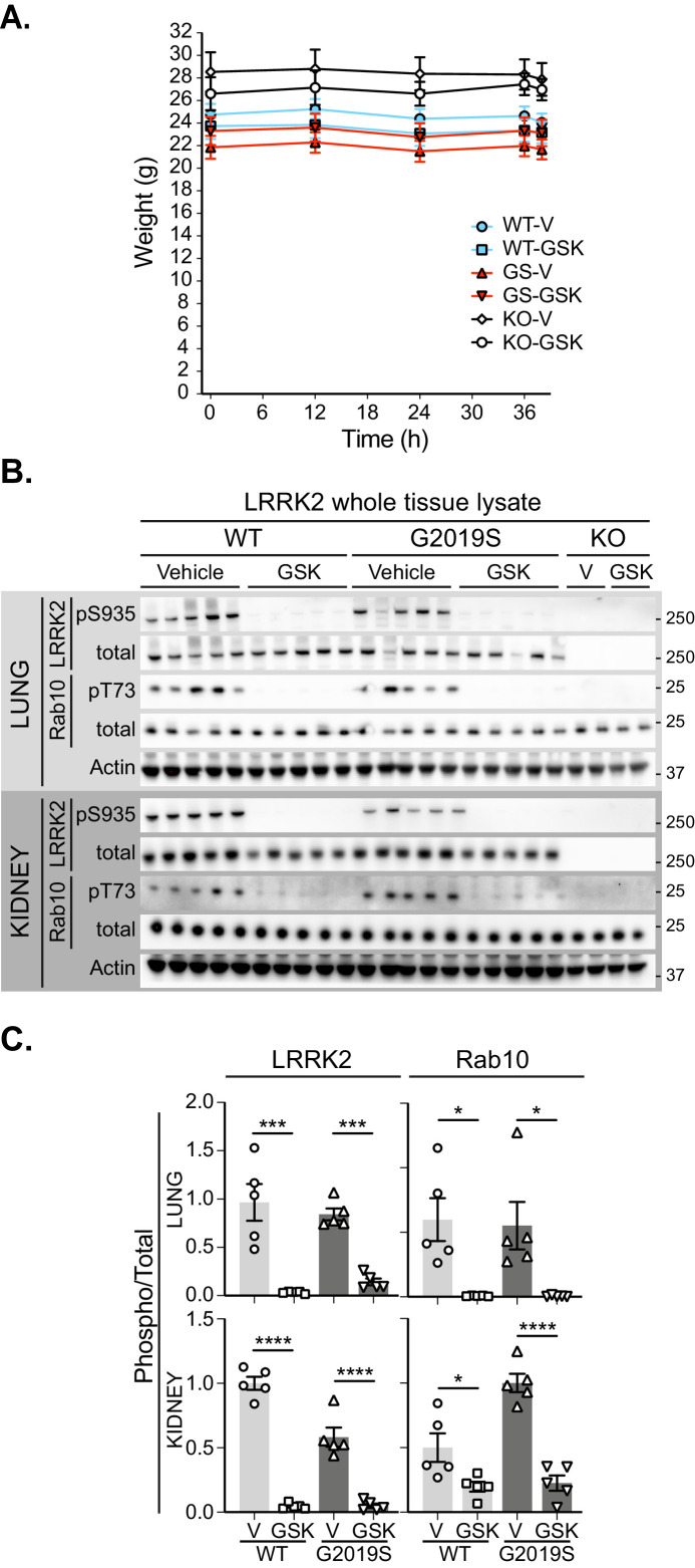
Figure 5—figure supplement 1. Pharmacological inhibition of LRRK2 kinase in the lung and kidney.

Figure 5—figure supplement 1.

(A) Body weights of mice treated over 36 hr with vehicle (V) or GSK3357679A (GSK). (B) Immunoblots of the indicated LRRK2-related, mitochondrial and autophagy proteins from lung and kidney lysates of LRRK2 WT, LRRK2 G2019S, and LRRK2 KO mito-QC mice treated with vehicle or GSK3357679A. (C) Quantitation of phosphorylation data from the LRRK2-related proteins displayed in B. *p<0.05, ***p<0.001, and ****p<0.0001.
Figure 5—figure supplement 1—source data 1. Numerical data for Figure 5—figure supplement 1.
Figure 5—figure supplement 1—source data 2. Western blot raw data files for Figure 5—figure supplement 1B.