Abstract
目的
比较背侧楔形截骨(dorsiflexion osteotomy,DO)及跖骨头置换(implant arthroplasty,IA)治疗晚期 Freiberg 病的疗效。
方法
回顾分析 2012 年 7 月—2016 年 7 月收治且符合选择标准的 25 例 Smillie Ⅳ、Ⅴ 型 Freiberg 病患者临床资料,其中 13 例行 DO 治疗(DO 组),12 例行 IA 治疗(IA 组)。两组患者性别、年龄、患足侧别、部位、Smillie 分型、病程以及术前疼痛视觉模拟评分(VAS)、患侧跖趾关节屈曲及背伸活动度、美国矫形足踝协会(AOFAS)评分比较,差异均无统计学意义(P>0.05)。比较两组患者住院费用及术后 VAS 评分、AOFAS 评分、患侧跖趾关节屈曲及背伸活动度,X 线片复查两组内固定或假体情况。
结果
两组术后切口均Ⅰ期愈合。患者均获随访,DO 组随访时间为 12~30 个月,平均 17 个月;IA 组为 12~24 个月,平均 16 个月。IA 组患者住院费用明显多于 DO 组(t=2.742,P=0.011)。末次随访时,两组 VAS 评分、AOFAS 评分、患侧跖趾关节屈曲及背伸活动度均较术前明显改善(P<0.05);两组间上述指标比较差异无统计学意义(P>0.05)。X 线片复查示,DO 组患者截骨均愈合,愈合时间为 8~12 周,平均 9.5 周;两组均无内固定及假体相关并发症发生。
结论
DO 及 IA 均可有效治疗晚期 Freiberg 病,明显缓解跖趾关节疼痛并改善活动度,但 DO 术式住院费用低于 IA。
Keywords: Freiberg 病, 跖骨头置换, 背侧楔形截骨, 跖趾关节
Abstract
Objective
To compare the dorsiflexion osteotomy (DO) and implant arthroplasty (IA) in terms of clinical and radiographic outcomes for patients with advaced Freiberg disease.
Methods
A clinical data of 25 cases of Freiberg disease, who were admitted between July 2012 and July 2016 and met selection criteria, was retrospectively reviewed. According to the Smillie classification, all patients were classified as stage Ⅳ-Ⅴ. Among them, 13 cases were treated with DO (DO group) and 12 cases were treated with IA (IA group). No significant difference was found between the two groups in gender, age, side of the affected metatarsophalangeal (MTP) joint, location, Smillie classification, disease duration, and preoperative visual analogue scale (VAS) score, range of motion of the affected MTP joints, and the American Orthopedic Foot and Ankle Society (AOFAS) score (P>0.05). Total costs for index admissions were compared between the two groups. Clinical outcomes were evaluated in accordance with the VAS score, AOFAS score, and the range of motion of the affected MTP joints.
Results
All incisions of the two groups healed by first intention. The follow-up time was 12-30 months (mean, 17 months) in DO group and 12-24 months (mean, 16 months) in IA group. The total cost of index admission was significantly higher in IA group than that n DO group (t=2.742, P=0.011). The AOFAS scores, VAS scores, and range of dorsiflexion and plantar flexion at last follow-up were significantly improved when compared with preoperative value in the two groups (P<0.05). There was no significant difference in all indexes between the two groups (P>0.05). X-ray film examination showed that the osteotomy healed within 8-12 weeks (mean, 9.5 weeks) after operation in DO group. None of the patients experienced internal fixator and implant related complications postoperatively.
Conclusion
DO and IA can provide significant improvement in pain and motion of the MTP joints for advanced Freiberg disease. But the DO may be the more economical method.
Keywords: Freiberg disease, implant arthroplasty, dorsiflexion osteotomy, metatarsophalangeal joint
Freiberg 病是指跖骨头骨软骨炎,主要临床表现为跖骨头处压痛、肿胀以及活动受限。临床常用 Smillie 分型标准对 Freiberg 病进行分型,根据跖骨头影像学表现将其分为 5 型。Ⅰ型为轻微软骨损伤,无明显影像学表现;Ⅱ型为跖骨头中央部软骨塌陷;Ⅲ型是在Ⅱ型基础上进一步进展,合并有内、外侧骨赘形成;Ⅳ型是指跖骨头中央塌陷,边缘不规则,伴游离体产生;Ⅴ 型为跖骨头明显变平变宽,关节间隙变窄,终末关节炎表现[1]。
Freiberg 病治疗方式包括保守治疗和手术治疗。保守治疗主要包括支具固定、限制关节活动、缓解疼痛,如保守治疗失败需考虑手术治疗。手术治疗大致分为保留关节与关节重建两种[2-3]。背侧楔形截骨(dorsiflexion osteotomy,DO)是一种经典的保留关节术式,跖骨头置换(implant arthroplasty,IA)是临床常用的关节重建方式,上述两种术式均可用于治疗 SmillieⅣ、Ⅴ 型 Freiberg 病[4-5]。但目前缺乏两种术式的比较研究。为此,我们回顾性分析了 2012 年 7 月—2016 年 7 月于我院接受 DO 或 IA 治疗的 Smillie Ⅳ、Ⅴ 型 Freiberg 病患者临床资料,比较两种术式疗效,以期为临床选择治疗方法提供参考。报告如下。
1. 临床资料
1.1. 患者选择标准
纳入标准:① Smillie Ⅳ、Ⅴ 型 Freiberg 病;② 2012 年 7 月—2016 年 7 月于我院接受 DO 或 IA 治疗;③ 患者均有长期主动活动跖趾关节时疼痛症状,负重行走时加重,且保守治疗无效;④ 患者无明显外伤史。排除标准:① 年龄>80 岁;② 存在严重基础疾病,不能耐受手术者。共 25 例患者符合选择标准纳入研究。术中根据软骨损伤情况,13 例行 DO 治疗(DO 组),12 例行 IA 治疗(IA 组)。本研究通过四川大学华西医院医学伦理委员会批准。
1.2. 一般资料
DO 组:男 2 例,女 11 例;年龄 16~31 岁,平均 23 岁。左足 9 例,右足 4 例。第 2 跖骨头 10 例,第 3 跖骨头 3 例。Smillie Ⅳ型 8 例,Ⅴ 型 5 例。病程 3~10 个月,平均 3.5 个月。
IA 组:男 1 例,女 11 例;年龄 19~42 岁,平均 29 岁。左足 9 例,右足 3 例。第 2 跖骨头 9 例,第 3 跖骨头 3 例。Smillie Ⅳ型 10 例,Ⅴ型 2 例。病程 3~8 个月,平均 4.2 个月。
两组患者性别、年龄、患足侧别、部位、Smillie 分型、病程以及术前疼痛视觉模拟评分(VAS)、患侧跖趾关节屈曲及背伸活动度、美国矫形足踝协会(AOFAS)评分比较,差异均无统计学意义(P>0.05)。见表 1、2。
表 1.
Comparison of VAS and AOFAS scores between the two groups at pre- and post-operation (
 )
)
两组患者手术前后 VAS 评分及 AOFAS 评分比较(
 )
)
| 组别
Group |
例数
n |
VAS 评分
VAS score |
AOFAS 评分
AOFAS score |
|||||
| 术前
Preoperative |
末次随访
Last follow-up |
统计值
Statistic |
术前
Preoperative |
末次随访
Last follow-up |
统计值
Statistic |
|||
| DO 组
DO group |
13 | 7.6±1.1 | 1.1±1.0 |
t=14.880
P= 0.000 |
47.4±8.6 | 79.7±5.5 |
t=10.570
P= 0.000 |
|
| IA 组
IA group |
12 | 7.9±1.0 | 1.0±0.9 |
t=17.104
P= 0.000 |
44.0±8.8 | 82.8±6.2 |
t=11.234
P= 0.000 |
|
| 统计值
Statistic |
t=0.891
P=0.382 |
t=1.012
P=0.322 |
t=0.969
P=0.342 |
t=1.338
P=0.194 |
||||
表 2.
Comparison of the range of motion of affected metatarsophalangeal joints between the two groups at pre- and post-operation (°,
 )
)
两组手术前后患侧跖趾关节活动度比较(°,
 )
)
| 组别
Group |
例数
n |
屈曲
Plantar flexion |
背伸
Dorsiflexion |
|||||
| 术前
Preoperative |
末次随访
Last follow-up |
统计值
Statistic |
术前
Preoperative |
末次随访
Last follow-up |
统计值
Statistic |
|||
| DO 组
DO group |
13 | 20.0±6.1 | 26.9±8.3 |
t=2.413
P=0.024 |
25.8±8.3 | 40.4±6.9 |
t=4.958
P=0.000 |
|
| IA 组
IA group |
12 | 25.4±5.8 | 41.3±9.1 |
t=3.880
P=0.000 |
19.0±5.2 | 27.1±5.0 |
t=4.846
P=0.000 |
|
| 统计值
Statistic |
t=0.443
P=0.662 |
t=0.059
P=0.954 |
t=0.125
P=0.901 |
t=0.266
P=0.792 |
||||
1.3. 手术方法
两组手术均由同一高年资术者完成。全麻后,在患侧大腿近端放置止血带,压力为 30 kPa。在患侧跖趾关节背侧作一长约 3 cm 切口,切开跖趾关节囊,清理病灶,包括关节内游离体和增生滑膜,暴露病变关节面。
DO 组:从跖骨头远端背侧向近端跖侧行楔形截骨,截骨大小根据跖骨头损伤面积决定,截骨完成后去除部分截骨块,将远端部分向近端及背侧翻转,完成翻转后利用可折断钉固定并进行埋头处理。生理盐水冲洗,逐层关闭切口。
IA 组:参考 Swanson 描述的手术过程进行假体置换[6]。根据跖骨头大小截取合适长度的跖骨头,利用扩髓钻进行扩大髓腔操作,完成关节间隙准备后,放入试模活动跖趾关节,选定合适大小的 Swanson 假体并植入关节间隙。生理盐水冲洗,逐层关闭切口。
1.4. 术后处理
DO 组患者以超跖趾关节的踝关节中立位石膏固定 4 周。拆除石膏后,在前足减压鞋保护下部分负重行走,主动活动足趾关节;6~8 周时影像学复查骨愈合情况,根据复查结果逐渐完全负重行走。IA 组患者术后视切口情况,早期进行跖趾关节屈伸锻炼,1 周后穿前足减压鞋负重行走,6~8 周时逐渐过渡至穿旅游鞋负重行走。所有患者术后 2 周视切口愈合情况拆除缝线,并在康复师指导下进行足趾关节康复锻炼。
1.5. 疗效评价指标
记录两组患者住院费用。采用 VAS 评分[7]评价主动活动跖趾关节时疼痛程度,测量患侧跖趾关节屈伸活动度,利用 AOFAS 评分[8]评价关节功能。随访时复查 X 线片,观察 DO 组截骨愈合情况。
1.6. 统计学方法
采用 Excel 软件进行统计分析。数据以均数±标准差表示,组间比较采用独立样本 t 检验,组内手术前后比较采用配对 t 检验;检验水准 α=0.05。
2. 结果
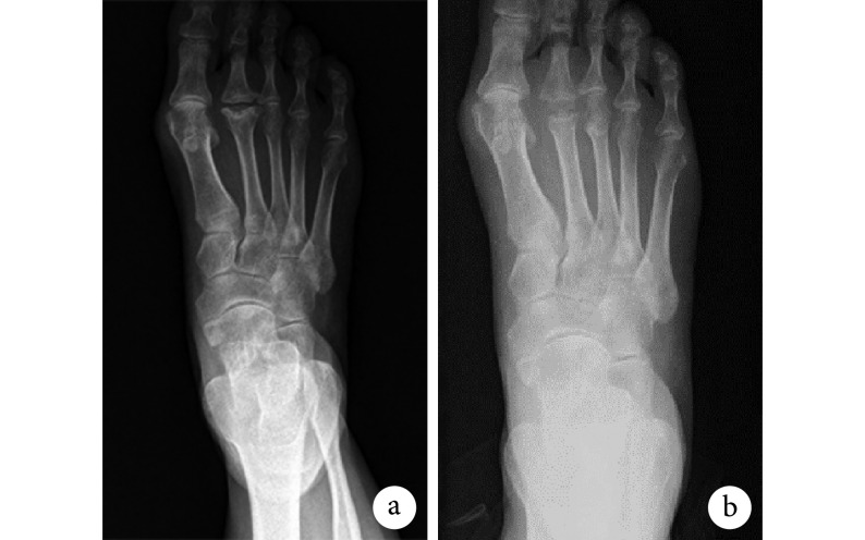
两组患者术后切口均Ⅰ期愈合。患者均获随访,DO 组随访时间为 12~30 个月,平均 17 个月;IA 组为 12~24 个月,平均 16 个月。IA 组患者住院费用为(19 141.9±1 273.3)元,DO 组为(14 230.8±1 609.5)元,差异有统计学意义(t=2.742,P=0.011)。所有患者疼痛症状明显改善,恢复正常生活。末次随访时,两组 VAS 评分、AOFAS 评分、患侧跖趾关节屈曲及背伸活动度均较术前明显改善,差异有统计学意义(P<0.05);两组间上述指标比较差异无统计学意义(P>0.05)。见表 1、2。X 线片复查示,DO 组患者截骨均愈合,无截骨不愈合或畸形愈合发生,愈合时间为 8~12 周,平均 9.5 周;末次随访时内固定在位,关节对位良好,无内固定失效迹象;未见明显跖骨头塌陷和缺血坏死。IA 组假体位置良好,无假体松动和脱落迹象。见图 1、2。
图 1.
X-ray films of a 21-year-old female patient with the 2nd metatarsophalangeal Freiberg disease (Smillie stage Ⅳ) in DO group
DO 组患者,女,21 岁,第 2 跖骨头 Freiberg 病(Smillie Ⅳ型)X 线片
a. 术前;b. 术后 12 个月
a. Before operation; b. At 12 months after operation
图 2.
X-ray films of a 50-year-old female patient with the 2nd metatarsophalangeal Freiberg disease (Smillie stage Ⅴ) in IA group
IA 组患者,女,50 岁,第 2 跖骨头(Smillie Ⅴ 型)Freiberg 病 X 线片
a. 术前;b. 术后 12 个月
a. Before operation; b. At 12 months after operation
3. 讨论
Freiberg 病好发于成年女性,男女比例为 1∶5[1],本研究患者也以女性为主。该病确切病因尚不明确,研究已证实创伤、供血不足、基因以及生物力学因素与该病发生有关[5],本研究患者均无明显外伤史。第 2 跖骨头是最易受累关节,约占所有病例的 68%,第 3、4 跖骨头的受累概率分别为 27% 和 3%[9];双侧受累概率为 6.6%[2]。本研究纳入的 25 例患者中,19 例为第 2 跖骨头受累。目前对于第 2 跖骨头为好发部位的原因尚不明确。有研究认为,第 2 跖骨相对长于其他跖骨,在负重时所承受的力最大,因此软骨下骨血供易受损,进而导致软骨坏死塌陷伴滑膜炎,持续的滑膜炎将导致关节肿胀、活动受限,最终引起跖骨头处骨软骨分离[9-10]。因此长期慢性跖趾关节疼痛是诊断该病的重要依据。
DO 是保留关节术式,术中将足跖侧完整关节面向后、向近端翻转来代偿损伤的关节面[11],已有大量文献报道该术式治疗 Freiberg 病可获得满意效果[12-14]。本研究结果显示,DO 组患者末次随访时 AOFAS 评分、患侧跖趾关节屈伸活动度均较术前明显改善。有研究认为该术式可以尽可能保留跖骨头血供,避免软骨塌陷,维持跖骨长度[15]。本研究中 DO 组患者术后均未见明显跖骨头塌陷和缺血坏死。
Swanson 等[6]最早报道采用硅胶胶体行 IA。目前关于硅胶胶体置换术后效果的报道结果存在较大差异,有文献报道患者术后功能长期改善,但也有文献报道该术式存在早期失败风险[16]。 温建民等[17]利用 Swanson 假体治疗 13 例 Freiberg 病患者,平均随访 11.3 个月,AOFAS 评分达(77.5±4.99)分。本研究获得了相似结果。IA 作为一种有效的治疗手段,不仅能缓解患者疼痛症状,还能改善患者活动度。一项纳入 26 例ル趾僵硬患者的研究发现,经 Swanson 假体置换后,末次随访时患者跖趾关节平均屈伸活动度>30°[16]。本研究置换术后患者的活动度较术前明显改善。值得注意的是,虽然 IA 术后跖趾关节活动度较术前明显改善,但不建议患者进行大范围活动,以避免假体脱位,影响假体寿命。
有研究发现,Swanson 假体置换术后存在假体脱位、转移性跖骨痛、继发性滑膜炎以及感染等风险[18-19]。Shankar 等[20]通过对 40 例患者进行平均 110 个月随访,发现 36% 患者末次随访时合并假体并发症,其中最常见并发症为假体断裂失效。另有研究发现假体硅胶材料的磨损颗粒是引起患者关节内肉芽肿性炎性反应的主要原因,最终导致假体周围骨质丢失,造成假体失败[21]。研究认为 IA 术后相关并发症多出现在置换后 2 年[20-23],本研究中 IA 组末次随访时患者均无相关并发症,但平均随访时间不足 2 年,远期疗效有待进一步观察。
本研究结果显示,DO 组与 IA 组患者术后关节功能以及疼痛缓解程度均相似,但 IA 组住院费用明显高于 DO 组。Silva 等[16]研究发现与其他手术方式(如关节融合等)相比,IA 治疗平均住院费用高出 1 000 欧元;而且 IA 术者学习曲线更长。
综上述,DO 及 IA 均能明显改善晚期 Freiberg 病患者跖趾关节疼痛及活动度,但前者治疗费用低于后者。此外,选择术式前应进行详细术前评估(年龄、生活质量等),并结合患者个体情况确定最适合术式,以期获得满意疗效[24]。
Funding Statement
四川省科技计划项目(2019YFS0270);建设世界一流大学专项师资队伍建设项目(2040204401005)
Supported by Sichuan Science and Technology Program (2019YFS0270); World-class University Construction Program (2040204401005)
References
- 1.Smillie IS Treatment of Freiberg’s infraction. Proc R Soc Med. 1967;60(1):29–31. doi: 10.1177/003591576706000117. [DOI] [PMC free article] [PubMed] [Google Scholar]
- 2.Carmont MR, Rees RJ, Blundell CM Current concepts review: Freiberg’s disease. Foot Ankle Int. 2009;30(2):167–176. doi: 10.3113/FAI-2009-0167. [DOI] [PubMed] [Google Scholar]
- 3.Schade VL Surgical management of Freiberg’s infraction: A systematic review. Foot Ankle Spec. 2015;8(6):498–519. doi: 10.1177/1938640015585966. [DOI] [PubMed] [Google Scholar]
- 4.Biz C, Zornetta A, Fantoni I, et al Freiberg’s infraction: A modified closing wedge osteotomy for an undiagnosed case. Int J Surg Case Rep. 2017;38:8–12. doi: 10.1016/j.ijscr.2017.07.013. [DOI] [PMC free article] [PubMed] [Google Scholar]
- 5.Talusan PG, Diaz-Collado PJ, Reach JS Jr Freiberg’s infraction: diagnosis and treatment. Foot Ankle Spec. 2014;7(1):52–56. doi: 10.1177/1938640013510314. [DOI] [PubMed] [Google Scholar]
- 6.Swanson AB, Biddulph SL, Hagert CG A silicone rubber implant to supplement the Keller toe arthroplasty. NY Univ Inter Clin Inform Bull. 1971;10:7–14. [Google Scholar]
- 7.Huskisson EC Measurement of pain. Lancet. 1974;2(7889):1127–1131. doi: 10.1016/s0140-6736(74)90884-8. [DOI] [PubMed] [Google Scholar]
- 8.Kitaoka HB, Alexander IJ, Adelaar RS, et al Clinical rating systems for the ankle-hindfoot, midfoot, hallux, and lesser toes. Foot Ankle Int. 1994;15(7):349–353. doi: 10.1177/107110079401500701. [DOI] [PubMed] [Google Scholar]
- 9.Cerrato RA Freiberg’s disease. Foot Ankle Clin. 2011;16(4):647–658. doi: 10.1016/j.fcl.2011.08.008. [DOI] [PubMed] [Google Scholar]
- 10.Ihedioha U, Sinha S, Campbell AC Surgery for symptomatic Freiberg’s disease: excision arthroplasty in eight patients. Foot. 2003;13(3):143–145. doi: 10.1016/S0958-2592(03)00030-0. [DOI] [Google Scholar]
- 11.Lee HS, Kim YC, Choi JH, et al Weil and dorsal closing wedge osteotomy for Freiberg’s disease. J Am Podiatr Med Assoc. 2016;106(2):100–108. doi: 10.7547/14-065. [DOI] [PubMed] [Google Scholar]
- 12.Özkul E, Gem M, Alemdar C, et al Results of two different surgical techniques in the treatment of advanced-stage Freiberg’s disease. Indian J Orthop. 2016;50(1):70–73. doi: 10.4103/0019-5413.173514. [DOI] [PMC free article] [PubMed] [Google Scholar]
- 13.Pereira BS, Frada T, Freitas D, et al Long-term follow-up of dorsal wedge osteotomy for pediatric Freiberg disease. Foot Ankle Int. 2016;37(1):90–95. doi: 10.1177/1071100715598602. [DOI] [PubMed] [Google Scholar]
- 14.Chao KH, Lee CH, Lin LC Surgery for symptomatic Freiberg’s disease: extra articular dorsal closing-wedge osteotomy in 13 patients followed for 2-4 years. Acta Orthop Scand. 1999;70(5):483–486. doi: 10.3109/17453679909000985. [DOI] [PubMed] [Google Scholar]
- 15.Petersen WJ, Lankes JM, Paulsen F, et al The arterial supply of the lesser metatarsal heads: a vascular injection study in human cadavers. Foot Ankle Int. 2002;23(6):491–495. doi: 10.1177/107110070202300604. [DOI] [PubMed] [Google Scholar]
- 16.Silva LF, Sousa CV, Rodrigues Pinto R, et al Preliminary results from the metis-newdeal(®) total metatarsophalangeal prosthesis. Rev Bras Ortop. 2015;46(2):200–204. doi: 10.1016/S2255-4971(15)30240-8. [DOI] [PMC free article] [PubMed] [Google Scholar]
- 17.温建民, 孙卫东, 桑志成, 等 Swanson 人工跖趾关节置换治疗 Freiberg 病近期疗效观察. 中国骨伤. 2009;11(6):423–425. [Google Scholar]
- 18.Shih AT, Quint RE, Armstrong DG, et al Treatment of Freiberg's infraction with the titanium hemi-implant. J Am Podiatr Med Assoc. 2004;94(6):590–593. doi: 10.7547/0940590. [DOI] [PubMed] [Google Scholar]
- 19.Squitieri L, Chung KC A systematic review of outcomes and complications of vascularized toe joint transfer, silicone arthroplasty, and PyroCarbon arthroplasty for posttraumatic joint reconstruction of the finger. Plast Reconstr Surg. 2008;121(5):1697–1707. doi: 10.1097/PRS.0b013e31816aa0b3. [DOI] [PubMed] [Google Scholar]
- 20.Shankar NS Silastic single-stem implants in the treatment of hallux rigidus. Foot Ankle Int. 1995;16(8):487–491. doi: 10.1177/107110079501600805. [DOI] [PubMed] [Google Scholar]
- 21.Moeckel BH, Sculco TP, Alexiades MM, et al The double-stemmed silicone-rubber implant for rheumatoid arthritis of the first metatarsophalangeal joint. Long-term results. J Bone Joint Surg (Am) 1992;74(4):564–570. doi: 10.2106/00004623-199274040-00012. [DOI] [PubMed] [Google Scholar]
- 22.Bales JG, Wall LB, Stern PJ Long-term results of Swanson silicone arthroplasty for proximal interphalangeal joint osteoarthritis. J Hand Surg (Am) 2014;39(3):455–461. doi: 10.1016/j.jhsa.2013.11.008. [DOI] [PubMed] [Google Scholar]
- 23.Sethu A, D’Netto DC, Ramakrishna B Swanson’s silastic implants in great toes. J Bone Joint Surg (Br) 1980;62-B(1):83–85. doi: 10.1302/0301-620X.62B1.7351441. [DOI] [PubMed] [Google Scholar]
- 24.朱敏, 冯凡哲 足踝外科新进展. 中国修复重建外科杂志. 2018;32(7):860–865. doi: 10.7507/1002-1892.201806032. [DOI] [PMC free article] [PubMed] [Google Scholar]


