Abstract
Background
Coronavirus disease 2019 (COVID-19) has caused more than 2 million deaths worldwide. Viral sepsis has been proposed as a description for severe COVID-19, and numerous therapies have been on trials based upon this hypothesis. However, whether the clinical characteristics of severe COVID-19 are similar to those of bacterial sepsis has not been elucidated.
Methods
We retrospectively compared the clinical data of non-surviving COVID-19 patients who were admitted to a 30-bed intensive care unit (ICU) in Wuhan Infectious Diseases Hospital (Wuhan, China) from 22 January 2020, to 28 February 2020, with those of non-surviving patients with bacterial sepsis who were admitted to the ICU in Zhongshan Hospital, Fudan University (Shanghai, China) from 3 July 2018, to 30 June 2020.
Results
A total of 53 COVID-19 patients and 26 septic patients were included in the analysis. The mean ages were 65.6 [standard deviation (SD): 11.1] and 70.4 (SD: 14.3) years in the COVID-19 cohort and sepsis cohort, respectively. The proportion of participants with hypertension was higher in non-survivors with COVID-19 than in non-survivors with sepsis (41.5% vs. 15.4%, P=0.020). The Sequential Organ Failure Assessment (SOFA) score of non-survivors with COVID-19 was lower than that of non-survivors with sepsis at ICU admission {4.0 [interquartile range (IQR): 3.0–6.0] vs. 7.5 [IQR: 5.8–11.0], P<0.001}. The clinical parameters at ICU admission assessed with principal component analysis and hierarchical cluster analysis showed that COVID-19 patients were distinct from bacterial septic patients. Compared with non-survivors with sepsis, non-survivors with COVID-19 had a higher neutrophil/lymphocyte ratio, total protein, globulin, lactate dehydrogenase (LDH), and D-dimer; a lower eosinophil count, procalcitonin, interleukin-6 (IL-6), total bilirubin, direct bilirubin, myohemoglobin, albumin/globulin ratio, activated partial thromboplastin time (APTT), prothrombin time (PT), and international normalization ratio (INR) at ICU admission. In addition, the levels of total protein, globulin, LDH, D-dimer, and IL-6 were significantly different between the two groups during the ICU stay.
Conclusions
Patients with critical COVID-19 have a phenotype distinct from that of patients with bacterial sepsis. Therefore, caution should be used when applying the previous experience of bacterial sepsis to patients with severe COVID-19.
Keywords: Coronavirus disease 2019 (COVID-19), viral sepsis, bacterial sepsis
Introduction
Since December 2019, there have been more than 110,000,000 cases of coronavirus disease 2019 (COVID-19) globally. The overall mortality rate of COVID-19 patients is 1.4% (1), while the mortality rate for severe patient cases is 50–60% (2,3). Sepsis is a life-threatening organ dysfunction caused by infection (4). The most common septic pathogen is bacteria, although it can also be viral, fungal, or protozoan. Many patients with severe or critical COVID-19 met the diagnostic criteria for sepsis according to the Third International Consensus Definitions for Sepsis and Septic Shock (Sepsis-3) definition (5). Several groups have reported that numerous critically ill COVID-19 patients developed multiple organ dysfunction in addition to acute lung injury (6,7), without evidence for bacterial or fungal infections (8). Therefore, viral sepsis has been proposed to describe severe COVID-19, and “cytokine storm” has been speculated as to the major cause of death among COVID-19 patients. An array of therapies targeting inflammatory responses or cytokine removal have been enthusiastically propelled based on this hypothesis (9). Despite advances in understanding and managing sepsis caused by bacterial infection, little is known about sepsis caused by severe acute respiratory syndrome coronavirus 2 (SARS-CoV-2). To test the hypothesis above, we compared the clinical characteristics of deceased patients, as representatives of the most severe cases, who were admitted to the intensive care unit (ICU) with either COVID-19 or bacterial sepsis. Our findings might suggest divergent host responses to bacteria and viruses and provide novel insights into further studies on the development of sepsis with underlying etiology of various pathogens and offer a new perspective in understanding the clinical pattern of severe COVID-19.
We present the following article in accordance with the STROBE reporting checklist (available at https://dx.doi.org/10.21037/atm-21-1291).
Methods
Study design and patients
We retrospectively compared deceased patients with COVID-19 and bacterial sepsis from 2 ICUs supervised by the same attending doctor during the study period. The attending doctor managed a 28-bed ICU in Zhongshan Hospital, Fudan University (Shanghai, China), and he temporarily worked as a chief physician in a 30-bed ICU in Wuhan Infectious Diseases Hospital (Wuhan, China) during the outbreak of the COVID-19 pandemic. Patients in the COVID-19 cohort were enrolled in the ICU of Wuhan Infectious Diseases Hospital from 22 January 2020 to 28 February 2020. The diagnosis of COVID-19 was confirmed by a positive result on a probe-based next-generation sequencing or real-time reverse-transcription-polymerase chain reaction (RT-PCR) test for SARS-CoV-2 from a nasopharyngeal swab or an anal swab (10). Patients in the bacterial sepsis cohort were admitted to the ICU in Zhongshan Hospital from 3 July 2018 to 30 June 2020. Sepsis was diagnosed according to the Sepsis-3 (5). Bacterial sepsis is defined as a sepsis diagnosis with affirmed bacterial infection (11,12). All eligible patients in both cohorts were at least 18 years of age and died in the ICU. Patients were excluded if there was insufficient data for analysis, as were COVID-19 patients with signs of bacterial infection at admission. The study was conducted following the Declaration of Helsinki (as revised in 2013). Participants in the bacterial septic cohort were recruited into a prospective study under informed consent guidelines approved by the Ethics Committee of Zhongshan Hospital, Fudan University (B2017-021R). Participants or their statutory surrogates provided written informed consent. This study on COVID-19 was approved by the Ethics Committee of Wuhan Infectious Diseases Hospital (KY-2020-56.01), and written informed consent was waived by the hospital's Ethics Committee due to the retrospective design of this study.
Data collection
The electronic clinical records of all eligible patients were reviewed at the Wuhan Infectious Diseases Hospital and Zhongshan Hospital. Demographic data, underlying diseases, clinical and laboratory findings, treatments (mechanical ventilation, antibiotics, antiviral therapies, corticosteroid therapy, immunoglobulin therapy, vasopressor treatment, renal replacement therapy), and outcomes (survival time in the ICU and overall survival time) were collected with standard data collection forms. Laboratory findings included complete blood count, serum biochemistry profile, coagulation tests, and the levels of cardiac biomarkers, procalcitonin (PCT), C-reactive protein (CRP), and interleukin-6 (IL-6). The Sequential Organ Failure Assessment (SOFA) score was determined on the day of ICU admission. The date of illness onset was defined as the day on which symptoms caused by the infection were first noticed. Overall survival time was defined as the time from illness onset to death. The data were independently reviewed and checked by 2 researchers (YW and SL).
Statistical analysis
Data were reported as percentages for categorical variables and as the means ± standard deviations (SDs) or medians with interquartile ranges (IQRs, 25–75%) for continuous variables. This study aimed to compare the clinical characteristics of non-surviving patients with COVID-19 and bacterial sepsis. Therefore, no formal hypothesis was used to calculate the necessary sample size, and we included all eligible patients in the analysis. Missing data were imputed with multiple imputations (predictive mean matching method). Categorical variables were compared with the chi-square (χ2) test or Fisher’s exact test, as appropriate, and continuous variables were compared by Student’s t tests or Mann-Whitney U tests. Repeated measures data were compared using the generalized linear mixed model. All tests of significance were 2-tailed, and a P value less than 0.05 was considered statistically significant. The results were analyzed with SPSS version 22.0 for Windows (SPSS Inc., Chicago, IL, USA). Principal component analysis (PCA) was performed to identify the major factors among the investigated clinical parameters that could be used to distinguish between patients with bacterial sepsis and those with COVID-19. Hierarchical cluster analysis was performed to categorize all participants into 2 categories. We performed PCA and hierarchical cluster analysis with the R package “factoextra” in R version 3.6.1 (https://www.R-project.org/).
Results
From 22 January 2020 to 28 February 2020, 58 patients with confirmed COVID-19 died in the ICU of Wuhan Infectious Diseases Hospital, Wuhan; in total, 5 (8.6%) were considered ineligible, including 3 who died within 24 h of admission and 2 who lacked sufficient data for analysis. A total of 53 patients were included in the COVID-19 cohort. From 3 July 2018 to 30 June 2020, 26 septic patients with bacterial infections died in the ICU of Zhongshan Hospital, Shanghai, and all of them were eligible for inclusion in the bacterial sepsis group. The primary causes of the infections in septic patients are shown in Table S1.
Demographics and clinical characteristics
Demographics and clinical characteristics of patients are shown in Table 1. The percentage of males was higher in the bacterial sepsis cohort than in the COVID-19 cohort (84.6% vs. 62.3%, P=0.042). Hypertension and diabetes were common in non-survivors with COVID-19 who had a much higher incidence rate than septic patients (Table 1). The SOFA scores of COVID-19 non-survivors were lower than those of bacterial sepsis non-survivors at ICU admission [4 (IQR: 3.0–6.0) vs. 7.5 (IQR: 5.8–11.0), P<0.001]. The median time from onset of illness to ICU admission was 13.0 days and 3.5 days in the COVID-19 and bacterial sepsis cohorts (P<0.001), respectively (Table 1).
Table 1. Demographics and clinical characteristics.
| Variable | Sepsis, N=26 (%) | COVID-19, N=53 (%) | P value |
|---|---|---|---|
| Age (years), mean (SD) | 70.4 (14.3) | 65.6 (11.1) | 0.102 |
| Male | 22 (84.6) | 33 (62.3) | 0.042 |
| Underlying disease | |||
| COPD | 3 (11.5) | 3 (5.7) | 0.389 |
| Hypertension | 4 (15.4) | 22 (41.5) | 0.020 |
| Diabetes | 3 (11.5) | 11 (20.8) | 0.367 |
| Cardiovascular disease | 2 (7.7) | 5 (9.4) | 1.000 |
| Chronic renal disease | 0 (0.0) | 4 (7.5) | 0.297 |
| Cerebrovascular disease | 1 (3.8) | 12 (22.6) | 0.050 |
| Carcinoma | 4 (15.4) | 5 (9.4) | 0.467 |
| Others | 3 (11.5) | 2 (3.8) | 0.324 |
| SOFA (score), median (IQR) | 7.5 (5.8, 11.0) | 4 .0 (3.0, 6.0) | <0.001 |
| Time from onset of illness to ICU admission (days), median (IQR) | 3.5 (2.0, 5.0) | 13.0 (10.0, 17.0) | <0.001 |
SD, standard deviation; COPD, chronic obstructive pulmonary disease; SOFA score, Sequential Organ Failure Assessment score; ICU, intensive care unit; IQR, interquartile range.
Laboratory findings
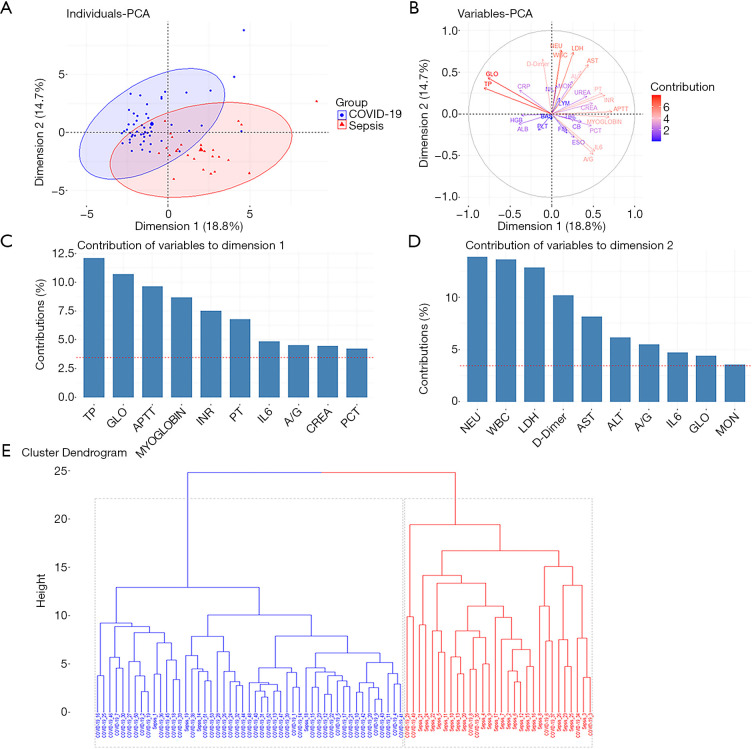
The PCA showed that bacterial septic patients and COVID-19 patients could be clearly distinguished in dimension 1 and dimension 2 (Figure 1A) when using the clinical parameters in Table 2 as variables. In dimension 1, the variables listed in descending order of contribution were: total protein, globulin, activated partial thromboplastin time (APTT), myoglobin, international normalization ratio (INR), and prothrombin time (PT) (Figure 1B,C); in dimension 2, the 5 most contributory variables were the neutrophil count, white blood cell count, lactate dehydrogenase (LDH) level, D-dimer level, and aspartate transaminase (AST) level (Figure 1B,D). Hierarchical cluster analysis was performed to divide all participants into 2 categories using the clinical parameters in Table 2 (Figure 1E). Most COVID-19 patients (45/53, 84.9%) and bacterial septic patients (22/26, 84.6%) were separately categorized into blue and red categories. Both PCA and hierarchical cluster analysis could distinguish non-survivors with COVID-19 from non-survivors with bacterial sepsis.
Figure 1.
Principal component analysis and hierarchical cluster analysis of clinical parameters in bacterial sepsis patients and COVID-19 patients. (A) PCA was performed with the R package “factoextra” to distinguish bacterial sepsis patients from COVID-19 patients. (B) Contributions of variables in PCA. (C) Top 10 most contributory variables in dimension 1. (D) Top 10 most contributory variables in dimension 2. (E) Hierarchical cluster analysis of bacterial sepsis patients and COVID-19 patients. Clusters computed by Ward's method according to the Euclidean distance between patients’ clinical parameters. PCA, principal component analysis; WBC, white blood cell count; NEU, neutrophil count; LYM, lymphocyte count; N/L, neutrophil/lymphocyte ratio; MON, monocyte count; ESO, eosinophil count; BAS, basophil count; PLT, platelet count; HGB, hemoglobulin; TP, total protein; ALB, albumin; GLO, globulin; A/G, albumin/globulin ratio; TBIL, total bilirubin, CB, direct bilirubin; ALT, glutamate-pyruvate transaminase; AST, aspartate transaminase; UREA, urea; CREA, creatine; LDH, lactate dehydrogenase; MYOGLOBULIN, myoglobulin; APTT, activated partial thromboplastin time; PT, prothrombin time; FIB, fibrinogen; INR, international normalized ratio; DD, D-dimer; PCT, procalcitonin; IL6, interleukin-6; CRP, C-reactive protein.
Table 2. Laboratory findings at ICU admission.
| Variable | Sepsis, N=26 (%) | COVID-19, N=53 (%) | P value |
|---|---|---|---|
| Immunological status | |||
| White blood cell count (×109/L), median (IQR) | 12.16 (5.31, 17.08) | 12.64 (8.41, 16.58) | 0.392 |
| Neutrophil count (×109/L), median (IQR) | 10.55 (4.88, 15.78) | 11.75 (7.48, 15.21) | 0.312 |
| Lymphocyte count (×109/L), median (IQR) | 0.70 (0.38, 1.00) | 0.53 (0.39, 0.77) | 0.358 |
| Neutrophil/lymphocyte ratio, median (IQR) | 13.11 (8.14, 26.88) | 19.94 (14.55, 32.73) | 0.025 |
| Monocyte count (×109/L), median (IQR) | 0.41 (0.23, 0.61) | 0.32 (0.21, 0.50) | 0.314 |
| Eosinophil count (×109/L), median (IQR) | 0.06 (0.01, 0.11) | 0.00 (0.00, 0.02) | <0.001 |
| Basophil count (×109/L), median (IQR) | 0.02 (0.01, 0.05) | 0.02 (0.00, 0.03) | 0.164 |
| IL-6 (pg/mL), median (IQR) | 820.00 (263.00, 1,000.00) | 10.76 (8.24, 16.68) | <0.001 |
| PCT (μg/L), median (IQR) | 8.45 (1.75, 21.02) | 0.14 (0.11, 0.60) | <0.001 |
| CRP (>10 mg/mL) | 26 (100.0) | 48 (90.6) | 0.165 |
| Organ function | |||
| Hemoglobin (g/L), median (IQR) | 112.00 (93.25, 135.50) | 122.00 (111.50, 134.00) | 0.129 |
| Total protein (g/L), median (IQR) | 51.50 (43.75, 56.25) | 64.10 (58.75, 68.45) | <0.001 |
| Albumin (g/L), median (IQR) | 27.50 (22.00, 31.00) | 28.20 (25.65, 29.95) | 0.498 |
| Globulin (g/L), median (IQR) | 22.00 (18.00, 27.00) | 36.10 (31.50, 41.95) | <0.001 |
| A/G, median (IQR) | 1.20 (0.99, 1.44) | 0.80 (0.70, 0.90) | <0.001 |
| Total bilirubin (μmol/L), median (IQR) | 23.95 (16.48, 46.90) | 16.80 (12.35, 24.75) | 0.004 |
| Direct bilirubin (μmol/L), median (IQR) | 12.70 (7.33, 26.15) | 7.10 (4.50, 10.15) | <0.001 |
| ALT (U/L), median (IQR) | 35.50 (22.25, 74.25) | 29.00 (19.00, 62.00) | 0.238 |
| AST (U/L), median (IQR) | 55.50 (37.50, 126.00) | 38.00 (31.00, 64.50) | 0.054 |
| Creatinine (μmol/L), median (IQR) | 98.00 (62.00, 175.25) | 76.70 (59.00, 119.65) | 0.431 |
| Urea (mmol/L), median (IQR) | 9.35 (5.43, 15.78) | 8.50 (6.70, 14.05) | 0.892 |
| LDH (U/L), median (IQR) | 260.00 (218.00, 457.25) | 527.00 (468.00, 763.50) | <0.001 |
| Myohemoglobin (ng/mL), median (IQR) | 380.80 (162.80, 2,527.50) | 123.00 (58.10, 428.30) | 0.002 |
| Coagulation | |||
| Platelet count (×109/L), median (IQR) | 142.50 (92.25, 267.00) | 159.00 (108.00, 237.50) | 0.950 |
| APTT (seconds), median (IQR) | 34.55 (31.80, 48.75) | 27.60 (23.90, 32.75) | <0.001 |
| PT (seconds), median (IQR) | 15.80 (13.78, 17.55) | 13.00 (11.95, 14.80) | <0.001 |
| Fibrinogen (g/L), median (IQR) | 3.50 (1.92, 5.32) | 4.50 (2.55, 5.70) | <0.001 |
| INR, median (IQR) | 1.45 (1.27, 1.62) | 1.10 (1.02, 1.26) | <0.001 |
| D-dimer (g/L), median (IQR) | 6.34 (3.14, 9.39) | 30.24 (5.63, 65.93) | 0.002 |
ICU, intensive care unit; IL-6, inteleukin-6; PCT, procalcitonin; CRP, C-reactive protein; A/G, albumin/globulin ratio; ALT, alanine aminotransferase; AST, aspartate aminotransferase; LDH, lactate dehydrogenase; APTT, activated partial thromboplastin time; PT, prothrombin time; INR, international normalized ratio; PCT, procalcitonin; IL-6, interleukin-6; IQR, interquartile range.
Table 2 shows the laboratory findings of patients at ICU admission. The normal reference ranges of these parameters are shown in Table S2. The neutrophil/lymphocyte ratio and levels of total protein, globulin, LDH, fibrinogen, and D-dimer were higher in non-survivors with COVID-19 than in non-survivors with sepsis; and non-survivors with COVID-19 had lower eosinophil counts, albumin/globulin ratios, APTTs, PTs, INRs, and lower levels of PCT, IL-6, total bilirubin, direct bilirubin, and myohemoglobin than non-survivors with sepsis.
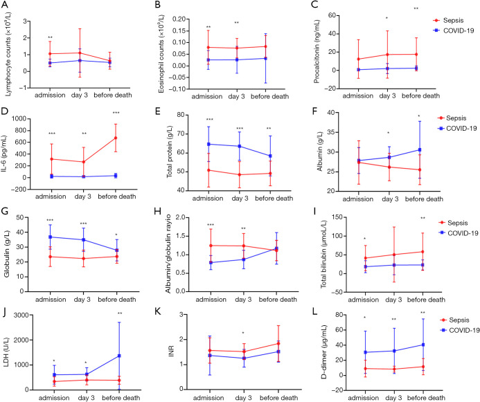
To further explore differences in the clinical course of disease between COVID-19 and bacterial sepsis, we compared laboratory results at admission, on day 3, and on the day before death in patients who survived the first 3 days in the ICU. A total of 18 COVID-19 patients and 19 bacterial septic patients with complete blood counts, serum biochemistry profiles, and coagulation test data were included in the analysis. The test for between-group effects showed that the lymphocyte counts, eosinophil counts, INR, albumin/globulin ratio, and levels of PCT, IL-6, total protein, albumin, globulin, total bilirubin, LDH, and D-dimer were different between the two groups (Figure 2). Compared with non-survivors with bacterial sepsis, non-survivors with COVID-19 had higher levels of total protein, globulin, LDH, and D-dimer and lower levels of IL-6 during their ICU stays (Figure 2).
Figure 2.
Dynamic laboratory findings. Laboratory findings of patients at ICU admission, on day 3, and on the day before death. (A) Lymphocyte counts (×109/L). (B) Eosinophil counts (×109/L). (C) Procalcitonin (ng/mL). (D) IL-6 (pg/mL). (E) Total protein (g/L). (F) Albumin (g/L). (G) Globulin (g/L). (H) Albumin/globulin ratio. (I) Total bilirubin (mol/L). (J) LDH (U/L). (K) INR. (L) D-dimer (µg/mL). ICU, intensive care unit; IL-6, interleukin-6; LDH, lactate dehydrogenase; INR, international normalized ratio. *, P<0.05; **, P<0.01; ***, P<0.001.
Treatments and outcomes
Mechanical ventilation was the main form of respiratory support provided to patients with COVID-19 and bacterial sepsis. More participants in the COVID-19 cohort received high-flow nasal cannula therapy than in the bacterial septic cohort (Table 3). Almost all participants were administered antibiotics, and antiviral therapy was only used in 15 (28.3%) COVID-19 patients. Corticosteroid and immunoglobulin were given to 29 (54.7%) and 31 (58.5%) COVID-19 patients, respectively, which were not routinely used for the treatment of bacterial sepsis. Vasopressors and renal replacement were more widely used in non-survivors with bacterial sepsis than in those with COVID-19 (Table 3). The survival time in ICU of non-survivors with COVID-19 was shorter than that of those with bacterial sepsis, while no difference was observed in the overall survival time between the two groups (Table 3).
Table 3. Treatments and outcomes.
| Variable | Sepsis, N=26 (%) | COVID-19, N=53 (%) | P value |
|---|---|---|---|
| HFNC | 2 (7.7) | 18 (34.0) | 0.012 |
| Mechanical ventilation | 23 (88.5) | 50 (94.3) | 0.389 |
| Antibiotic therapy | 26 (100.0) | 52 (98.1) | 1.000 |
| Anti-viral therapy | 0 (0.0) | 15 (28.3) | 0.002 |
| Corticosteroids | 1 (3.8) | 29 (54.7) | 0.000 |
| Immunoglobulin therapy | 0 (0.0) | 31 (58.5) | 0.000 |
| Vasopressor therapy | 23 (88.5) | 16 (30.2) | 0.000 |
| CRRT | 12 (46.2) | 11 (20.8) | 0.020 |
| Overall survival time (days), mean (SD) | 28.0 (32.3) | 22.1 (8.2) | 0.255 |
HFNC, high-flow nasal cannula; CRRT, continuous renal replacement therapy; ICU, intensive care unit; SD, standard deviation.
Discussion
Numerous patients with severe COVID-19 have clinical manifestations that meet the Sepsis-3 diagnostic criteria for sepsis with negative blood or respiratory sample cultures for bacteria or fungi. It has been speculated that viral sepsis may be a proper description of severe COVID-19 (6,13). Here, we compared the clinical characteristics of 53 deceased patients with confirmed COVID-19 with those of 26 with bacterial sepsis after ICU admission. Our analyses showed that non-survivors with severe COVID-19 and bacterial sepsis had distinct clinical characteristics regarding susceptibility, organ function, immunologic status, and coagulation. Based on the PCA and hierarchical cluster analysis of the laboratory test results, non-survivors with COVID-19 and non-survivors with bacterial sepsis belong to 2 separate categories, suggesting that severe COVID-19 is a unique category of sepsis that is different from bacterial sepsis.
Older males with comorbidities are more likely to be affected by COVID-19 (14). Hypertension, diabetes mellitus, cardiovascular disease, and cerebrovascular disease are major risk factors for mortality in patients with COVID-19 and in those with sepsis (14-18). In our study, the proportion of hypertension was higher in patients with COVID-19 than in those with bacterial sepsis. This suggests that patients with hypertension might be more predisposed to become severe cases after SARS-Cov-2 infection. However, several observational studies have shown no substantial increase in the susceptibility to COVID-19 or in the risk of severe COVID-19 among patients treated with anti-hypertension agents renin-angiotensin-aldosterone system (RAAS) blockers (19,20).
Patients with severe COVID-19 often presented heart, kidney, or liver dysfunction in addition to respiratory failure (6). These features of COVID-19 seem to resemble multiorgan dysfunction in sepsis, which accounts for clinical deterioration and death in severe cases. We detected a striking elevation of troponin in the COVID-19 cohort; however, we could not directly compare their levels between the two groups because different troponin isoforms were measured in the 2 hospitals (Table S3). In addition, vasopressor was less used in patients with COVID-19 (Table 2). Regarding kidney function, although the levels of creatinine and urea were comparable between the two groups from the time of ICU admission to the time of death, renal replacement therapy was less used in the COVID-19 cohort than in the sepsis cohort, suggesting that fewer severe cases with COVID-19 developed kidney dysfunction during their ICU stays. In addition, liver injury, as indicated by increased bilirubin levels, also occurred less often in the COVID-19 cohort than in the sepsis cohort. These results suggest that respiratory failure is the primary cause of death in patients with severe COVID-19 who experience rapid deterioration, although virus-induced organ dysfunction also contributes to mortality. The SOFA score was deemed as a reliable indicator for predicting in-hospital and 28-day mortality in sepsis. Nevertheless, the deceased COVID-19 patients in our study had a much lower SOFA score than septic patients, which implied that SOFA might not be sensitive for these "viral sepsis" patients due to SARS-CoV-2.
The immune response is essential for resolving an infection, but a dysregulated immune response can also result in immunopathology. A cytokine storm is a harmful immune reaction involving swiftly elevated levels of circulating cytokines and immune-cell hyperactivation that leads to multiorgan failure and early mortality (21). It has been revealed that pro-inflammatory cytokine levels are consistently elevated above the normal range in patients with severe or critical COVID-19, which was associated with poor outcomes (22,23). Based on these clinical results and previous experience with SARS-COV, cytokine storms have been proposed as a pathophysiological mechanism underlying the poor outcomes of severe COVID-19 (7). Anti-inflammatory strategies, such as an anti-IL-6 receptor, anti-tumor necrosis factor (TNF) antibody, and corticosteroids, had been suggested as potential treatments for COVID-19 based on this theory (7,24-26). Recently, the use of the term cytokine storm in the context of COVID-19 has been challenged (27). Sinha et al. reported that plasma cytokine levels in most COVID-19 patients were lower than those in previous cohorts of patients with acute respiratory distress syndrome (ARDS) (27). In particular, the median levels of IL-6, a key pro-inflammatory cytokine in patients with acute infections, were found to be 10- to 200-fold lower in patients with severe COVID-19 than those with the hyperinflammatory phenotype of ARDS (27). Consistently, we also found that the level of IL-6 was 65-fold lower in non-survivors with COVID-19 than in those with sepsis (Table 2). All the above suggest that the elevation of cytokine levels in patients with COVID-19 is limited, which differs from the excessive inflammatory cytokine release observed in patients with bacterial sepsis (21). Further evidence supporting this claim is that tocilizumab, an anti-IL-6 receptor monoclonal antibody, has been approved to treat multiple inflammatory diseases like sepsis and arthritis. Nevertheless, in the latest randomized controlled trials, tocilizumab failed to show significant effects on the risk of intubation or a survival benefit in hospitalized patients with COVID-19 (28-32).
Coagulation data from published studies have shown that patients with severe COVID-19 were susceptible to microvascular thrombosis and disseminated intravascular coagulation (DIC) without evidence of bleeding (33). We found that D-dimer levels in the COVID-19 cohort were 5 times as much as those in the bacterial sepsis cohort, while the PT, APTT, and INR were less disturbed in participants with COVID-19 than in those with sepsis. In bacterial sepsis, coagulopathy is triggered by the activation of tissue factors and is potently enhanced by immune cell activation and cytokine release. As discussed above, the cytokine levels were not as high in COVID-19 participants as in those with bacterial sepsis, indicating that cytokine release might not be the primary cause of coagulation dysfunction. Recently, a postmortem report revealed that severe lung injury was observed in patients with ARDS due to COVID-19. Alveolar microthrombi were 9 times more prevalent in patients with ARDS due to COVID-19 than in patients with ARDS due to influenza (34). Therefore, it is reasonable to presume that the SARS-CoV-2 virus impaired endothelial cells via the angiotensin-converting enzyme 2 (ACE2) receptor, switching the vascular milieu to procoagulant activity. A recent pathological report confirmed the presence of viral elements within endothelial cells and an accumulation of inflammatory cells (35).
There were several limitations to this study. First, our COVID-19 cohort and bacterial sepsis cohort involved patients admitted to 2 different hospitals. There might have been some systemic bias due to differences in laboratory protocols or managing strategies. However, all participants were supervised by the same attending physician, which might have minimized such institutional heterogeneity. Second, we only compared the clinical characteristics between patients with bacterial sepsis and patients with severe COVID-19, limiting the generalizability of the findings to sepsis caused by other viral infections.
According to the analysis of susceptibility, organ function, immunologic status, and coagulation between the COVID-19 cohort and bacterial sepsis cohort, non-surviving patients with COVID-19 were distinguished from non-surviving patients with bacterial sepsis.
Conclusions
In conclusion, this comparative study demonstrated that severe COVID-19 patients had distinct phenotypes from those with bacterial sepsis. Therefore, it is prudent to acquire more evidence before applying previous experience for bacterial sepsis to patients with severe COVID-19.
Supplementary
The article’s supplementary files as
Acknowledgments
Funding: This work was supported by the National Natural Science Foundation of China (81971807) and he National Key Research and Development Program of China (2020YFC0848100).
Ethical Statement: The authors are accountable for all aspects of the work in ensuring that questions related to the accuracy or integrity of any part of the work are appropriately investigated and resolved. The study was conducted in accordance with the Declaration of Helsinki (as revised in 2013). The study involving patients in the sepsis cohort was conducted according to the informed consent guidelines approved by the Ethics Committee of Zhongshan Hospital, Fudan University (B2017-021R). Written informed consent was provided by participants or their statutory surrogates. This study on COVID-19 was approved by the Ethics Commission of Wuhan Infectious Diseases Hospital (KY-2020-56.01), and written informed consent was waived by the Ethics Commission of the hospital due to the retrospective design of the study.
Footnotes
Reporting Checklist: The authors have completed the STROBE reporting checklist. Available at https://dx.doi.org/10.21037/atm-21-1291
Data Sharing Statement: Available at https://dx.doi.org/10.21037/atm-21-1291
Conflicts of Interest: All authors have completed the ICMJE uniform disclosure form (available at https://dx.doi.org/10.21037/atm-21-1291). The authors have no conflicts of interest to declare.
References
- 1.Guan WJ, Ni ZY, Hu Y, et al. Clinical Characteristics of Coronavirus Disease 2019 in China. N Engl J Med 2020;382:1708-20. 10.1056/NEJMoa2002032 [DOI] [PMC free article] [PubMed] [Google Scholar]
- 2.Yang X, Yu Y, Xu J, et al. Clinical course and outcomes of critically ill patients with SARS-CoV-2 pneumonia in Wuhan, China: a single-centered, retrospective, observational study. Lancet Respir Med 2020;8:475-81. 10.1016/S2213-2600(20)30079-5 [DOI] [PMC free article] [PubMed] [Google Scholar]
- 3.Bhatraju PK, Ghassemieh BJ, Nichols M, et al. Covid-19 in Critically Ill Patients in the Seattle Region - Case Series. N Engl J Med 2020;382:2012-22. 10.1056/NEJMoa2004500 [DOI] [PMC free article] [PubMed] [Google Scholar]
- 4.Seymour CW, Liu VX, Iwashyna TJ, et al. Assessment of Clinical Criteria for Sepsis: For the Third International Consensus Definitions for Sepsis and Septic Shock (Sepsis-3). JAMA 2016;315:762-74. 10.1001/jama.2016.0288 [DOI] [PMC free article] [PubMed] [Google Scholar]
- 5.Singer M, Deutschman CS, Seymour CW, et al. The Third International Consensus Definitions for Sepsis and Septic Shock (Sepsis-3). JAMA 2016;315:801-10. 10.1001/jama.2016.0287 [DOI] [PMC free article] [PubMed] [Google Scholar]
- 6.Li H, Liu L, Zhang D, et al. SARS-CoV-2 and viral sepsis: observations and hypotheses. Lancet 2020;395:1517-20. 10.1016/S0140-6736(20)30920-X [DOI] [PMC free article] [PubMed] [Google Scholar]
- 7.Moore JB, June CH. Cytokine release syndrome in severe COVID-19. Science 2020;368:473-4. 10.1126/science.abb8925 [DOI] [PubMed] [Google Scholar]
- 8.Zhou F, Yu T, Du R, et al. Clinical course and risk factors for mortality of adult inpatients with COVID-19 in Wuhan, China: a retrospective cohort study. Lancet 2020;395:1054-62. 10.1016/S0140-6736(20)30566-3 [DOI] [PMC free article] [PubMed] [Google Scholar]
- 9.Mahmudpour M, Roozbeh J, Keshavarz M, et al. COVID-19 cytokine storm: The anger of inflammation. Cytokine 2020;133:155151. 10.1016/j.cyto.2020.155151 [DOI] [PMC free article] [PubMed] [Google Scholar]
- 10.Huang C, Wang Y, Li X, et al. Clinical features of patients infected with 2019 novel coronavirus in Wuhan, China. Lancet 2020;395:497-506. 10.1016/S0140-6736(20)30183-5 [DOI] [PMC free article] [PubMed] [Google Scholar]
- 11.Chirico G, Cortinovis S, Fonte C, et al. Bacterial sepsis. J Chemother 2007;19 Suppl 2:28-30. 10.1080/1120009X.2007.11782440 [DOI] [PubMed] [Google Scholar]
- 12.Reyes M, Filbin MR, Bhattacharyya RP, et al. An immune-cell signature of bacterial sepsis. Nat Med 2020;26:333-40. 10.1038/s41591-020-0752-4 [DOI] [PMC free article] [PubMed] [Google Scholar]
- 13.Lin GL, McGinley JP, Drysdale SB, et al. Epidemiology and Immune Pathogenesis of Viral Sepsis. Front Immunol 2018;9:2147. 10.3389/fimmu.2018.02147 [DOI] [PMC free article] [PubMed] [Google Scholar]
- 14.Chen N, Zhou M, Dong X, et al. Epidemiological and clinical characteristics of 99 cases of 2019 novel coronavirus pneumonia in Wuhan, China: a descriptive study. Lancet 2020;395:507-13. 10.1016/S0140-6736(20)30211-7 [DOI] [PMC free article] [PubMed] [Google Scholar]
- 15.Wang D, Hu B, Hu C, et al. Clinical Characteristics of 138 Hospitalized Patients With 2019 Novel Coronavirus-Infected Pneumonia in Wuhan, China. JAMA 2020;323:1061-9. 10.1001/jama.2020.1585 [DOI] [PMC free article] [PubMed] [Google Scholar]
- 16.Wang Y, Lu X, Li Y, et al. Clinical Course and Outcomes of 344 Intensive Care Patients with COVID-19. Am J Respir Crit Care Med 2020;201:1430-4. 10.1164/rccm.202003-0736LE [DOI] [PMC free article] [PubMed] [Google Scholar]
- 17.Grasselli G, Zangrillo A, Zanella A, et al. Baseline Characteristics and Outcomes of 1591 Patients Infected With SARS-CoV-2 Admitted to ICUs of the Lombardy Region, Italy. JAMA 2020;323:1574-81. 10.1001/jama.2020.5394 [DOI] [PMC free article] [PubMed] [Google Scholar]
- 18.Wang HE, Shapiro NI, Griffin R, et al. Chronic medical conditions and risk of sepsis. PLoS One 2012;7:e48307. 10.1371/journal.pone.0048307 [DOI] [PMC free article] [PubMed] [Google Scholar]
- 19.Reynolds HR, Adhikari S, Pulgarin C, et al. Renin–Angiotensin–Aldosterone System Inhibitors and Risk of Covid-19. N Engl J Med 2020;382:2441-8. 10.1056/NEJMoa2008975 [DOI] [PMC free article] [PubMed] [Google Scholar]
- 20.Mancia G, Rea F, Ludergnani M, et al. Renin–Angiotensin–Aldosterone System Blockers and the Risk of Covid-19. N Engl J Med 2020;382:2431-40. 10.1056/NEJMoa2006923 [DOI] [PMC free article] [PubMed] [Google Scholar]
- 21.van der Poll T, van de Veerdonk FL, Scicluna BP, et al. The immunopathology of sepsis and potential therapeutic targets. Nat Rev Immunol 2017;17:407-20. 10.1038/nri.2017.36 [DOI] [PubMed] [Google Scholar]
- 22.Gong J, Dong H, Xia SQ, et al. Correlation Analysis Between Disease Severity and Inflammation-related Parameters in Patients with COVID-19: a retrospective study. BMC Infect Dis 2020;20:963. 10.1186/s12879-020-05681-5 [DOI] [PMC free article] [PubMed] [Google Scholar]
- 23.Liu J, Li S, Liu J, et al. Longitudinal characteristics of lymphocyte responses and cytokine profiles in the peripheral blood of SARS-CoV-2 infected patients. EBioMedicine 2020;55:102763. 10.1016/j.ebiom.2020.102763 [DOI] [PMC free article] [PubMed] [Google Scholar]
- 24.Mehta P, McAuley DF, Brown M, et al. COVID-19: consider cytokine storm syndromes and immunosuppression. Lancet 2020;395:1033-4. 10.1016/S0140-6736(20)30628-0 [DOI] [PMC free article] [PubMed] [Google Scholar]
- 25.Conti P, Ronconi G, Caraffa A, et al. Induction of pro-inflammatory cytokines (IL-1 and IL-6) and lung inflammation by Coronavirus-19 (COVI-19 or SARS-CoV-2): anti-inflammatory strategies. J Biol Regul Homeost Agents 2020;34:327-31. [DOI] [PubMed] [Google Scholar]
- 26.Feldmann M, Maini RN, Woody JN, et al. Trials of anti-tumour necrosis factor therapy for COVID-19 are urgently needed. Lancet 2020;395:1407-9. 10.1016/S0140-6736(20)30858-8 [DOI] [PMC free article] [PubMed] [Google Scholar]
- 27.Sinha P, Matthay MA, Calfee CS. Is a "Cytokine Storm" Relevant to COVID-19? JAMA Intern Med 2020;180:1152-4. 10.1001/jamainternmed.2020.3313 [DOI] [PubMed] [Google Scholar]
- 28.Stone JH, Frigault MJ, Serling-Boyd NJ, et al. Efficacy of Tocilizumab in Patients Hospitalized with Covid-19. N Engl J Med 2020;383:2333-44. 10.1056/NEJMoa2028836 [DOI] [PMC free article] [PubMed] [Google Scholar]
- 29.Hermine O, Mariette X, Tharaux P-L, et al. Effect of Tocilizumab vs Usual Care in Adults Hospitalized With COVID-19 and Moderate or Severe Pneumonia: A Randomized Clinical Trial. JAMA Intern Med 2021;181:32-40. 10.1001/jamainternmed.2020.6820 [DOI] [PMC free article] [PubMed] [Google Scholar]
- 30.Salama C, Han J, Yau L, et al. Tocilizumab in Patients Hospitalized with Covid-19 Pneumonia. N Engl J Med 2021;384:20-30. 10.1056/NEJMoa2030340 [DOI] [PMC free article] [PubMed] [Google Scholar]
- 31.Salvarani C, Dolci G, Massari M, et al. Effect of Tocilizumab vs Standard Care on Clinical Worsening in Patients Hospitalized With COVID-19 Pneumonia: A Randomized Clinical Trial. JAMA Intern Med 2021;181:24-31. 10.1001/jamainternmed.2020.6615 [DOI] [PMC free article] [PubMed] [Google Scholar]
- 32.Veiga VC, Prats JAGG, Farias DLC, et al. Effect of tocilizumab on clinical outcomes at 15 days in patients with severe or critical coronavirus disease 2019: randomised controlled trial. BMJ 2021;372:n84. [DOI] [PMC free article] [PubMed] [Google Scholar]
- 33.Connors JM, Levy JH. COVID-19 and its implications for thrombosis and anticoagulation. Blood 2020;135:2033-40. 10.1182/blood.2020006000 [DOI] [PMC free article] [PubMed] [Google Scholar]
- 34.Ackermann M, Verleden SE, Kuehnel M, et al. Pulmonary Vascular Endothelialitis, Thrombosis, and Angiogenesis in Covid-19. N Engl J Med 2020;383:120-8. 10.1056/NEJMoa2015432 [DOI] [PMC free article] [PubMed] [Google Scholar]
- 35.Varga Z, Flammer AJ, Steiger P, et al. Endothelial cell infection and endotheliitis in COVID-19. Lancet 2020;395:1417-8. 10.1016/S0140-6736(20)30937-5 [DOI] [PMC free article] [PubMed] [Google Scholar]
Associated Data
This section collects any data citations, data availability statements, or supplementary materials included in this article.
Supplementary Materials
The article’s supplementary files as


