Abstract
Purpose
To compare optical coherence tomography angiography (OCTA)–derived flux with conventional OCTA measures of retinal vascular density in assessment of physiological changes in retinal blood flow.
Methods
Healthy subjects were recruited, and 3 × 3-mm2 fovea-centered scans were acquired using commercially available swept-source OCTA (SS-OCTA) while participants were breathing room air, 100% O2, or 5% CO2. Retinal perfusion was quantified using vessel area density (VAD) and vessel skeleton density (VSD), as well as novel measures of retinal perfusion, vessel area flux (VAF) and vessel skeleton flux (VSF). Flux is proportional to the number of red blood cells moving through a vessel segment per unit time. The percentage change in each measure was compared between the O2 and CO2 gas conditions for images of all vessels (arterioles, venules, and capillaries) and capillary-only images. Statistical significance was determined using paired t-tests and a linear mixed-effects model.
Results
Eighty-four OCTA scans from 29 subjects were included (age, 45.9 ± 19.5 years; 14 male, 48.3%). In capillary-only images, the change under the CO2 condition was 168% greater in VAF than in VAD (P = 0.002) and 124% greater in VSF than in VSD (P = 0.004). Similarly, under the O2 condition, the change was 94% greater in VAF than in VAD (P = 0.004) and 57% greater in VSF than in VSD (P = 0.01). Flux measures showed significantly greater change in capillary-only images compared with all-vessels images.
Conclusions
OCTA-derived flux measures quantify physiological changes in retinal blood flow at the capillary level with a greater effect size than conventional vessel density measures.
Translational Relevance
OCTA-derived flux is a useful measure of subclinical changes in retinal capillary perfusion.
Keywords: retinal blood flow, flux, capillary, retinal vascular reactivity, optical coherence tomography angiography
Introduction
Optical coherence tomography angiography (OCTA) is a US Food and Drug Administration (FDA)–approved technology that provides rapid in vivo visualization of red blood cell (RBC) movements.1,2 In clinical and research settings, en face OCTA images are very useful to detect and quantify frank capillary loss.1,3–10 Nevertheless, OCTA has provided relatively limited quantitative information about retinal blood flow (rather than vascular density). For example, commercially available OCTA devices are typically used to create en face maps demonstrating vascular segments with RBC movement occurring within a range of 0.3 to 3 mm/s.1,11 Absolute measurement of RBC velocity has been elusive. The application of OCTA-derived measures to assess relative quantitative changes in capillary blood flow has been only possible with custom-designed OCTA systems.12 Therefore, there is a major unmet clinical and research need to more accurately quantify subclinical changes in blood flow that precede frank pathological changes in capillary structure. For example, improving the accuracy and sensitivity of measures of subclinical changes in blood flow that precede capillary loss would be highly relevant to early interventions in retinal vascular diseases such as diabetic retinopathy.13,14
Previous in vitro microfluidic modeling of blood flow suggests that the intensity of the optical microangiography (OMAG)15,16 decorrelation signal is likely proportional to the number of RBCs moving within a capillary segment at the time of the OCTA scan, a concept otherwise known as “flux.”17 These phantom studies suggest that flux may be a more accurate and sensitive measure of subclinical changes in capillary perfusion (i.e., in the absence of frank capillary loss) than conventional skeleton density or vessel density measures.
The aim of this study was to compare conventional OCTA-derived measures of retinal vascular density with OCTA-derived measures of flux to quantify changes in retinal perfusion in the absence of capillary pathology. We compared how well these OCTA-derived metrics can quantify the magnitude of retinal vascular reactivity in response to well-characterized physiological stimuli in capillaries versus larger caliber retinal vessels (arterioles and venules) of normal human subjects.
Methods
The study was designed and conducted at the University of Southern California (USC) Roski Eye Institute between October 2017 and February 2020. The study was approved by the USC Institutional Review Board and conformed to the tenets of the Declaration of Helsinki. The study was explained to and written informed consent was obtained from all subjects prior to recruitment.
Healthy subjects older than 18 years of age without any history of ophthalmic or systemic vascular disease were recruited from volunteers or USC Roski Eye Institute clinic patients who agreed to participate in the study. Exclusion criteria included inability to consent, pregnancy, and history of any of the following: macular edema, age-related macular degeneration, retinal vein occlusion, glaucoma, media opacities (vitreous hemorrhage or visually significant cataract), intraocular surgery, systemic vascular diseases (diabetes and hypertension), and vasculitis. Subjects with a history of pulmonary disease or recent hospitalization were also excluded. All subjects completed a study questionnaire with self-reported information regarding medical history, caffeine consumption, and medication history. This information was verified and supplemented by information from available medical records.
For each subject, one eye was selected based on the exclusion criteria above. If both eyes were eligible for the study, one eye was randomly selected. Then, 3 × 3-mm2 fovea-centered OCTA images were acquired using a commercially available swept-source OCTA (SS-OCTA) device (PLEX Elite 9000; Carl Zeiss Meditec, Dublin, CA). The device used a swept-source tunable laser with a center wavelength of 1040 to 1060 nm and operated at a 100-kHz A-scan rate. It had an optical axial resolution of 6.3 µm and a transverse resolution of ∼16 µm, and it used OMAG intensity and phase processing to differentiate static tissue from moving particles, particularly RBCs.15,16
OCTA images were acquired while subjects were breathing three different gas mixtures in the following order and duration: (1) atmospheric room air, (2) 1 minute after starting to breathe hypercapnic gas mixture (5% CO2, 21% O2, and 74% N2), (3) 10-minute break, and (4) 1 minute after starting to breathe 100% O2. A custom-designed non-rebreathing apparatus that has been described previously was used to deliver the gas mixtures during retinal imaging. The order of gas inhalation was optimized to prevent the carry-over effect from one condition to the next, as previously demonstrated.18,19 This protocol was previously described in studies of cerebrovascular reactivity and retinal vascular reactivity and was reported to produce a negligible change in breathing rate and heart rate.20,21 An opportunity to rest was also provided to each subject to ensure that fatigue was not a factor during gas inhalation conditions. During the gas trials, the subjects’ heart rates and blood oxygen saturation levels were monitored with finger-tip pulse oximeter probes, and the testing was halted if any subject's saturation dropped below 94%, if the subject's heart rate demonstrated a change of more than 10% from the baseline, or if the participant experienced any unexpected adverse effect. The acquired OCTA images were automatically segmented using the device manufacturer's software (PLEX Elite Version 1.7), and full-thickness en face OCTA images were created. Images were included if they had a device manufacturer–reported signal strength of 8 or higher. Images were excluded if they had major artifacts, including segmentation errors, shadow, motion, tilt, or decentration artifacts. The best quality image was selected when the subject had multiple images available.
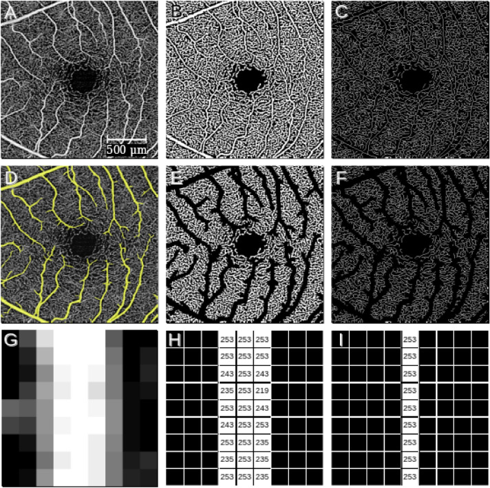
A custom semiautomated program (MATLAB R2019a; The MathWorks, Inc., Natick, MA) was used to calculate the OCTA measures, including vessel area density (VAD), vessel skeleton density (VSD), vessel area flux (VAF), and vessel skeleton flux (VSF), which are illustrated in Figure 1. VAD and VSD have been described in the past.1,5–8,22 Briefly, for VAD and VSD each OCTA image was exported as a grayscale image in bitmap format in which each pixel had a value of 0 to 255 (Fig. 1A). This image was converted to a binary image (Fig. 1B) where each pixel had either a value of 1 if that pixel covered a vessel or a value of 0 otherwise using a combined approach of global thresholding, Hessian filtering, and adaptive thresholding.1,20 VAD was calculated as the number of pixels with a value of 1 in the binarized image divided by the total number of pixels in the image. VSD was calculated by symmetrically reducing the binary image to a 1-pixel-wide skeletonized representation of each vessel (Fig. 1C), using the “skel” operation of the MATLAB bwmorph function. VSD was then defined as the number of pixels with a value of 1 in the skeletonized image divided by the total number of pixels in the image.
Figure 1.
(A) Original grayscale OCTA bitmap image. (B) All-vessels binarized image derived from previous panel. (C) All-vessels skeletonized image derived from previous panel. (D) Automatic detection of arterioles and venules (highlighted in yellow). (E) Capillary-only binary image (after removal of arterioles and venules). (F) Capillary-only skeletonized image. (G) To visually illustrate the calculation of OCTA-derived perfusion measures, we present a 9 × 9-pixel sample region that covers a larger vessel from A. (H) Binarized image derived from previous panel with raw decorrelation intensity pixel values overlaid. (I) Skeletonized image derived from previous panel with raw decorrelation intensity pixel values overlaid. For the 9 × 9-pixel image in G, VAD is the number of white pixels in H divided by the total number of pixels: 27/81 = 0.33. VSD is the number of white pixels in I divided by the total number of pixels: 9/81 = 0.11. VAF is the average of pixel values in H: 6667/27 = 246.92. VSF is the average of pixel values in I: 2277/9 = 253.
OCTA-derived flux measures were calculated from the grayscale (non-binarized) bitmap exports of the en face OCTA image (Fig. 1A). VAF was calculated by using the binarized image created for the calculation of VAD as a mask to select pixels in the grayscale bitmap image. VAF was defined as the average value of these masked pixels (Fig. 1H). All flux values reported in the manuscript represent this VAF measure unless otherwise stated. VSF was similarly calculated by using the skeletonized binary image created for the calculation of VSD as a mask to select pixels in the grayscale bitmap image. VSF was defined as the average value of these masked pixels in the grayscale bitmap exports (Fig. 1I).
Because capillary blood flow is highly correlated with flux (capillaries only pass one RBC at a time),17 OCTA-derived measures of capillary flux may be more accurate measures of capillary perfusion than measures derived from binarized decorrelation intensities such as VSD or VAD. Therefore, to isolate capillaries for our analyses, capillary-only bitmap images were created by using a large vessel mask to remove arterioles and venules from the OCTA image. This was achieved using a custom-made, semiautomated MATLAB program with a manually determined threshold for vessel size that has been previously described23 and includes end-arteriolar capillary segments (Figs. 1D–1F). All perfusion measures were calculated both with the original all-vessels image and the corresponding capillary-only image.
To assess the reliability of OCTA-derived measures, repeated OCTA images were acquired in the same imaging session during the room-air breathing condition. OCTA measures were calculated from the images, and intraclass correlation coefficient (ICC) agreement between the measures from repeated images from the same participant was evaluated using two-way absolute agreement parameters.
Statistical analyses were performed in Python 3.8.0 using SciPy 1.4.1 and statsmodels 0.11.1, or in R 4.0.2 (R Foundation for Statistical Computing, Vienna, Austria) using lme4 package version 1.1-23, and irr package version 0.84.1. Paired t-test was used to compare the percentage change in each measure between all-vessels and capillary-only images. Paired t-test was used for pairwise comparison of perfusion measures. A linear mixed-effects model analysis was used to evaluate the relationship between percentage change in OCTA perfusion measures and the condition under which the measure was calculated. Fixed effects were vessel method (all-vessels vs. capillary-only), type of perfusion measure (VAD, VSD, or VAF vs. VSF), breathing condition (O2 vs. CO2), gender, age, and caffeine consumption. Subject ID was modeled as a random effect. All values for the OCTA perfusion measures are reported as a percentage change from the room-air condition. Variables are presented as mean ± SD, and the level of significance was set at 0.05.
Results
In total, 84 SS-OCTA images from 29 subjects were included in the study. The characteristics of the subjects are shown in Table 1. Three subjects had CO2-condition images that did not pass the image quality criteria. Twenty-eight subjects were excluded based on exclusion criteria or image quality criteria.
Table 1.
Characteristics of Study Subjects
| Variable | Value |
|---|---|
| Subjects, n | 29 |
| Age (y), mean ± SD | 45.9 ± 19.5 |
| Male gender, n (%) | 14 (48.3) |
| OCTA images per breathing condition, n | |
| Room air | 29 |
| CO2 | 26 |
| O2 | 29 |
| Right eye imaged, n (%) | 15 (51.7) |
| Systolic blood pressure, mean ± SD | 123.2 ± 14.0 |
| Diastolic blood pressure, mean ± SD | 79.4 ± 12.0 |
| Hyperlipidemia, n (%) | 4 (16.0) |
| Caffeine consumption, n (%) | 13 (48.1) |
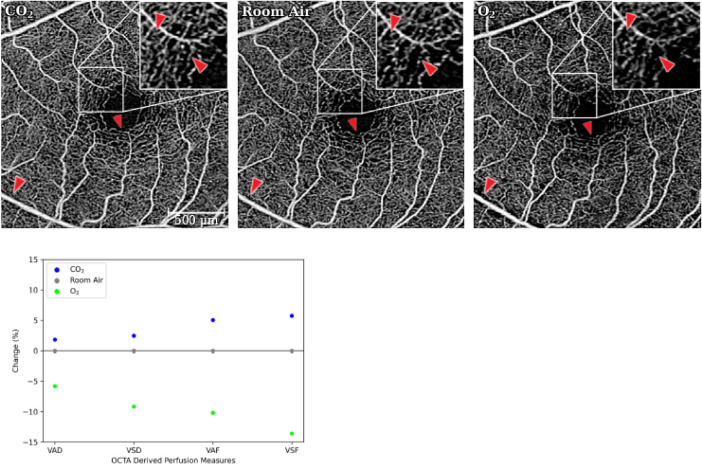
Figure 2 shows en face OCTA images acquired from a single representative subject under each gas breathing condition and illustrates the relative change in retinal perfusion measures seen in these images in response to the breathing conditions.
Figure 2.
OCTA scans of a representative subject under three breathing conditions. Arrows highlight the change in blood perfusion between scans. This change is most evident comparing the CO2 and O2 images. OCTA-derived retinal perfusion measures were calculated for each scan. The bottom plot illustrates the percentage change seen in each perfusion measure in response to the CO2 and O2 breathing conditions relative to the room-air breathing condition.
Comparison of Capillary-Only Versus All-Vessel Images
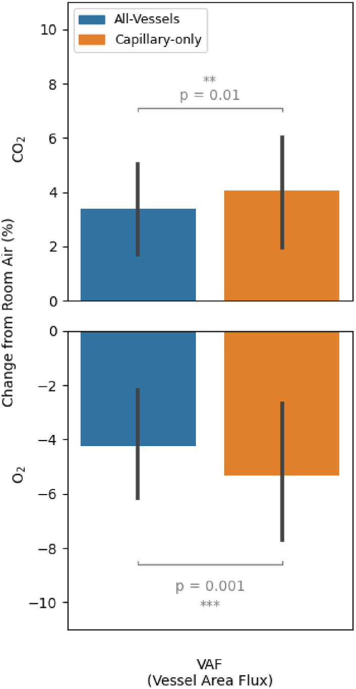
Perfusion measures showed a greater magnitude of change in capillary-only images compared with all-vessels images. Figure 3 shows the change in VAF in all-vessels images (including arterioles and venules) versus capillary-only images. In response to the CO2 breathing condition, the increase in VAF was 20.2% greater (P = 0.01) in capillary-only images. Similarly, in the O2 condition, the decrease in VAF was 25.3% greater (P = 0.001) in capillary-only images (Fig. 3, Table 2). VSF results were similar to the VAF results and are presented in Table 2 and Supplementary Figure S1. Absolute values for OCTA measures are provided in Supplementary Table S1.
Figure 3.

Comparison of the change in OCTA-derived VAF in all-vessels (including arterioles and venules) versus capillary-only images. Means and 95% confidence intervals of the mean are shown.
Table 2.
Comparison of the Percentage Change (± SD) in Retinal Perfusion Measures in Capillary-Only Versus All-Vessels Images Under CO2 and O2 Breathing Conditions
| CO2 | O2 | |||||
|---|---|---|---|---|---|---|
| Capillary Only | All Vessels | P | Capillary Only | All Vessels | P | |
| VAD | 1.51 ± 2.04 | 1.31 ± 1.76 | 0.056 | −2.76 ± 3.22 | −2.47 ± 2.69 | 0.025 |
| VSD | 2.01 ± 3.24 | 1.77 ± 2.85 | 0.092 | −3.94 ± 4.38 | −3.72 ± 3.95 | 0.119 |
| VAF | 4.05 ± 5.4 | 3.37 ± 4.57 | 0.010 | −5.35 ± 6.97 | −4.27 ± 5.67 | 0.001 |
| VSF | 4.5 ± 7.0 | 3.84 ± 6.22 | 0.020 | −6.18 ± 8.44 | −5.25 ± 7.48 | 0.002 |
Significant P-values are indicated in bold face.
Comparison of OCTA-Derived Flux Versus Density Measures in Capillary-Only Images
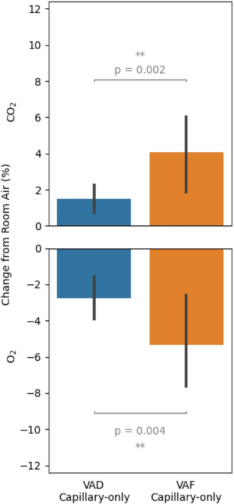
VAF showed a significantly greater magnitude of change compared with its vessel density counterpart VAD. In the CO2 condition, the increase in VAF was 168.2% greater than that in VAD (4.05% ± 5.4 vs. 1.51% ± 2.04; P = 0.002). Similarly, in the O2 condition, the decrease in VAF was 93.8% greater than that in VAD (–5.35% ± 6.97 vs. –2.76% ± 3.22; P = 0.004). These results are illustrated in Figure 4. Comparing VSF to its vessel density counterpart VSD showed very similar results (Supplementary Fig. S2).
Figure 4.

Comparison of the change in OCTA-derived VAD and VAF measures in capillary-only images. Means and 95% confidence intervals of the mean are shown.
Comparison of OCTA-Derived Flux Versus Density Measures in All-Vessels Images
In all-vessels images, flux measures had significantly greater magnitude of change compared with density measures, as well. These results are presented in Supplementary Figures S3 and S4.
Linear Mixed-Effects Model
An additional analysis using a linear mixed-effects model was used to evaluate the effect of demographic characteristics of the subjects on the change in retinal perfusion measures. In this analysis, vessel method (capillary-only > all-vessels; P = 0.007), perfusion measure (VSF > VAF > VSD > VAD; P < 0.001), and gas condition (O2 > CO2; P < 0.001) all significantly affected the percentage change. Gender (P = 0.17), age (P = 0.43), and caffeine consumption (P = 0.22) did not significantly affect the percentage change of perfusion measures.
Reliability
In total, 25 subjects had repeated images available during the room-air condition taken during the same imaging session. The ICC agreement values for VAD, VSD, VAF, and VSF were, respectively, 0.973, 0.962, 0.939, and 0.919 for capillary-only images and 0.972, 0.954, 0.954, and 0.915 for all-vessels images (Supplementary Fig. S5).
Discussion
We investigated the application of a novel OCTA-derived measure of retinal blood flow called flux in quantifying physiological changes in retinal blood flow during O2 and CO2 inhalation and compared these results to conventional OCTA-derived vessel density measures. Conventional vessel density and skeleton density measures are widely used and are based on binarized decorrelation signal intensity, which indicates only the presence or absence of blood flow within the retina.1 In contrast, flux measurements are based on the nonbinarized OCT signals, relating to information about both RBC velocity and concentration.17 In vitro modeling of RBC flow using commercially available OCTA device parameters suggests that flux is proportional to RBC concentration and likely a more accurate measure of blood flow.17 Our results confirm that flux has a significantly greater dynamic range than VAD and VSD for detecting physiological changes in blood flow. This finding suggests that flux is a more useful (and likely sensitive) measure for the assessment of subclinical blood flow changes that occur in the absence of gross capillary loss.
Our findings were most pronounced when capillary-only data were considered and larger vessels such as arterioles and venules were removed from the analysis. This suggests that measurement of OCTA-derived capillary flux is sufficient to capture significant changes in vascular autoregulatory responses in the retina and provides a powerful new research tool in the study of retinal vascular physiology and pathophysiology. Our results are supported by data from research groups using custom-designed (non-FDA approved) research instruments to study human subjects. For example, Duan et al.24 reported a higher percentage of change in capillaries compared to pre-capillary arterioles and post-capillary venules in vessels responding to hyperoxia and hypercapnia. The same group also reported that a higher proportion of retinal capillaries reacted to flicker stimulation and showed a significant change in diameter compared with pre-capillary arterioles and post-capillary venules.25 Kornfield et al.26 showed a higher percentage change in capillary flux when short-duration flicker stimulus was used in rat retina. Jeppesen et al.27 also reported that smaller diameter arterioles showed a higher degree of reaction to isometric exercise, although their data only compared arterioles of difference sizes and not capillaries.
Our data show that, in response to gas provocations, the relative magnitude of change in retinal blood flow is greater than that of retinal vessel density measures and is in agreement with previous reports. Gilmore et al.28 showed that, in reaction to hyperoxic provocation, blood flow increased more than diameter in retinal arterioles. Luksch et al.29 similarly reported a higher percentage change in retinal blood flow than retinal vessel diameter in reaction to hyperoxia. This greater magnitude of change in blood flow was also reported in reaction to hypercapnia. Dorner et al.30 reported a greater change in retinal blood flow than vessel diameter in reaction to hypercapnia. Similarly, Venkataraman et al.31 reported that, in reaction to hypercapnia, retinal blood flow increased more than the arteriolar caliber. This finding is not surprising considering Poiseuille's law, which states that the flow rate is proportional to the radius of the cannula to the fourth power. This relationship of vessel diameter and blood flow has been shown in the retina previously.32 There are several reasons why OCTA-based studies such as this study and others23 do not seem to reliably detect autoregulatory responses in larger caliber vessels elicited by gas inhalation. First, larger caliber vessels are prone to cardiac pulsation, which may wash out any incremental changes in caliber or flow. Second, unlike capillaries that pass individual RBCs, larger caliber vessels have laminar flow patterns with different flow velocities that likely confound OCTA-based measurements. Finally, the increased speed of blood flow in larger caliber vessels saturates the optical microangiography OCTA signal.
Our study had some limitations. We used a fixed inspired concentration of O2 and CO2 using a gas non-rebreathing apparatus. This method is easier to implement, but, due to between-subject variability of alveolar ventilation, it does not guarantee a constant end-tidal partial pressure of O2 and CO2.33,34 However, our results agree with the findings of studies that have used a more controlled breathing environment. Another limitation of our study was that we did not correct for the image magnification differences between the subjects due to the variation in refractive error of the eyes. However, because we excluded subjects with high magnification errors and only compared the images within individual subjects, we do not believe that this limitation would significantly affect our results or change our conclusions.
In conclusion, our study demonstrates a novel and useful tool for assessing subclinical changes in retinal perfusion at the capillary level using a commercially available SS-OCTA system and a clinically feasible, gas-breathing provocation that is of minimal risk but sufficient to induce physiological changes in retinal blood flow. Our study evaluated the change in the OCTA-derived quantitative perfusion measures in full-thickness en face OCTA images and can be expanded by assessing layer-specific changes and studying non-macular regions of the retina. Our study can also be further expanded by using other methods of provoking retinal vascular reactivity, such as flickering light stimulation, isometric exercise, or the use of other gas mixtures.
Supplementary Material
Acknowledgments
Supported by grants from the National Institutes of Health (K08EY027006 and R01EY030564 to AHK; R21EY028721 to XJ; and R01EY028753 to RW), unrestricted departmental funding from Research to Prevent Blindness, and research grants from Carl Zeiss Meditec. Carl Zeiss Meditec was not consulted in the design, implementation, or analysis of the study data.
Disclosure: F. Abdolahi, None; X. Zhou, None; B.S. Ashimatey, None; Z. Chu, None; X. Jiang, None; R.K. Wang, Carl Zeiss Meditec (P, C), Kowa Pharmaceuticals America (P), Insight Phototonic Solutions (C); A.H. Kashani, Carl Zeiss Meditec (F, R)
References
- 1. Kashani AH, Chen C-L, Gahm JK, et al.. Optical coherence tomography angiography: a comprehensive review of current methods and clinical applications. Prog Retin Eye Res. 2017; 60: 66–100. [DOI] [PMC free article] [PubMed] [Google Scholar]
- 2. Spaide RF, Fujimoto JG, Waheed NK, Sadda SR, Staurenghi G.. Optical coherence tomography angiography. Prog Retin Eye Res. 2018; 64: 1–55. [DOI] [PMC free article] [PubMed] [Google Scholar]
- 3. Matsunaga D, Yi J, Puliafito CA, Kashani AH.. OCT angiography in healthy human subjects. Ophthalmic Surg Lasers Imaging Retina. 2014; 45(6): 510–515. [DOI] [PubMed] [Google Scholar]
- 4. Kashani AH, Lee SY, Moshfeghi A, Durbin MK, Puliafito CA.. Optical coherence tomography angiography of retinal venous occlusion. Retina. 2015; 35(11): 2323–2331. [DOI] [PubMed] [Google Scholar]
- 5. Green KM, Toy BC, Ashimatey BS, et al.. Quantifying subclinical and longitudinal microvascular changes following episcleral plaque brachytherapy using spectral domain–optical coherence tomography angiography. J Vitreoretin Dis. 2020; 4(6): 499–508. [DOI] [PMC free article] [PubMed] [Google Scholar]
- 6. Kim AY, Chu Z, Shahidzadeh A, Wang RK, Puliafito CA, Kashani AH.. Quantifying microvascular density and morphology in diabetic retinopathy using spectral-domain optical coherence tomography angiography. Invest Ophthalmol Vis Sci. 2016; 57(9): OCT362–OCT370. [DOI] [PMC free article] [PubMed] [Google Scholar]
- 7. Kim AY, Rodger DC, Shahidzadeh A, et al.. Quantifying retinal microvascular changes in uveitis using spectral-domain optical coherence tomography angiography. Am J Ophthalmol. 2016; 171: 101–112. [DOI] [PMC free article] [PubMed] [Google Scholar]
- 8. Koulisis N, Kim AY, Chu Z, et al.. Quantitative microvascular analysis of retinal venous occlusions by spectral domain optical coherence tomography angiography. PLoS One. 2017; 12(4): e0176404. [DOI] [PMC free article] [PubMed] [Google Scholar]
- 9. Agemy SA, Scripsema NK, Shah CM, et al.. Retinal vascular perfusion density mapping using optical coherence tomography angiography in normals and diabetic retinopathy patients. Retina. 2015; 35(11): 2353–2363. [DOI] [PubMed] [Google Scholar]
- 10. Adhi M, Filho MAB, Louzada RN, et al.. Retinal capillary network and foveal avascular zone in eyes with vein occlusion and fellow eyes analyzed with optical coherence tomography angiography. Invest Ophthalmol Vis Sci. 2016; 57(9): OCT486–OCT494. [DOI] [PubMed] [Google Scholar]
- 11. Tokayer J, Jia Y, Dhalla A-H, Huang D.. Blood flow velocity quantification using split-spectrum amplitude-decorrelation angiography with optical coherence tomography. Biomed Opt Express. 2013; 4(10): 1909–1924. [DOI] [PMC free article] [PubMed] [Google Scholar]
- 12. Choi W, Moult EM, Waheed NK, et al.. Ultrahigh-speed, swept-source optical coherence tomography angiography in nonexudative age-related macular degeneration with geographic atrophy. Ophthalmology. 2015; 122(12): 2532–2544. [DOI] [PMC free article] [PubMed] [Google Scholar]
- 13. Bursell SE, Clermont AC, Kinsley BT, Simonson DC, Aiello LM, Wolpert HA.. Retinal blood flow changes in patients with insulin-dependent diabetes mellitus and no diabetic retinopathy. Invest Ophthalmol Vis Sci. 1996; 37(5): 886–897. [PubMed] [Google Scholar]
- 14. Higashi S, Clermont AC, Dhir V, Bursell SE.. Reversibility of retinal flow abnormalities is disease-duration dependent in diabetic rats. Diabetes. 1998; 47(4): 653–659. [DOI] [PubMed] [Google Scholar]
- 15. Wang RK, Jacques SL, Ma Z, Hurst S, Hanson SR, Gruber A.. Three dimensional optical angiography. Opt Express. 2007; 15(7): 4083–4097. [DOI] [PubMed] [Google Scholar]
- 16. Wang RK, An L, Francis P, Wilson DJ.. Depth-resolved imaging of capillary networks in retina and choroid using ultrahigh sensitive optical microangiography. Opt Lett. 2010; 35(9): 1467–1469. [DOI] [PMC free article] [PubMed] [Google Scholar]
- 17. Choi WJ, Qin W, Chen C-L, et al.. Characterizing relationship between optical microangiography signals and capillary flow using microfluidic channels. Biomed Opt Express. 2016; 7(7): 2709–2728. [DOI] [PMC free article] [PubMed] [Google Scholar]
- 18. Ashimatey BS, Green KM, Chu Z, Wang RK, Kashani AH.. Impaired retinal vascular reactivity in diabetic retinopathy as assessed by optical coherence tomography angiography. Invest Ophthalmol Vis Sci. 2019; 60(7): 2468–2473. [DOI] [PMC free article] [PubMed] [Google Scholar]
- 19. Kushner-Lenhoff S, Ashimatey BS, Kashani AH.. Retinal vascular reactivity as assessed by optical coherence tomography angiography. J Vis Exp. 2020;(157): 10.3791/60948. [DOI] [PMC free article] [PubMed] [Google Scholar]
- 20. Yezhuvath US, Lewis-Amezcua K, Varghese R, Xiao G, Lu H.. On the assessment of cerebrovascular reactivity using hypercapnia BOLD MRI. NMR Biomed. 2009; 22(7): 779–786. [DOI] [PMC free article] [PubMed] [Google Scholar]
- 21. Tayyari F, Venkataraman ST, Gilmore ED, Wong T, Fisher J, Hudson C.. The relationship between retinal vascular reactivity and arteriolar diameter in response to metabolic provocation. Invest Ophthalmol Vis Sci. 2009; 50(10): 4814–4821. [DOI] [PubMed] [Google Scholar]
- 22. Chu Z, Lin J, Gao C, et al.. Quantitative assessment of the retinal microvasculature using optical coherence tomography angiography. J Biomed Opt. 2016; 21(6): 66008. [DOI] [PMC free article] [PubMed] [Google Scholar]
- 23. Singer M, Ashimatey BS, Zhou X, Chu Z, Wang R, Kashani AH.. Impaired layer specific retinal vascular reactivity among diabetic subjects. PLoS One. 2020; 15(9): e0233871. [DOI] [PMC free article] [PubMed] [Google Scholar]
- 24. Duan A, Bedggood PA, Metha AB, Bui BV.. Reactivity in the human retinal microvasculature measured during acute gas breathing provocations. Sci Rep. 2017; 7(1): 2113. [DOI] [PMC free article] [PubMed] [Google Scholar]
- 25. Duan A, Bedggood PA, Bui BV, Metha AB.. Evidence of flicker-induced functional hyperaemia in the smallest vessels of the human retinal blood supply. PLoS One. 2016; 11(9): e0162621. [DOI] [PMC free article] [PubMed] [Google Scholar]
- 26. Kornfield TE, Newman EA.. Regulation of blood flow in the retinal trilaminar vascular network. J Neurosci. 2014; 34(34): 11504–11513. [DOI] [PMC free article] [PubMed] [Google Scholar]
- 27. Jeppesen P, Sanye-Hajari J, Bek T.. Increased blood pressure induces a diameter response of retinal arterioles that increases with decreasing arteriolar diameter. Invest Ophthalmol Vis Sci. 2007; 48(1): 328–331. [DOI] [PubMed] [Google Scholar]
- 28. Gilmore ED, Hudson C, Preiss D, Fisher J.. Retinal arteriolar diameter, blood velocity, and blood flow response to an isocapnic hyperoxic provocation. Am J Physiol Heart Circ Physiol. 2005; 288(6): H2912–H2917. [DOI] [PubMed] [Google Scholar]
- 29. Luksch A, Garhöfer G, Imhof A, et al.. Effect of inhalation of different mixtures of O(2) and CO(2) on retinal blood flow. Br J Ophthalmol. 2002; 86(10): 1143–1147. [DOI] [PMC free article] [PubMed] [Google Scholar]
- 30. Dorner GT, Garhoefer G, Zawinka C, Kiss B, Schmetterer L.. Response of retinal blood flow to CO2-breathing in humans. Eur J Ophthalmol. 2002; 12(6): 459–466. [DOI] [PubMed] [Google Scholar]
- 31. Venkataraman ST, Hudson C, Fisher JA, Rodrigues L, Mardimae A, Flanagan JG.. Retinal arteriolar and capillary vascular reactivity in response to isoxic hypercapnia. Exp Eye Res. 2008; 87(6): 535–342. [DOI] [PubMed] [Google Scholar]
- 32. Feke GT, Tagawa H, Deupree DM, Goger DG, Sebag J, Weiter JJ.. Blood flow in the normal human retina. Invest Ophthalmol Vis Sci. 1989; 30(1): 58–65. [PubMed] [Google Scholar]
- 33. Fisher JA. The CO2 stimulus for cerebrovascular reactivity: fixing inspired concentrations vs. targeting end-tidal partial pressures. J Cereb Blood Flow Metab. 2016; 36(6): 1004–1011. [DOI] [PMC free article] [PubMed] [Google Scholar]
- 34. Gilmore ED, Hudson C, Venkataraman ST, Preiss D, Fisher J.. Comparison of different hyperoxic paradigms to induce vasoconstriction: implications for the investigation of retinal vascular reactivity. Invest Ophthalmol Vis Sci. 2004; 45(9): 3207–3212. [DOI] [PubMed] [Google Scholar]
Associated Data
This section collects any data citations, data availability statements, or supplementary materials included in this article.


