PLOS ONE logoLink to PLOS ONE
. 2021 Aug 5;16(8):e0254421. doi: 10.1371/journal.pone.0254421

Impaired memory B-cell recall responses in the elderly following recurrent influenza vaccination

Rodrigo B Abreu 1, Greg A Kirchenbaum 1,2,¤, Giuseppe A Sautto 1, Emily F Clutter 1, Ted M Ross 1,2,*
Editor: Victor C Huber3
PMCID: PMC8341655  PMID: 34351920

Abstract

Influenza is a highly contagious viral respiratory disease that affects million of people worldwide each year. Annual vaccination is recommended by the World Health Organization with the goal of reducing influenza severity and limiting transmission through elicitation of antibodies targeting the hemagglutinin (HA) glycoprotein. The antibody response elicited by current seasonal influenza virus vaccines is predominantly strain-specific, but pre-existing influenza virus immunity can greatly impact the serological antibody response to vaccination. However, it remains unclear how B cell memory is shaped by recurrent annual vaccination over the course of multiple seasons, especially in high-risk elderly populations. Here, we systematically profiled the B cell response in young adult (18–34 year old) and elderly (65+ year old) vaccine recipients that received annual split inactivated influenza virus vaccination for 3 consecutive seasons. Specifically, the antibody serological and memory B-cell compartments were profiled for reactivity against current and historical influenza A virus strains. Moreover, multiparametric analysis and antibody landscape profiling revealed a transient increase in strain-specific antibodies in the elderly, but with an impaired recall response of pre-existing memory B-cells, plasmablast (PB) differentiation and long-lasting serological changes. This study thoroughly profiles and compares the immune response to recurrent influenza virus vaccination in young and elderly participants unveiling the pitfalls of current influenza virus vaccines in high-risk populations.

Introduction

Seasonal influenza virus infection remains a major public health concern with significant social and economic impact. During the 2018–2019 northern hemisphere influenza season, more than 30 million people were sick with influenza with >50% seeking healthcare services. Influenza is classified as of moderate severity disease by U.S. Centers for Disease Control and Prevention (CDC), with influenza viruses causing ~500,000 hospitalizations and ~30,000 deaths annually [1]. The World Health Organization (WHO) recommends annual vaccination to prevent seasonal influenza virus infection and transmission. Nonetheless, vaccination effectiveness is low (generally below 50%) and highly variable between influenza virus subtypes. In the U.S. each year, ≅50% of the population is vaccinated each season, which is far from the Healthy People 2020 goal of 70% coverage [2]. This results in a large proportion of the population at risk of influenza virus infection each year [3]. Furthermore, the young and the old are disproportionately impacted by influenza virus induced disease [4], with vaccinations having a lower effectiveness in these high-risk populations [5].

Influenza viruses undergo change (drift) from season to season forcing continued updating of the vaccine to include novel seasonal antigenic variants [6]. The current quadrivalent, inactivated influenza virus vaccines (QIV) mainly induce humoral immune responses, eliciting strain-specific receptor-blocking antibodies with a narrow breadth of neutralizing activity [7]. Recently, it was reported that there is antigenic competition between the four vaccine strains included in QIV, leading to a subdominant H3N2 immune response during the 2016–2017 influenza season [8]. Previous studies have hinted at the possibility of skewed immune responses to influenza virus vaccination as a consequence of past influenza virus exposures. Early-life exposure is generally described as original antigenic sin or imprinting [911]. This is particularly evident when comparing the responses to H1N1 and H3N2 influenza A viruses (IAV) in participants born when only one of these subtypes circulated in the human population [8, 1214]. However, the impact of recurrent vaccination on the immune response to QIV remains controversial [1517]. In elderly populations, recurrent vaccination was first reported to enhance neutralizing antibodies to influenza B viruses [18]. In contrast, recurrent influenza virus vaccination might hinder neutralizing antibody responses and decrease vaccine effectiveness, particularly against H3N2 IAV strains [16].

Inactivated influenza virus vaccines are poorly immunogenic and mainly rely on pre-existing immune memory. Recent developments in single cell sequencing technologies have begun to unravel the complex process of memory B-cell (Bmem) recall, clonal expansion, affinity maturation and plasmablast (PB) expansion that follows influenza virus vaccination [1922]. Still, it remains unclear how recurrent influenza virus vaccination shapes the memory B-cell compartment and the influenza-reactive serological antibody profile. In this report, the composition of serum and B cell memory polyclonal antibodies in young adult and elderly participants was tracked following recurrent vaccination for three consecutive influenza seasons. Through systematic analysis of serological antibody binding and hemagglutination-inhibition (HAI) activity against current and past influenza strains, the polyclonal antibody reactivity in young and elderly participants was profiled. Furthermore, unlike young adults, elderly participants have transient rises in antibody with HAI activity to the current influenza strains, but with minimal long-term changes in the influenza-reactive antibody profile. Mechanistically, these seems to be associated with inefficient differentiation of pre-existing vaccine-reactive Bmems into antibody-secreting PB following influenza virus vaccination.

Results

Recurrent vaccination redirects serological repertoire to receptor-blocking antibodies

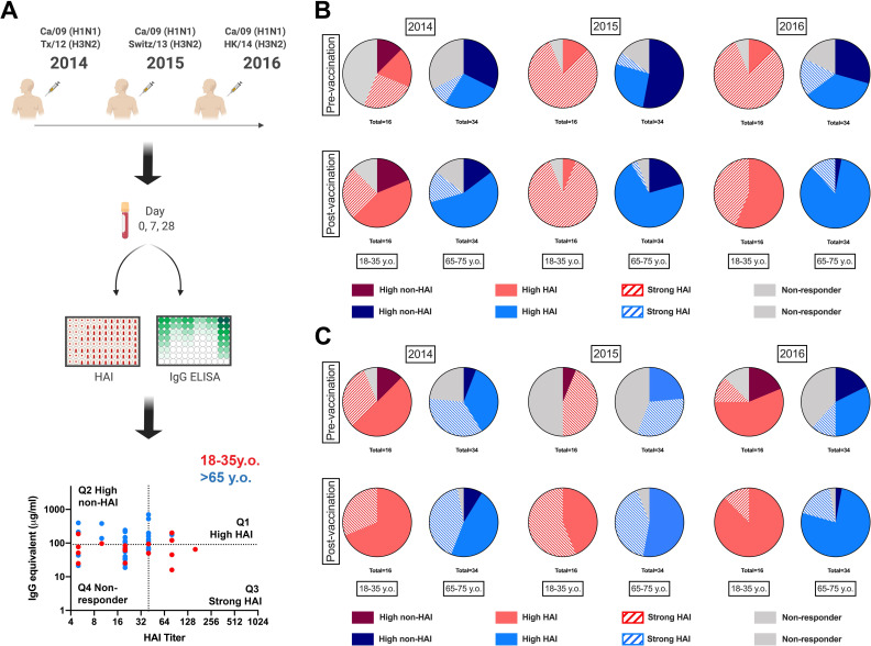
Immunological changes following influenza virus vaccination are generally assessed through serological hemagglutination inhibition (HAI) activity as a surrogate of receptor-blocking antibodies [2325]. Influenza virus vaccines strongly induce receptor-blocking antibodies in healthy young adults, but less efficiently in the elderly [24, 26, 27]. In contrast, the long-term impact of vaccination, particularly in the context of recurrent influenza virus vaccination is controversial and less-well understood [16, 18, 28]. To measure changes in the serological antibody response to yearly recurrent influenza virus vaccination in the young and elderly, 50 participants (16 young adults and 34 elderly) were vaccinated in 3 consecutive influenza seasons (2014–2015 through 2016–2017) with the split inactivated influenza virus vaccine (Fluzone®, Sanofi Pasteur, Swiftwater, PA, USA). Sera was collected pre- and post-vaccination and anti-HA specific antibodies were measured against each of the HA vaccine components. In addition, the HAI titers against both H1N1 and H3N2 vaccine components were assessed (Fig 1A). With a biparametric analysis of the anti-HA elicited antibodies, participants were categorized as high-HAI (Q1), high-non-HAI (Q2), strong-HAI (Q3) serological profiles against both IAV vaccine components. Influenza virus vaccination efficiently elicited HAI activity ~28 days post-vaccination in young and elderly participants (S1 Fig). In young adult participants, recurrent vaccination with the exact same vaccine strain (i.e. H1N1) induced long-term persistent changes in the serological profile towards receptor-binding epitopes (χ2 2016D0_2014D0 p = 0.007, Fig 1B). In contrast, elderly participants had a transient increase in HAI activity, but insignificant long-term changes in the overall antibody profile (χ2 2016D0_2014D0 p = 0.19, Fig 1B). In parallel, recurrent vaccination with antigenically distinct strains resulted in ≅45% of young participants acquiring serological cross-reactive HAI activity to the new H3N2 vaccine strain in 2015 prior to vaccination and this number increased to greater than 65% in 2016 (Fig 1C). Again, elderly participants can transiently adapt their serological antibody repertoires to the new H3N2 antigenically drifted HA proteins, but only 45% of elderly participants had cross-reactive HAI activity to the new H3N2 vaccine strain in 2016 (Fig 1C).

Fig 1. Changes in serological antibody profile following recurrent influenza vaccination.

Fig 1

A) General experimental approach for serological profiling. 50 participants (16 young-adult and 34 elderly) were vaccinated for three consecutive years with standard of care inactivated influenza vaccine and serum samples collected prior to and 21–28 days post-vaccination. Serum samples were tested for hemagglutination inhibition (HAI) activity against the H1N1 and H3N2 vaccine virus strains as described in M&M section. In parallel total rHA-reactive IgG-antibodies were quantified by ELISA as described in the M&M analysis. Biparametric quadrant analysis of each subject’s HAI titer and rHA-specific IgG (μg/ml) identified participants with High-HAI antibodies in Q1, high non-HAI in Q2, strong-HAI in Q3 and non-responders in Q4. B) Changes in H1N1-reative serological antibodies in young-adult (red) and elderly (blue) participants vaccinated for three consecutive years, measured as in A. C) Changes in H3N12-reative serological antibodies in young-adult (red) and elderly (blue) participants vaccinated for three consecutive years, measured as in A. Changes in the proportion of participants in each quadrant over time were assess by Chi-square test (χ2).

Subdominant response to H3N2 vaccine component

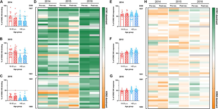
Previously, we reported a significant subdominant immune response to the H3N2 vaccine component in 2016 [8]. To understand the impact of recurrent influenza vaccination on the immunogenicity of individual H3N2 vaccine strains, the percentage of anti-H3 binding antibodies was compared to the total response against all the IAV vaccine components over the three consecutive seasons (Fig 2A–2D). Young participants had an overall balanced antibody response to both H1N1 and H3N2 vaccine components in 2014 and 2015 (Fig 2A and 2B). In contrast, elderly participants had a significantly subdominant antibody response to the H3N2 vaccine component (% H3 binding ≠ 50% p<0.0001) (Fig 2A and 2B). As previously reported [8, 17], pre-immunity against the H3N2 vaccine component in 2016 was significantly subdominant compared to the humoral response against the H1N1 vaccine component in both young and elderly participants and did not change following influenza virus vaccination (Fig 2C and 2D). Interestingly, there were overall balanced HAI activity against H3N2 and H1N1 vaccine components in both young and elderly participants to both IAV vaccine components.

Fig 2. H3N2 vaccine immunogenicity during recurrent vaccination.

Fig 2

A-C) Percentage of H3N2 rHA-binding relative to the total serum IgG antibodies against IAV vaccine strains of 50 participants (16 young adult and 36 elderly) in 2014 (A), 2015 (B) and 2016 (C). The dashed-line represents the hypothetical balanced response to both IAV (H1N1 and H3N2) vaccine components. D) IAV subtype immunodominance, based in total rHA-reactive antibodies, in 50 participants organized by age (oldest to youngest from top to bottom) vaccinated for three consecutive years. Dark green represents 100% rHA binding to H1N1 vaccine component. Bright orange represents 100% rHA binding to H3N2 vaccine component. E-G) Percentage of serum HAI activity against the H3N2 vaccine strain relative to total serological HAI activity against IAV vaccine components of 50 participants (16 young adult and 36 elderly) in 2014 (E), 2015 (F) and 2016 (G). The dashed-line represents the hypothetical balanced response to both IAV (H1N1 and H3N2) vaccine components. D) IAV subtype immunodominance, based in serological HAI activity, in 50 participants organized by age (oldest to youngest from top to bottom) vaccinated for three consecutive years. Dark green represents 100% HAI titer response to H1N1 vaccine component. Bright orange represents 100% HAI titer responses to H3N2 vaccine component. Gray boxes represent missing values.

Vaccination elicits a vaccine-specific plasmablast response

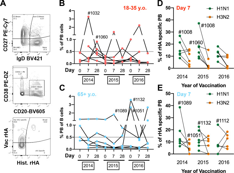
Vaccine-induced changes in the serological antibody repertoire derive from PB expansion and differentiation following vaccination [29]. To understand why the elderly had such transient changes in theirs serological repertoires compared to young participants, we selected 12 participants (6 young and 6 elderly) and quantified the frequency of PBs (CD27+/CD38++/CD20-) in peripheral blood 7 days after influenza virus vaccination (Fig 3A). Young adult participants had a prominent increase in B-cell PBs every year following vaccination (Figs 3B and S2). In contrast, elderly participants exhibited a minimal increase in the frequency of circulating PBs 7 days post-vaccination. Interestingly, 3 of the 6 elderly participants analyzed possessed elevated frequencies of circulating PBs across the multiple time-points, regardless of vaccination (D#1089, D#1132 and D#1132) (Fig 3C).

Fig 3. Plasmablast response in young and elderly participants vaccinated for three consecutive years.

Fig 3

A) Representative gating strategy to quantify rHA-specific plasmablast (CD27+/CD38++/CD20-) in the peripheral blood of vaccinated participants. B-C) Changes in frequency of plasmablast B-cells in the peripheral blood of young-adult (B) and elderly (C) participants vaccinated over three consecutive years. D-E) Frequency of vaccine-specific (H1N1 in green and H3N2 in orange) plasmablast 7 days post-vaccination in young adult (D) and elderly (E) participants vaccinated for three consecutive years. Participants of interest are identified with the corresponding ID numbers. H1N1 Vac rHA is CA/09 for 2014–16, and H1N1 Hist. rHA are NC/99 and Sing/86 (pooled at half concentration); H3N2 Vac rHA is TX/12 for 2014, Switz/13 for 2015 and HK/14 for 2016; H3N2 Hist rHA are Pan/99 and Wisc/05 for 2014–16.

To assess the frequency of antigen-specific PBs in circulation 7 days after vaccination, PBMCs were stained with fluorochrome-conjugated rHA probes, as previously described [8, 30]. Furthermore, to distinguish vaccine-specific from past cross-reactive PB responses, PBMCs were stained with both the current vaccine and a pool of historical influenza strains rHA probes (Fig 3A). Both H1N1 and H3N2 PB responses are highly vaccine-specific (S3 Fig) in young and elderly participants, while the frequency of broadly-reactive PBs is generally much lower (except D#1089 and D#1108). As extensively reported previously, influenza virus vaccination fails to recall historical-specific (and non-cross-reactive) PB responses. The serological responses to H3N2 influenza vaccine component are highly subdominant compared to H1N1 vaccine strain (Fig 2). When the frequencies of vaccine-specific PBs against H1N1 and H3N2 vaccine components were assessed, again a subdominant PB response was observed against the H3N2 vaccine component in young and elderly participants (Figs 3D, 3E and S3).

Young adult serological profile in response to recurrent influenza vaccination

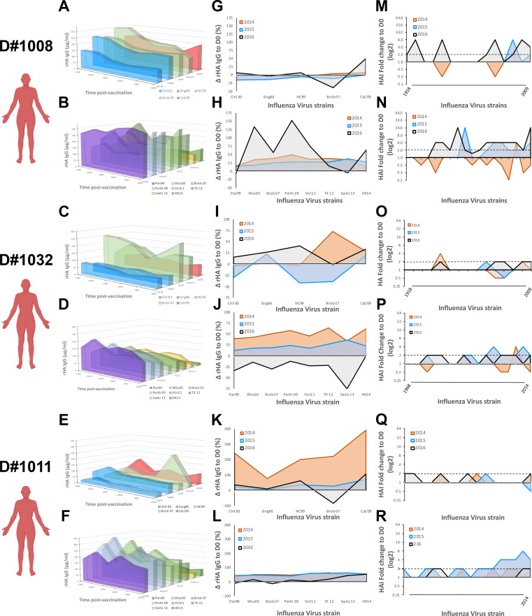
The impact of past influenza virus exposures on the immune response to influenza virus vaccination has been previously reported [8, 1214]. To profile the serological antibody repertoire, we measured the levels of antibodies reactive against a panel of historical rHAs in 6 young adult participants (Figs 4 and S4). In parallel, to profile the specificity of HAI activity, serological HAI activity was screened against an extensive panel of current and historical IAV. While some participants (D#1008 and D#1032) show signs of imprinting against both H1N1 and H3N2 strains (Figs 4A–4D and S4) from mid to late 1990’s, other participants (D#1011 ad D#1137) had signs of imprinting to just one of the IAV subtypes (Figs 4E, 4F and S4). Finally, one last young subject had signs of imprinting to an H3N2 strain from the late 1990’s (S4 Fig).

Fig 4. Serological antibody landscape in young participants vaccinated for three consecutive years.

Fig 4

A,C,E) Serological IgG antibodies against rHA from current H1N1 vaccine strain and 4 historical seasonal H1N1 virus strains (1983–2007) in three young participants vaccinated for three consecutive years. Colors represent antigenically similarity between H1 rHA. B,D,F) Serological IgG antibody levels against rHA the current H3N2 vaccine strains and 5 historical seasonal H3N2 virus strains (1999–2011) in three young participants vaccinated for three consecutive years. Colors represent antigenically similarity between H3 rHA. G,I,K) Changes in serological antibody levels against rHA from different H1N1 virus strains, measured as in A, 21–28 days after vaccination in young participants vaccinated for three consecutive years. H,J,L) Changes in serological antibody levels against rHA from different H3N2 virus strains, measured as in B, 21–28 days after vaccination in young participants vaccinated for three consecutive years. M,O,Q) Changes in serological HAI activity titer against different H1N1 virus strains (1918–2009) 21–28 days after vaccination in young participants vaccinated for three consecutive years. N,P,R) Changes in serological HAI activity titer against different H3N2 virus strains (1968–2016) 21–28 days after vaccination in young participants vaccinated for three consecutive years.

Overall, vaccination does not drastically change the serological antibody repertoire (Fig 4A–4F). To understand which antibodies are recalled and adapted towards the vaccine strain, the rise in antibody levels was calculated (ΔD21-D0) against the vaccine strains and historical influenza strains 21–28 days after vaccination, over three consecutive years (Fig 4G–4L). Again, despite such close age-range, we observe tremendously different responses to influenza virus vaccination (Fig 4G–4J). Participants imprinted with both H1N1 and H3N2 influenza strains (D#1008 and 1032) had a moderate increase in cross-reactive antibodies against both current and historical IAV strains following vaccination in 2014 and 2015. Strikingly, in 2016, these two participants had contrasting responses to influenza virus vaccination. While D#1008 mainly recalled cross-reactive antibodies against the current and past H3N2 influenza strains, D#1032 demonstrated a pronounced decrease in their overall H3N2-reactive serological antibody repertoire (Fig 4G–4J). In parallel, a participant originally imprinted with an H3N2 IAV (D#1011) had a significant increase in their cross-reactive H1N1 antibody repertoire following the first vaccination in 2014 (p = 0.032), with continuous adaptation of their antibody repertoire to the H1N1 vaccine strain in subsequent seasons (Fig 4K). The anti-H3N2 immune response was also marked by a moderate increase in serological antibodies against both current and historical strains, without pronounced changes in the overall repertoire (Fig 4L). Finally, the impact of influenza virus vaccination on antibodies with HAI activity is diverse amongst young participants (S5 Fig). While some participants had a marked increase in broadly-reactive antibodies with HAI activity (Fig 4M and 4N), others showed minimal changes in their HAI activity, even against the vaccine strain (Fig 4O and 4R).

Elderly participants show transient rises in HA-specific antibody titer

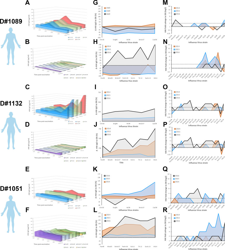
Elderly participants had impaired PB responses and long-term adaption of their serological antibody repertoire to the “new” drifted vaccine strains (Figs 13). To profile the serological antibody repertoire in 6 elderly participants, antibody titers were measured for reactivity against a panel of historical (1980–2016) rHAs (Figs 5 and S6). Elderly participants have similar levels of rHA-specific antibodies against current and past IAV strains; however, extremely polarized serological signatures were also observed, generally directed against H1N1 (Figs 5A–5F and S6). To assess imprinting in elderly participants, serological HAI activity was screened against an extensive panel of current and historical IAV strains (1918–2016) (S7 Fig). The oldest participants tested were born in 1934 (D#1089) and 1937 (D#1132). Each of these participants had increased HAI titers against H1N1 strains that were circulating in the 1940s (S7 Fig). Interestingly, the remaining elderly participants that were born in 1939 and 1940, had high HAI activity against H1N1 strains from 1940’s, but also higher HAI titers against H1N1 and H3N2 IAV strains from 1970–1980’s (S7 Fig).

Fig 5. Serological antibody landscape in elderly participants vaccinated for three consecutive years.

Fig 5

A,C,E) Serological IgG antibodies against rHA from current H1N1 vaccine strain and 4 historical seasonal H1N1 virus strains (1983–2007) in three elderly participants vaccinated for three consecutive years. Colors represent antigenically similarity between H1 rHA. B,D,F) Serological IgG antibody levels against rHA the current H3N2 vaccine strains and 5 historical seasonal H3N2 virus strains (1999–2011) in three elderly participants vaccinated for three consecutive years. Colors represent antigenically similarity between H3 rHA. G,I,K) Changes in serological antibody levels against rHA from different H1N1 virus strains, measured as in A, 21–28 days after vaccination in elderly participants vaccinated for three consecutive years. H,J,L) Changes in serological antibody levels against rHA from different H3N2 virus strains, measured as in B, 21–28 days after vaccination in elderly participants vaccinated for three consecutive years. M,O,Q) Changes in serological HAI activity titer against different H1N1 virus strains (1918–2009) 21–28 days after vaccination in elderly participants vaccinated for three consecutive years. N,P,R) Changes in serological HAI activity titer against different H3N2 virus strains (1968–2016) 21–28 days after vaccination in elderly participants vaccinated for three consecutive years.

To understand if pre-existing antibody immunity is recalled and adapted towards the vaccine strain, the rise in antibody titers (ΔD21-D0) was calculated against the vaccine strains and historical influenza strains 21 days after vaccination, over three consecutive seasons (Fig 5G–5L). Despite a prominent increase in antibody levels with broad cross-reactivity following vaccination (Fig 5G–5L), these waned significantly by the subsequent year (S6 Fig). In the elderly, this transient increase in HA-specific antibodies often translated into increased serological HAI activity against current and historical IAV strains (Figs 5M–5R and S7), particularly after three consecutive vaccinations.

Impaired memory recall responses in the elderly

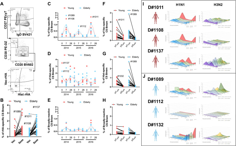
Inactivated influenza virus vaccines heavily rely on memory B-cell (Bmem) responses for protective immunity [31]. To measure the impact of recurrent influenza virus vaccination on the memory B-cell compartment in young and elderly participants, the frequency of HA-specific amongst class-switched memory B-cells (CS-Bmems) was tracked by flow cytometry (Fig 6A). Overall, the frequency of historical-reactive memory B-cells was higher than vaccine-specific (Fig 6B). However, when tracking the dynamics of HA-specific CS-Bmems over time, there was an increase in vaccine-specific CS-Bmems relative to cells reactive to historical strains 7–9 days after vaccination (Fig 6C and 6D). In contrast, recurrent influenza virus vaccination had minimal impact on broadly-reactive CS-Bmems (Fig 6E).

Fig 6. Class-switched memory B-cell (CS-Bmem) responses in young and elderly participants vaccinated for three consecutive years.

Fig 6

A) Representative gating strategy to quantify rHA-specific CS-Bmem in the peripheral blood of vaccinated participants. B) Percentage of vaccine-specific and historical-specific CS-Bmem in the peripheral blood of young (red) and elderly (blue) vaccinated participants. C-E) Percentage of vaccine-specific (C), historical-specific (D) and broadly-reactive (E) CS-Bmem in young (red) and elderly (blue) participants vaccinated for three consecutive years. Lines show cubic spline interpolation model for young adult (red) and elderly (blue) participants. F-H) Percentage of H1N1 and H3N2 vaccine-specific (F), historical-specific (G), and broadly reactive (H) CS-Bmem in young adult(red) and elderly participants vaccinated for three consecutive years I) Bmem-derived IgG antibodies against rHA from current vaccine strain and historical seasonal virus strains, as in Figs 4 and 5, in three young adult (red) and three elderly (blue) participants vaccinated for three consecutive years.

Influenza virus vaccination elicits subdominant PB and serological H3N2 responses compared to immune responses against the H1N1 vaccine component (Fig 2). Similarly, the frequency of H3N2-reactive CS-Bmems was lower than the corresponding H1N1-reactive compartment (Fig 6F and 6H). Despite their low frequency, broadly-reactive CS-Bmems were well-balanced between H1N1 and H3N2 IAV strains.

Overall, there was no difference in the frequency of vaccine-specific CS-Bmems in young and elderly participants. To ascertain the potential and binding profile of Bmem-derived antibodies, unfractionated PBMC were subjected to in vitro differentiation and conditioned supernatant samples were screened for reactivity against a panel of rHA proteins representing current and past IAV strains (Figs 6I, S8 and S9). Each season, young participants had increased HA-specific Bmem-derived antibodies 21–28 days post-vaccination. Interestingly, most young participants had a Bmem-derived antibody repertoire highly skewed towards the recent H1N1 vaccine strains with decreased reactivity against historical strains (Figs 6I and S8). In contrast, H3N2 Bmem-derived antibodies were generally lower and exhibited a broader binding profile (Figs 6I and S8).

Half of the tested elderly participants had no increase in Bmem-derived antibodies after vaccination (Figs 6J and S9). The oldest participants tested (D#1089) showed a significant increase in Bmem-derived antibodies 21–28 days after vaccination in 2014 and 2015. However, in this subject, Bmem-derived antibodies had higher reactivity against historical IAV than the current vaccine strain (Fig 6J). Furthermore, the prominent increase in Bmem-derived antibodies was followed by an almost complete depletion of the rHA-reactive Bmem compartment in 2016. In contrast, two other elderly participants had a significant rise in Bmem-derived antibodies in 2016, occurring after their third consecutive vaccination (Figs 6H and S9). Noticeably, both participants had an increase in Bmem-derived antibodies with broad reactivity against the H1N1 historical viruses, but with higher specificity towards the vaccine strain. The opposite was observed in regards to the immune responses against the H3N2 vaccine component, with an increase in their reactivity towards historical H3N2 IAV strains (Figs 6H and S9).

Elderly participants have increased frequencies of vaccine-specific atypical B-cells

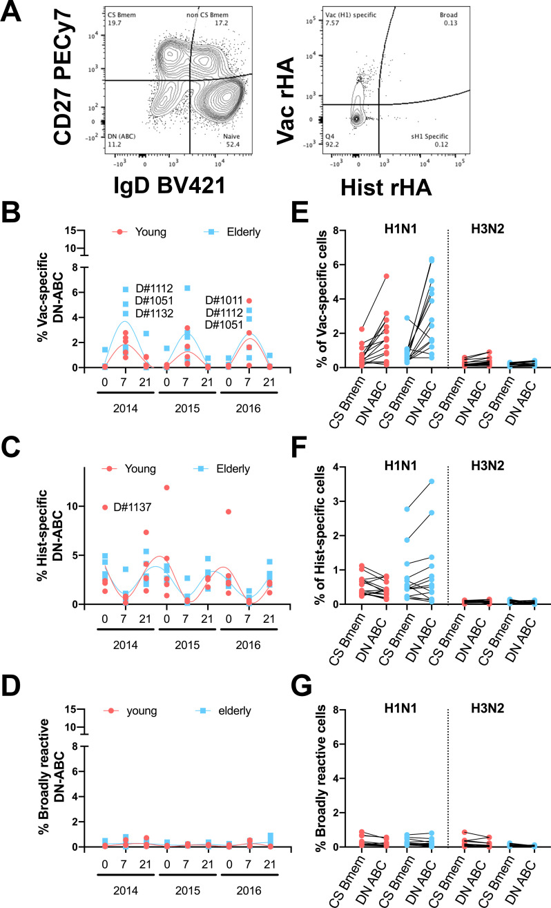
Recent reports exposed the biological relevance of double negative (CD27-/IgD-) B-cells (DN-C) [3235]. To measure the impact of influenza virus vaccination on the DN-C compartment in young and elderly participants, changes in the frequency of HA-specific DN-C were tracked by flow cytometry after staining with rHA-probes from past and current IAV strains. Overall, the dynamics of vaccine-specific DN-C is similar to that of CS-Bmems with an increase 7 days post-vaccination and concurrent with a decrease in historical IAV HA-specific DN-C (Fig 7A and 7B). However, particularly during the 2014 and 2015 influenza seasons, elderly participants had higher frequencies of vaccine-specific DN-C than young adult participants (Fig 7A). Also, in this case, the frequency of broadly-reactive DN-C is lower (Fig 7C). Moreover, the frequency of H1N1 vaccine and historical HA-specific DN-C is generally higher than the frequency of cognate CS-Bmems, especially in the elderly (Fig 7E and 7F). Interestingly, despite the lower frequencies of H3N2 rHA-B-cells, these seem to be equally distributed between the CS-Bmem and DN-C compartments (Fig 7E and 7F). Finally, the frequency of H1N1 and H3N2 broadly-reactive B-cells was similarly low in both CS-Bmem and DN-C compartments (Fig 7G).

Fig 7. Frequency of rHA-reactive DN-C in young and elderly participants.

Fig 7

A) Representative gating strategy to quantify rHA-specific DN-C in the peripheral blood of vaccinated participants. B-D) Percentage of vaccine-specific (B), historical-specific (C) and broadly reactive (D) DN-C in young adult (red) and elderly (blue) participants vaccinated for three consecutive years. E-G) Frequency of H1N1 and H3N2 vaccine-specific (E), historical-specific (F), and broadly reactive (G) B-cells in the CS-Bmem or DN-C compartment in young adult and elderly participants 7 days after vaccination.

Discussion

Shortly after Wilson Smith and colleagues first identified the etiological agent of influenza disease in 1933 and proved the induction of strong neutralizing humoral responses following influenza A virus (IAV) infection [36], they quickly recognized that antigenically drifted strains could evade the host pre-immunity and cause subsequent infections [37]. Since then, over 50 different IAV strains have been used in vaccine formulations to control seasonal influenza virus epidemics [38]. Over the past ten years the H3N2 vaccine strain included in the northern hemisphere has been were updated 7 times, while during this same interval the H1N1 vaccine strain component, representing the swine-origin influenza virus (SOIV) causing the 2009 pandemic, was not updated until the 2016–2017 season. In parallel, the past decade was marked by extensive efforts to increase vaccine coverage in high-risk populations, especially in infants and elderly subjects. Nonetheless, there is a need for comprehensive longitudinal studies assessing the impact of recurrent influenza virus vaccination in the elderly. While previous studies often lack in-depth immunological analyses, in this study the serological and memory B-cell responses were systemically characterized following recurrent influenza virus vaccination over three consecutive years, 2014–2015 to 2016–2017.

From October 2014 to March 2017, the U.S. experienced three influenza seasons of low to mild influenza virus activity. The 2014–2015 and 2016–2017 seasons were dominated by H3N2 influenza viruses with higher infection and hospitalization rates than the 2015–2016 season, which was dominated by H1N1 influenza viruses [3941]. During these three seasons, the H3N2 vaccine strain was updated each year [42]. Vaccine effectiveness across all ages and against all vaccine strains ranged from 20 to 50%, with the lowest in 2014 against H3N2 influenza viruses (5%) and the highest in 2015 against H1N1 influenza viruses (45%) [39, 4345]. In high-risk populations, such as the elderly, vaccine effectiveness was higher than the overall average against H1N1 IAV in 2015, and consistently lower against H3N2 IAV in 2014 and 2016. Interestingly, the immediate subsequent influenza season (2017–2018) was marked by an extremely severe disease outcome [45], even in the absence of significant antigenic drift between concurrent vaccines strains [42].

Our group described the impact of influenza virus vaccination between 2013 to 2017 on IAV vaccine-specific serological antibodies across different ages groups [24]. During this period, influenza virus vaccination elicited vaccine-specific neutralizing antibodies in 18–85 year old participants and back-boosted cross-reactive neutralizing antibodies to historical IAV strains from the past 30 years [24]. This was particularly evident in participants born after 1975, when both H1N1 and H3N2 IAV strains were circulating in the human population. At the time, it was not clear if this reflected a change in the influenza-reactive antibody repertoire or the result of continuous increases in influenza-specific antibody titers. Comparing serological HAI activity with total HA-reactive IgG antibodies over three consecutive seasons showed that recurrent vaccination redirects the serological influenza-reactive antibody repertoires towards antigenic sites involved in receptor binding. These changes were retained up to three seasons in young adults, but not in the elderly participants (Fig 1). Yearly updates with antigenically distinct vaccine strains required adaptation of the antibody profile to drifted epitopes, but again these changes persisted for more than a year in young adults (Fig 1C).

Recent studies explored the impact of influenza virus vaccination on the PB and memory B-cell repertoires through single-cell next generation sequencing. Despite the small subset of donors, these previous reports showed highly oligoclonal responses that originated from expansion of pre-existing memory B-cells [21]. In parallel, vaccination with dramatically different influenza virus vaccine strains requires adaptation through somatic hypermutation and affinity maturation of pre-existing memory B-cells in young adults. Furthermore, this process seems to be impaired in the elderly, but the mechanism is still not well understood [46]. Here, elderly participants showed impaired PB expansion following influenza virus vaccination (Fig 3). In contrast, young adults had increased vaccine-specific PB responses. Interestingly, elderly participants with abnormally high PB frequencies prior to vaccination showed stronger vaccine-specific PB responses and stronger serological changes following influenza virus vaccination (Figs 3 and 5). Unfortunately, no further information regarding participant chronic or acute inflammatory status is available, but this observation is aligned with the recent theory of “inflammaging” and how it can impact the response to infectious agents in the elderly [4749]. Many have speculated that immunosenescence and impaired innate immune responses are the main reasons behind decreased vaccine effectiveness in the elderly [50]. It is therefore possible that elderly participants with chronic or acute inflammatory diseases at the time of vaccination may have improved immune responses to influenza virus vaccination as observed in this study.

B-cell repertoire single-cell sequencing or serum Ig-seq elegantly depicts the molecular evolution of the immune response to influenza virus vaccination or infection [20, 22, 5155], but fails to capture the complex synergistic and competitive interactions of a polyclonal antibody mixture. Vaccination does not drastically change the influenza virus-reactive antibody landscapes (Figs 4 and 5). Young adults in this study have significantly higher antibody reactivity against historical IAV strains in circulation during the first ten years of life (1990’s), reflective of original antigenic sin. In contrast, elderly participants have similar antibody titers against H1N1 influenza virus strains isolated in 1990’s to present (Fig 5). Obviously, these IAV strains are not representative of those in circulation during early-life of elderly participants, however the significant H3N2 subdominant antibody profile in the elderly is likely a result of early-life exposure to H1N1 IAV strains (Figs 2, 3 and 5). This phenomenon is further supported by increased serological activity against H1N1 IAV strains in circulation in the 1940’s (S7 Fig). Moreover, recurrent influenza virus vaccination significantly back-boosted the serological response against historical vaccine strains, but these responses were tremendously variable even between individuals born in the same year and a with similar pre-immunity background (Fig 4). While speculative, this is likely the result of divergent Bmem-repertoire recall following vaccination (Figs 6 and 7).

Since the first identification of the two separate lymphocyte lineages (B and T-cells) in 1965 [56], many new discoveries have clarified aspects of B-cell development and different activation stages [57]. The mechanism of T-cell interactions, memory B-cell differentiation, GC-reactions, and affinity maturation are now well understood [5861]. In contrast, peripheral Bmem-cell fate is less clear and the intrinsic signals determining Bmem cell proliferation, longevity and PB differentiation are still convoluted [6264]. Moreover, recent studies in the elderly, shed light on the possibility that individuals with autoimmune or chronic infection/inflammation related disorders exhibited an exhausted B-cell compartment [34, 35, 65]. Half of the elderly participants tested in this study had negligible Bmem-derived antibody responses following influenza virus vaccination, despite similar frequencies of influenza virus-reactive memory B-cells in the periphery 7 and 21–28 days after vaccination. Vaccine-specific Bmem significantly increased 7–9 days after vaccination at the cost of recalling historical strain-reactive Bmem cells. This is most likely reflective of rapid recall and proliferation of circulating memory B-cells through direct cognate BCR-signaling and may be independent of GC reactions. Interestingly, aside from 7 days after vaccine administration, the frequency of historical-reactive Bmem cells were consistently higher than that of vaccine-reactive Bmem cells in both young and elderly participants (Fig 6). It is likely that the majority of vaccine-specific Bmem cells continue to differentiate into PBs throughout the course of the 7–10 days post-vaccination window. The fact that the PB compartment is highly enriched with vaccine-specific cells gives further supports to this hypothesis (Fig 3). Alternatively, these cells may migrate into the mucosa tissue and reside in situ awaiting cognate BCR stimulation upon subsequent infection. In this study, the distribution of IgA vs IgG Bmem cells was not assessed, but, in some participants, IgA+ cells can represent up to 50% of the memory B-cell compartment (CD27+/IgD-) that are particularly prone to populate mucosal sites.

Class-switched memory B-cells have long been defined by the expression of CD27 and negative for the IgD surface markers [8, 6668]. Recent reports highlighted the biological relevance of CD27-/IgD- double negative B-cells cells (DN-C) due to their higher frequencies in elderly participants and patients affected by autoimmune disorders [3335, 65, 69, 70]. First associated with an exhausted phenotype, this compartment is now characterized by tremendous heterogeneity [71]. Further resolution of this compartment by CD38, CD21, CD11c and T-bet transcription factor expression reveals three different memory B-cell fates: CD38+/CD21+ memory precursors; CD38-/CD21-/CD11c+/T-bet+ extrafollicular precursors; and CD38-/CD21-/FcRL4/5+ exhausted B-cells [32, 35, 70, 7274]. Here, vaccine-specific DN-ABCs kinetics resembled those of CS-Bmem, with an increase 7–9 days post-vaccination followed by a decrease 28 days after vaccination. However, this increase is significantly higher in elderly participants vaccinated in 2014 and 2015 than young adults. This difference between elderly and young adult participants was no longer detectable in 2016 after three consecutive recurrent vaccinations (Fig 7).

Perhaps the main limitation of this study relies on its small sample size, preventing definitive conclusions based on robust statistical inference. Participant recruitment for multi-year-long longitudinal studies with sufficient sample for multiparametric immunological profiling is challenging, and with such small numbers, individual variation stands-out. Nonetheless, this and other reports seem to point towards a common trend; that despite all variation, young vaccinees seem to develop and adapt their repertoire to newly drifted strains, while elderly vaccinees, recall pre-existing non-neutralizing antibodies. This study deeply profiles the immune responses to influenza virus vaccination in young and elderly participants, but it has deeper immunological implications to other multivalent vaccines and vaccinations or infections in the context of pre-existing immunity. The here reported antigenic competition between influenza virus vaccine components may explain the incomplete effectiveness of other polyvalent formulations, such as the pneumococcus vaccine. This study also highlights the relevant impact of pre-existing immunity in the response to subsequent influenza vaccinations. This is also likely to be pertinent for other infectious agents, such as the current SARS-CoV-2 pandemic which has motivated intense vaccine development efforts. In fact, it is important to recognize that any vaccine testing and assessments of efficacy performed using immunologically naïve models may not accurately reflect the magnitude and/or fine-specificity of the antibody responses elicited in humans endowed with pre-existing immunity against influenza or other coronaviruses.

Materials and methods

Study design

Ethics statement and role of the funding source

The study procedures, informed consent, and data collection documents were reviewed and approved by the University of Georgia Institutional Review Board. Volunteers were recruited at medical facilities in two sites: Pittsburgh, Pennsylvania and Stuart, Florida. All were enrolled with written, informed consent.

The funding source had no role in sample collection, nor decision to submit the manuscript for publication.

Participants and vaccine

Eligible volunteers between the ages of 18 to 35 and 65 to 85 years old (y.o.), who had not yet received the seasonal influenza vaccine, were enrolled beginning in September of each year, from 2014 to 2016. All vaccine formulations are based on World Health Organization recommendations for the Northern Hemisphere influenza seasons beginning in the Fall, and as such, all vaccinations and sample collections occurred each year between September 1st to December 15th. Influenza virus did not circulate widely in the community during the time periods that the volunteers participated, and as such, participants were not monitored for influenza virus infection during that time-period; they were however asked during each visit if they had flu symptoms, and those who did were excluded from the study. Volunteers were recruited at medical facilities in two sites: Pittsburgh, Pennsylvania and Stuart, Florida. All were enrolled with written, informed consent. Exclusion criteria included documented contraindications to Guillain-Barré syndrome, dementia or Alzheimer disease, allergies to eggs or egg products, estimated life expectancy <2 years, medical treatment causing or diagnosis of an immunocompromising condition, or concurrent participation in another influenza vaccine research study. These two cohorts spanned for four years from 2013 to 2016 [24, 25]. However, for this study only the 50 (16 young and 34 elderly) repeatedly vaccinated participants from 2014 to 2016 were selected for serological antibody profiling. Serological hemagglutination inhibition (HAI) responses from recurrent vaccinated participants were similar to matching age groups of the original cohorts. Blood (70–90 mL) was collected from each subject at the time of vaccination (D0) and 21–28 days (D21) post-vaccination. Blood samples were processed for sera and peripheral blood mononuclear cells (PBMC). For PBMC isolation, blood was collected in CPT tubes (Becton, Dickinson and Company, Franklin Lakes, NJ, USA) at D0 and D21. These samples were processed immediately, within 1–24 hours of collection, and stored at -150°C for future analysis. Sera was collected in SST tubes (Becton, Dickinson and Company) and processed within 24–48 hours, storing at 4°C until separated and aliquoted for long-term storage at -30°C. These serum samples were tested for the ability to mediate HAI and HA-specific IgG antibodies against the matching and past vaccine strains (S1 Table). Throughout the study, the H1N1 strain (A/California/7/2009) in the vaccine remained constant for three seasons, whereas the H3N2 (A/Texas/50/2012 in 2014, A/Switzerland/9715293/2013 in 2015, and A/Hong Kong/4801/2014 in 2016) vaccine strains were updated and changed each season.

Viruses and HA antigens

Influenza viruses were obtained through the Influenza Reagents Resource (IRR), BEI Resources, the Centers for Disease Control and Prevention (CDC), or were provided by Sanofi Pasteur and Virapur, LLC (San Diego, CA, USA). Viruses were passaged once in the same growth conditions as they were received, in 10-day old embryonated, specific pathogen-free (SPF) chicken eggs per the protocol provided by the WHO. Titrations were performed with turkey erythrocytes and virus was standardized to 8 HAU/50 μL for use in HAI assays. A complete list of the virus strains used is provided in S1 Table.

Recombinant HA proteins

Full-length HA proteins were developed for a panel of H1N1 and H3N2 IAV strains (S1 Table). Wild type and chimeric recombinant HA (rHA) proteins were expressed in EXPI293F cells and purified via a C-terminal histidine tag using HisTrap excel nickel-affinity chromatography columns (GE Healthcare Life Sciences, Marlborough, MA, USA) as previously described [8, 17, 7577]. Purified rHA proteins were dialyzed against PBS and total protein concentration adjusted to ~1 mg/mL after BCA estimation.

Enzyme linked immunosorbent assay (ELISA)

Hemagglutinin-specific IgG-antibodies were quantified by ELISA as previously described [8]. Briefly, Immulon® 4HBX plates (Thermo Fisher Scientific, Waltham, MA, USA) were coated with 50 ng/well of rHA in carbonate buffer (pH 9.4) with 250 ng/mL bovine serum albumin (BSA) for ~16 h at 4°C in humidified chambers. Plates were blocked with blocking buffer (2% BSA, 1% gelatin in PBS/0.05% Tween20) at 37°C for 2 h. Serum samples collected from participants prior to and 21–28 days following vaccination were initially diluted 1:500 and then further serially diluted 1:2 in blocking buffer to generate 7-point binding curves. Serially diluted serum samples were added to the assay plate in duplicate and incubated ~16 h overnight at 4°C in humidified chambers. Plates were washed 4 times with phosphate buffered saline (PBS) and HA-specific IgG detected using horseradish peroxidase (HRP)-conjugated goat anti-human IgG (Southern Biotech, Birmingham, AL, USA) at a 1:4,000 dilution and incubated for 2 h at 37°C. Plates were then washed PBS prior to development with 100 μL of 0.1% 2,2’-azino-bis(3-ethylbenzothiazoline-6-sulphonic acid) (ABTS) solution with 0.05% H2O2 for 20 min at 37°C. The reaction was terminated with 1% (w/v) sodium dodecyl sulfate (SDS). Colorimetric absorbance at 414nm was measured using a PowerWaveXS (Biotek, Winooski, VT, USA) plate reader. HA-specific IgG equivalent concentration was calculated based on an 8-point standard curve generated using a human IgG reference protein (Athens Research and Technology, Athens, GA, USA). Cumulative IAV HA binding was calculated by adding the IgG-equivalent of the both IAV vaccine components (H1N1 + H3N2).

Flow cytometry

Human PBMC (~5 x 10⁶ live cells) were stained on ice for 30 min with fluorochrome conjugated rHA probes (180–350 pM) in 100 μL of staining buffer (PBS/2% fetal bovine serum [FBS]) as previously described [8, 30, 54, 78, 79]. Human PBMC were first treated with Fc receptor blocking solution (BioLegend, Dedham, MA, USA) then stained for 30 min on ice using titrated quantities of fluorescently conjugated monoclonal antibodies (S1 Table). After completion of surface labeling, human PBMC were washed extensively with staining buffer prior to fixation with 1.6% paraformaldehyde in staining buffer for 15 min at RT. Following fixation, cells were pelleted by centrifugation at 400xg for 5 min, resuspended in staining buffer and maintained at 4˚C protected from light until acquisition. Data acquisition was performed using the BD FACSARIA Fusion and analysis performed using FlowJo (FlowJo LLC, Ashland, OR, USA). Compensation values were established prior to acquisition using appropriate single stain controls. PBs were defined as CD3/CD14neg CD19+, CD27+, CD38++ cells as previously described [67, 80].

In vitro differentiation of B cells

PBMC were cultured (2 x 10⁶ viable cells/mL) in complete media containing Roswell Park Memorial Institute (RPMI) 1640 medium (Sigma, St. Loius, MO, USA) with 10% FBS (Atlanta Biologicals, Flowery Branch, GA, USA), 23.8mM sodium bicarbonate (Fisher Scientific, Waltham, MA, USA), 7.5 mM HEPES (Amresco, Framingham, MA, USA), 170 μM Penicillin G (Tokyo Chemical Industry, Portland, OR, USA), 137 μM Streptomycin (Sigma, Burlington, MA, USA), 50 μM β-mercaptoethanol (Sigma, Burlington, MA, USA), 1 mM sodium pyruvate (Thermo Fisher Scientific, Waltham, MA, USA), essential amino acid solution (Thermo Fisher Scientific, Waltham, MA, USA), non-essential amino acid solution (Thermo Fisher Scientific, Waltham, MA, USA), 500 ng/mL R848 (Invivogen, San Diego, CA, USA) and 5 ng/mL rIL-2 (R&D, Minneapolis, MN, USA) for 7–9 days at 37°C in 5% CO2 [63, 81]. Conditioned medium supernatants were harvested and evaluated for total and rHA-specific IgG abundance by ELISA starting at a 1:5 dilution. Frequency of B cells amongst total viable PBMC was assessed by CD19 surface labeling and flow cytometry analysis.

Statistical methods

Participants were grouped by age as previously described [25] and the response to each individual vaccine component was categorized as per the WHO and European Committee for Medicinal Products to evaluate influenza vaccines [82]. Minimal seroprotection was defined as HAI titer of 1:40 to 1:80 and participants were considered seronegative with a titer below 1:40. Statistical significance between groups was calculated using one-way ANOVA Friedman test and Dunns multiple comparisons. Values were considered significant for p<0.05. Unless otherwise stated, data is presented from at least three independent experiments.

Percentage of HA binding to each vaccine strain was calculated from the cumulative IgG binding to the both IAV vaccine components for each subject individually (H1+H3). For percentage of HAI activity, serum titers were transformed to a Log2 scale prior to calculation, to avoid skewness. Significant immunodominance in a group was calculated by One-sample Wilcoxon Signed rank test (%HA≠25) and 1-way ANOVA Friedman test and Dunn’s multiple comparisons (H1≠H3). Statistical significance (p<0.05) must be reached in both tests and the highest p value is represented. Differences between pre- and post-vaccination were calculated by one-way ANOVA multiple comparisons. Percentage of rHA binding to the H3N2 vaccine component heatmap analysis was performed with GraphPad for each subject. All statistical analyses were performed using GraphPad Prism V.8.01 software.

Landscape analysis was performed on excel with average IgG antibody levels reactive to rHAs from a broad panel of current and historical IAV. Vaccine induced antibodies were calculated as the difference in rHA-reactive antibodies prior and 21–28 days post-vaccination (S10 Fig) normalized to pre-existing antibody levels as follows: ΔD28D0D0X100. Vaccine-specific HAI antibodies were determined against current and historical IAV and vaccine induced changes calculated as the fold increase in HAI prior and 21 to 28 days post-vaccination, represented in a log2 scale.

Supporting information

S1 Fig. Biparametric quadrant analysis of HAI titer and rHA-specific IgG (μg/mL) from 50 subjects (16 young-adult and 34 elderly) vaccinated for three consecutive years with standard of care inactivated influenza vaccine.

A-F) Profile response to the H1N1 vaccine strain. G-L) Profile response to the H3N2 vaccine strains. High-HAI antibodies in Q1, high non-HAI in Q2, strong HAI-Abs in Q3 and non-responders in Q4. Young-adult participants are depicted as red dots and elderly in blue. Doted lines represent the cohort’s average for rHA-specific IgG pre-vaccination (horizontal) and the generally correlated 1:40 protective serum HAI titer (vertical). Changes in the proportion of participants in each quadrant over time were assess by a Chi-square test (χ2).

(DOCX)

S2 Fig

Changes in frequency of plasmablasts of total B-cells 7 and 21–28 days after vaccination in young-adult (A) and elderly (B) participants.

(DOCX)

S3 Fig. Frequency of rHA-reactive plasmablasts against H1N1 and H3N2 vaccine strains in young (red) and elderly (blue) subjects vaccinated over three consecutive years.

(DOCX)

S4 Fig. Serological antibody landscape in young-adult participants vaccinated for three consecutive years.

A-C) Serological IgG antibodies against rHA from current H1N1 vaccine strain and 4 historical seasonal H1N1 virus strains (1983–2007) in three young subjects vaccinated for three consecutive years. Colors represent antigenic similarities between H1 rHA. D-F) Serological IgG antibody levels against rHA from the current H3N2 vaccine strains and 5 historical seasonal H3N2 virus strains (1999–2011) in three young subjects vaccinated for three consecutive years. Colors represent antigenic similarities between H3 rHA.

(DOCX)

S5 Fig. HAI antibody landscape against a broad panel of H1N1 (A-C and G-I) or H3N2 (D-F and J-L) in 6 young-adult participants vaccinated for three consecutive years.

(DOCX)

S6 Fig. Serological antibody landscape in young-adult participants vaccinated for three consecutive years.

A-C) Serological IgG antibodies against rHA from current H1N1 vaccine strain and 4 historical seasonal H1N1 virus strains (1983–2007) in three elderly subjects vaccinated for three consecutive years. Colors represent antigenic similarities between H1 rHA. D-F) Serological IgG antibody levels against rHA from the current H3N2 vaccine strains and 5 historical seasonal H3N2 virus strains (1999–2011) in three elderly subjects vaccinated for three consecutive years. Colors represent antigenic similarities between H3 rHA.

(DOCX)

S7 Fig. HAI antibody landscape against a broad panel of H1N1 (A-C and G-I) or H3N2 (D-F and J-L) in 6 elderly participants vaccinated for three consecutive years.

(DOCX)

S8 Fig. Bmem-derived IgG antibodies against rHA from current vaccine and historical seasonal influenza virus strains in three young adult participants vaccinated for three consecutive years.

(DOCX)

S9 Fig. Bmem-derived IgG antibodies against rHA from current vaccine and historical seasonal influenza virus strains in three elderly participants vaccinated for three consecutive years.

(DOCX)

S10 Fig. Illustrative approach to calculate vaccine induced rHA-reactive antibodies every year in each analyzed participant (D#1132 in 2014 shown).

Resulting transformed data was used for panels G-L in Figs 4 and 5.

(DOCX)

S1 Table. Key resources and reagents.

(DOCX)

Acknowledgments

The authors thank NIH Biodefense and Emerging Infections Research Resources Repository, NIAID, NIH for providing crucial reagents for this work. The authors would like to thank the members of the CVI protein production core, Jeffrey Ecker, Spencer Pierce, and Ethan Cooper for expression and purification of the recombinant proteins. We also thank Jonathan Murrow, Brad Phillips, Kim Schmitz, and the entire members of the UGA Clinical Trials Evaluation Unit, and give a special thanks and appreciation to the volunteer participants in the study.

Data Availability

All relevant data are within the manuscript and its Supporting Information files.

Funding Statement

This work was funded, in part, by the University of Georgia (UGA) (UGA-001) and the Emory-UGA Center of Excellence of Influenza research and Surveillance (Emory-UGA CEIRS) contract grant (HHSN272201400004C). The content is solely the responsibility of the authors and does not necessarily represent the official views of the NIH. In addition, TMR is supported by the Georgia Research Alliance as an Eminent Scholar. GAK's contributions to this manuscript preceded his current position at Cellular Technology Limited (CTL), and CTL was not involved in any part of the study. The funding organizations did not play a role in the study design, data collection and analysis, decision to publish, or preparation of the manuscript.

References

  • 1.Chung JR, Rolfes MA, Flannery B, Prasad P, O’Halloran A, Garg S, et al. Effects of Influenza Vaccination in the United States During the 2018–2019 Influenza Season. Clin Infect Dis. 2020; 1–9. doi: 10.1093/cid/ciz1244 [DOI] [PMC free article] [PubMed] [Google Scholar]
  • 2.CDC/NCIRD. FluVaxView—2010–11 through 2018–19 Influenza Seasons Vaccination Coverage Trend Report. 2020.
  • 3.Flannery B, Kondor RJG, Chung JR, Gaglani M, Reis M, Zimmerman RK, et al. Spread of Antigenically Drifted Influenza A(H3N2) Viruses and Vaccine Effectiveness in the United States During the 2018–2019 Season. J Infect Dis. 2020;221: 8–15. doi: 10.1093/infdis/jiz543 [DOI] [PMC free article] [PubMed] [Google Scholar]
  • 4.Cromer D, Van Hoek AJ, Jit M, Edmunds WJ, Fleming D, Miller E. The burden of influenza in England by age and clinical risk group: A statistical analysis to inform vaccine policy. J Infect. 2014;68: 363–371. doi: 10.1016/j.jinf.2013.11.013 [DOI] [PubMed] [Google Scholar]
  • 5.Baselga-Moreno V, Trushakova S, McNeil S, Sominina A, Nunes MC, Draganescu A, et al. Influenza epidemiology and influenza vaccine effectiveness during the 2016–2017 season in the Global Influenza Hospital Surveillance Network (GIHSN). BMC Public Health. 2019;19. doi: 10.1186/s12889-019-6713-5 [DOI] [PMC free article] [PubMed] [Google Scholar]
  • 6.Petrova VN, Russell CA. The evolution of seasonal influenza viruses. Nat Rev Microbiol. 2018;16: 47–60. doi: 10.1038/nrmicro.2017.118 [DOI] [PubMed] [Google Scholar]
  • 7.Sautto GA, Kirchenbaum GA, Ross TM. Towards a universal influenza vaccine: Different approaches for one goal. Virol J. 2018;15: 1–12. doi: 10.1186/s12985-017-0913-3 [DOI] [PMC free article] [PubMed] [Google Scholar]
  • 8.Abreu RB, Kirchenbaum GA, Clutter EF, Sautto GA, Ross TM. Preexisting subtype immunodominance shapes memory B cell recall response to influenza vaccination. JCI Insight. 2020;5. doi: 10.1172/jci.insight.132155 [DOI] [PMC free article] [PubMed] [Google Scholar]
  • 9.Francis T. On the Doctrine of Original Antigenic Sin. Proc Am Philos Soc. 1953;104: 572–578. Available: https://www.jstor.org/stable/985534 [Google Scholar]
  • 10.Vatti A, Monsalve DM, Pacheco Y, Chang C, Anaya JM, Gershwin ME. Original antigenic sin: A comprehensive review. J Autoimmun. 2017;83: 12–21. doi: 10.1016/j.jaut.2017.04.008 [DOI] [PubMed] [Google Scholar]
  • 11.Cobey S, Hensley SE. Immune history and influenza virus susceptibility. Curr Opin Virol. 2017;22: 105–111. doi: 10.1016/j.coviro.2016.12.004 [DOI] [PMC free article] [PubMed] [Google Scholar]
  • 12.Gostic KM, Ambrose M, Worobey M, Lloyd-Smith JO. Potent protection against H5N1 and H7N9 influenza via childhood hemagglutinin imprinting. Science (80-). 2016;354: 722–726. doi: 10.1126/science.aag1322 [DOI] [PMC free article] [PubMed] [Google Scholar]
  • 13.Fonville JM, Wilks SH, James SL, Fox A, Ventresca M, Aban M, et al. Antibody landscapes after influenza virus infection or vaccination. Science (80-). 2014;346: 996–1000. doi: 10.1126/science.1256427 [DOI] [PMC free article] [PubMed] [Google Scholar]
  • 14.Nachbagauer R, Choi A, Hirsh A, Margine I, Iida S, Barrera A, et al. Defining the antibody cross-reactome directed against the influenza virus surface glycoproteins. Nat Immunol. 2017;18: 464–473. doi: 10.1038/ni.3684 [DOI] [PMC free article] [PubMed] [Google Scholar]
  • 15.Bartoszko JJ, McNamara IF, Aras OAZ, Hylton DA, Zhang YB, Malhotra D, et al. Does consecutive influenza vaccination reduce protection against influenza: A systematic review and meta-analysis. Vaccine. 2018. doi: 10.1016/j.vaccine.2018.04.049 [DOI] [PubMed] [Google Scholar]
  • 16.Belongia EA, Skowronski DM, McLean HQ, Chambers C, Sundaram ME, De Serres G. Repeated annual influenza vaccination and vaccine effectiveness: review of evidence. Expert Rev Vaccines. 2017;16: 723–736. doi: 10.1080/14760584.2017.1334554 [DOI] [PubMed] [Google Scholar]
  • 17.Abreu RB, Clutter EF, Attari S, Sautto GA, Ross TM. IgA Responses Following Recurrent Influenza Virus Vaccination. Front Immunol. 2020;11: 1–13. doi: 10.3389/fimmu.2020.00001 [DOI] [PMC free article] [PubMed] [Google Scholar]
  • 18.de Bruijn IA, Remarque EJ, Beyer WEP, Cessie S le, Masurel N, Ligthart GJ. Annually repeated influenza vaccination improves humoral responses to several influenza virus strains in healthy elderly. Vaccine. 1997;15: 1323–1329. doi: 10.1016/s0264-410x(97)00019-4 [DOI] [PubMed] [Google Scholar]
  • 19.Setliff I, Shiakolas AR, Pilewski KA, Murji AA, Mapengo RE, Janowska K, et al. High-Throughput Mapping of B Cell Receptor Sequences to Antigen Specificity. Cell. 2019;179: 1636–1646.e15. doi: 10.1016/j.cell.2019.11.003 [DOI] [PMC free article] [PubMed] [Google Scholar]
  • 20.Lee J, Boutz DR, Chromikova V, Joyce MG, Vollmers C, Leung K, et al. Molecular-level analysis of the serum antibody repertoire in young adults before and after seasonal influenza vaccination. Nat Med. 2016;22: 1456–1464. doi: 10.1038/nm.4224 [DOI] [PMC free article] [PubMed] [Google Scholar]
  • 21.Lee J, Paparoditis P, Horton AP, Frühwirth A, McDaniel JR, Jung J, et al. Persistent Antibody Clonotypes Dominate the Serum Response to Influenza over Multiple Years and Repeated Vaccinations. Cell Host Microbe. 2019;0: 1–10. doi: 10.1016/j.chom.2019.01.010 [DOI] [PMC free article] [PubMed] [Google Scholar]
  • 22.Neu KE, Guthmiller JJ, Huang M, La J, Vieira MC, Kim K, et al. Spec-seq unveils transcriptional subpopulations of antibody-secreting cells following influenza vaccination. J Clin Invest. 2019;129: 93–105. doi: 10.1172/JCI121341 [DOI] [PMC free article] [PubMed] [Google Scholar]
  • 23.Kaufmann L, Syedbasha M, Vogt D, Hollenstein Y, Hartmann J, Linnik JE, et al. An optimized hemagglutination inhibition (HI) assay to quantify influenzaspecific antibody titers. J Vis Exp. 2017;2017: 1–10. doi: 10.3791/55833 [DOI] [PMC free article] [PubMed] [Google Scholar]
  • 24.Nuñez IA, Carlock MA, Allen JD, Owino SO, Moehling KK, Nowalk P, et al. Impact of age and pre-existing influenza immune responses in humans receiving split inactivated influenza vaccine on the induction of the breadth of antibodies to influenza A strains. PLoS One. 2017;12: 1–23. doi: 10.1371/journal.pone.0185666 [DOI] [PMC free article] [PubMed] [Google Scholar]
  • 25.Carlock MA, Ingram JG, Clutter EF, Cecil NC, Ramgopal M, Zimmerman RK, et al. Impact of age and pre-existing influenza on the induction of human antibody responses against influenza B viruses. Hum Vaccin Immunother. 2019; 21645515.2019.1642056. doi: 10.1080/21645515.2019.1642056 [DOI] [PMC free article] [PubMed] [Google Scholar]
  • 26.Goodwin K, Viboud C, Simonsen L. Antibody response to influenza vaccination in the elderly: A quantitative review. Vaccine. 2006;24: 1159–1169. doi: 10.1016/j.vaccine.2005.08.105 [DOI] [PubMed] [Google Scholar]
  • 27.Sasaki S, Sullivan M, Narvaez CF, Holmes TH, Furman D, Zheng NY, et al. Limited efficacy of inactivated influenza vaccine in elderly individuals is associated with decreased production of vaccine-specific antibodies. J Clin Invest. 2011;121: 3109–3119. doi: 10.1172/JCI57834 [DOI] [PMC free article] [PubMed] [Google Scholar]
  • 28.Kosikova M, Li L, Radvak P, Ye Z, Wan XF, Xie H. Imprinting of repeated influenza A/H3 exposures on antibody quantity and antibody quality: Implications for seasonal vaccine strain selection and vaccine performance. Clin Infect Dis. 2018;67: 1523–1532. doi: 10.1093/cid/ciy327 [DOI] [PMC free article] [PubMed] [Google Scholar]
  • 29.Palm A-KE, Henry C. Remembrance of Things Past: Long-Term B Cell Memory After Infection and Vaccination. Front Immunol. 2019;10: 1–13. doi: 10.3389/fimmu.2019.00001 [DOI] [PMC free article] [PubMed] [Google Scholar]
  • 30.Whittle JRR, Wheatley AK, Wu L, Lingwood D, Kanekiyo M, Ma SS, et al. Flow Cytometry Reveals that H5N1 Vaccination Elicits Cross-Reactive Stem-Directed Antibodies from Multiple Ig Heavy-Chain Lineages. J Virol. 2014;88: 4047–4057. doi: 10.1128/JVI.03422-13 [DOI] [PMC free article] [PubMed] [Google Scholar]
  • 31.Abreu R, Ross TMTM. Influenza–A new pathogen every year. Curr Opin Syst Biol. 2018;12: 12–21. doi: 10.1016/j.coisb.2018.08.011 [DOI] [Google Scholar]
  • 32.Johnson JL, Rosenthal RL, Knox JJ, Myles A, Naradikian MS, Madej J, et al. The Transcription Factor T-bet Resolves Memory B Cell Subsets with Distinct Tissue Distributions and Antibody Specificities in Mice and Humans. Immunity. 2020;52: 842–855.e6. doi: 10.1016/j.immuni.2020.03.020 [DOI] [PMC free article] [PubMed] [Google Scholar]
  • 33.Blanco E, Pérez-Andrés M, Arriba-Méndez S, Contreras-Sanfeliciano T, Criado I, Pelak O, et al. Age-associated distribution of normal B-cell and plasma cell subsets in peripheral blood. J Allergy Clin Immunol. 2018;141: 2208–2219.e16. doi: 10.1016/j.jaci.2018.02.017 [DOI] [PubMed] [Google Scholar]
  • 34.Zou Y-R. Age-associated B cells acquire a new wrinkle. 2018;19: 317–318. doi: 10.1038/s41590-018-0072-8 [DOI] [PubMed] [Google Scholar]
  • 35.Phalke S, Marrack P. Age (autoimmunity) associated B cells (ABCs) and their relatives. Curr Opin Immunol. 2018;55: 75–80. doi: 10.1016/j.coi.2018.09.007 [DOI] [PubMed] [Google Scholar]
  • 36.Smith W, Andrewes CH, Laidlaw PP. A VIRUS OBTAINED FROM INFLUENZA PATIENTS. Lancet. 1933;222: 66–68. doi: 10.1016/S0140-6736(00)78541-2 [DOI] [Google Scholar]
  • 37.Smith W, Andrewes CH. Epidemic Influenza Virus. 293 Serological Races of Influenza Virus. Aust J Exp Biol Med Sci. 1937;15: 291. Available: https://europepmc.org/backend/ptpmcrender.fcgi?accid=PMC2065290&blobtype=pdf [Google Scholar]
  • 38.Barberis I, Myles P, Ault SK, Bragazzi NL, Martini M. History and evolution of influenza control through vaccination: From the first monovalent vaccine to universal vaccines. J Prev Med Hyg. 2016;57: E115–E120. doi: 10.15167/2421-4248/jpmh2016.57.3.642 [DOI] [PMC free article] [PubMed] [Google Scholar]
  • 39.Flannery B, Clippard J, Zimmerman RK, Nowalk MP, Jackson ML, Jackson LA, et al. Early Estimates of Seasonal Influenza Vaccine Effectiveness—United States, January 2015. Morb Mortal Wkly Rep. 2015;64: 10–15. Available: http://www.ncbi.nlm.nih.gov/pubmed/25590680%0Ahttp://www.pubmedcentral.nih.gov/articlerender.fcgi?artid=PMC4584793 [PMC free article] [PubMed] [Google Scholar]
  • 40.Davlin SL, Blanton L, Kniss K, Mustaquim D, Smith S, Kramer N, et al. Influenza activity—United States, 2015–16 season and composition of the 2016–17 influenza vaccine. Morb Mortal Wkly Rep. 2016;65: 567–575. doi: 10.15585/mmwr.mm6522a3 [DOI] [PubMed] [Google Scholar]
  • 41.Pivette M, Nicolay N, de Lauzun V, Hubert B. Characteristics of hospitalizations with an influenza diagnosis, France, 2012–2013 to 2016–2017 influenza seasons. Influenza Other Respi Viruses. 2020;14: 340–348. doi: 10.1111/irv.12719 [DOI] [PMC free article] [PubMed] [Google Scholar]
  • 42.World Health Organization. WHO recommendations on the composition of influenza virus vaccines. 2020. Available: https://www.who.int/influenza/vaccines/virus/recommendations/en/
  • 43.Souty C, Blanchon T, Bonmarin I, Lévy-Bruhl D, Behillil S, Enouf V, et al. Early estimates of 2014/15 seasonal influenza vaccine effectiveness in preventing influenza-like illness in general practice using the screening method in France. Hum Vaccines Immunother. 2015;11: 1621–1625. doi: 10.1080/21645515.2015.1046661 [DOI] [PMC free article] [PubMed] [Google Scholar]
  • 44.Jackson ML, Chung JR, Jackson LA, Phillips CH, Benoit J, Monto AS, et al. Influenza vaccine effectiveness in the United States during the 2015–2016 season. N Engl J Med. 2017;377: 534–543. doi: 10.1056/NEJMoa1700153 [DOI] [PMC free article] [PubMed] [Google Scholar]
  • 45.Rolfes MA, Flannery B, Chung JR, O’Halloran A, Garg S, Belongia EA, et al. Effects of Influenza Vaccination in the United States during the 2017–2018 Influenza Season. Clin Infect Dis. 2019;69: 1845–1853. doi: 10.1093/cid/ciz075 [DOI] [PMC free article] [PubMed] [Google Scholar]
  • 46.Henry C, Zheng N-Y, Huang M, Cabanov A, Rojas KT, Kaur K, et al. Influenza Virus Vaccination Elicits Poorly Adapted B Cell Responses in Elderly Individuals. Cell Host Microbe. 2019; 1–10. doi: 10.1016/j.chom.2019.01.002 [DOI] [PMC free article] [PubMed] [Google Scholar]
  • 47.Fulop T, Larbi A, Dupuis G, Page A Le, Frost EH, Cohen AA, et al. Immunosenescence and inflamm-aging as two sides of the same coin: Friends or Foes? Front Immunol. 2018;8. doi: 10.3389/fimmu.2017.01960 [DOI] [PMC free article] [PubMed] [Google Scholar]
  • 48.Franceschi C, Garagnani P, Parini P, Giuliani C, Santoro A. Inflammaging: a new immune–metabolic viewpoint for age-related diseases. Nat Rev Endocrinol. 2018;14: 576–590. doi: 10.1038/s41574-018-0059-4 [DOI] [PubMed] [Google Scholar]
  • 49.Accardi G, Caruso C. Immune-inflammatory responses in the elderly: An update. Immun Ageing. 2018;15: 4–7. doi: 10.1186/s12979-017-0106-3 [DOI] [PMC free article] [PubMed] [Google Scholar]
  • 50.Crooke SN, Ovsyannikova IG, Poland GA, Kennedy RB. Immunosenescence and human vaccine immune responses. Immun Ageing. 2019;16: 1–16. doi: 10.1186/s12979-018-0141-8 [DOI] [PMC free article] [PubMed] [Google Scholar]
  • 51.Wu YC, Kipling D, Leong HS, Martin V, Ademokun AA, Dunn-Walters DK. High-throughput immunoglobulin repertoire analysis distinguishes between human IgM memory and switched memory B-cell populations. Blood. 2010;116: 1070–1078. doi: 10.1182/blood-2010-03-275859 [DOI] [PMC free article] [PubMed] [Google Scholar]
  • 52.Shah HB, Smith K, Wren JD, Webb CF, Ballard JD, Bourn RL, et al. Insights from analysis of human antigen-specific memory B cell repertoires. Front Immunol. 2019;10: 1–15. doi: 10.3389/fimmu.2019.00001 [DOI] [PMC free article] [PubMed] [Google Scholar]
  • 53.Holmes AB, Corinaldesi C, Shen Q, Kumar R, Compagno N, Wang Z, et al. Single-cell analysis of germinal-center B cells informs on lymphoma cell of origin and outcome. J Exp Med. 2020;217. doi: 10.1084/jem.20200483 [DOI] [PMC free article] [PubMed] [Google Scholar]
  • 54.Ellebedy AH, Jackson KJL, Kissick HT, Nakaya HI, Davis CW, Roskin KM, et al. Defining antigen-specific plasmablast and memory B cell subsets in human blood after viral infection or vaccination. Nat Immunol. 2016;17: 1226–1234. doi: 10.1038/ni.3533 [DOI] [PMC free article] [PubMed] [Google Scholar]
  • 55.Cortina-Ceballos B, Godoy-Lozano EE, Téllez-Sosa J, Ovilla-Muñoz M, Sámano-Sánchez H, Aguilar-Salgado A, et al. Longitudinal analysis of the peripheral B cell repertoire reveals unique effects of immunization with a new influenza virus strain. Genome Med. 2015;7: 1–17. doi: 10.1186/s13073-014-0122-2 [DOI] [PMC free article] [PubMed] [Google Scholar]
  • 56.COOPER MD, PETERSON RDA, GOOD RA. Delineation of the Thymic and Bursal Lymphoid Systems in the Chicken. Nature. 1965;205: 143–146. doi: 10.1038/205143a0 [DOI] [PubMed] [Google Scholar]
  • 57.Pieper K, Grimbacher B, Eibel H. B-cell biology and development. J Allergy Clin Immunol. 2013;131: 959–971. doi: 10.1016/j.jaci.2013.01.046 [DOI] [PubMed] [Google Scholar]
  • 58.Cooper MD. The early history of B cells. Nat Rev Immunol. 2015;15: 191–197. doi: 10.1038/nri3801 [DOI] [PubMed] [Google Scholar]
  • 59.Whitlock C, Denis K, Robertson D, Witte O. In Vitro Analysis of Murine B-Cell Development. Annu Rev Immunol. 1985;3: 213–235. doi: 10.1146/annurev.iy.03.040185.001241 [DOI] [PubMed] [Google Scholar]
  • 60.Toboso-Navasa A, Gunawan A, Morlino G, Nakagawa R, Taddei A, Damry D, et al. Restriction of memory B cell differentiation at the germinal center B cell positive selection stage. J Exp Med. 2020;217. doi: 10.1084/jem.20191933 [DOI] [PMC free article] [PubMed] [Google Scholar]
  • 61.Adachi Y, Onodera T, Yamada Y, Daio R, Tsuiji M, Inoue T, et al. Distinct germinal center selection at local sites shapes memory B cell response to viral escape. J Exp Med. 2015;212: 1709–1723. doi: 10.1084/jem.20142284 [DOI] [PMC free article] [PubMed] [Google Scholar]
  • 62.Fink K. Origin and function of circulating plasmablasts during acute viral infections. Front Immunol. 2012;3: 1–5. doi: 10.3389/fimmu.2012.00001 [DOI] [PMC free article] [PubMed] [Google Scholar]
  • 63.Frasca D, Diaz A, Romero M, Blomberg BB. The generation of memory B cells is maintained, but the antibody response is not, in the elderly after repeated influenza immunizations. Vaccine. 2016;34: 2834–2840. doi: 10.1016/j.vaccine.2016.04.023 [DOI] [PMC free article] [PubMed] [Google Scholar]
  • 64.Rothaeusler K, Baumgarth N. B-cell fate decisions following influenza virus infection. Eur J Immunol. 2010;40: 366–377. doi: 10.1002/eji.200939798 [DOI] [PMC free article] [PubMed] [Google Scholar]
  • 65.Blaeser A, McGlauchlen K, Vogel LA. Aged B lymphocytes retain their ability to express surface markers but are dysfunctional in their proliferative capability during early activation events. Immun Ageing. 2008;5: 1–10. doi: 10.1186/1742-4933-5-1 [DOI] [PMC free article] [PubMed] [Google Scholar]
  • 66.Wu YB, Kipling D, Dunn-Walters DK. The Relationship between CD27 Negative and Positive B Cell Populations in Human Peripheral Blood. Front Immunol. 2011;2: 1–12. doi: 10.3389/fimmu.2011.00001 [DOI] [PMC free article] [PubMed] [Google Scholar]
  • 67.Sanz I, Wei C, Lee FE-H, Anolik J. Phenotypic and functional heterogeneity of human memory B cells. Semin Immunol. 2008;20: 67–82. doi: 10.1016/j.smim.2007.12.006 [DOI] [PMC free article] [PubMed] [Google Scholar]
  • 68.Allman D, Pillai S. Peripheral B cell subsets. Curr Opin Immunol. 2008;20: 149–157. doi: 10.1016/j.coi.2008.03.014 [DOI] [PMC free article] [PubMed] [Google Scholar]
  • 69.Rosser EC, Mauri C. Regulatory B Cells: Origin, Phenotype, and Function. Immunity. 2015;42: 607–612. doi: 10.1016/j.immuni.2015.04.005 [DOI] [PubMed] [Google Scholar]
  • 70.Thorarinsdottir K, Camponeschi A, Cavallini N, Grimsholm O, Jacobsson L, Gjertsson I, et al. CD21–/low B cells in human blood are memory cells. Clin Exp Immunol. 2016;185: 252–262. doi: 10.1111/cei.12795 [DOI] [PMC free article] [PubMed] [Google Scholar]
  • 71.Sanz I, Wei C, Jenks SA, Cashman KS, Tipton C, Woodruff MC, et al. Challenges and opportunities for consistent classification of human b cell and plasma cell populations. Front Immunol. 2019;10: 1–17. doi: 10.3389/fimmu.2019.00001 [DOI] [PMC free article] [PubMed] [Google Scholar]
  • 72.Lau D, Lan LY-L, Andrews SF, Henry C, Rojas KT, Neu KE, et al. Low CD21 expression defines a population of recent germinal center graduates primed for plasma cell differentiation. Sci Immunol. 2017;2: eaai8153. doi: 10.1126/sciimmunol.aai8153 [DOI] [PMC free article] [PubMed] [Google Scholar]
  • 73.Nipper AJ, Smithey MJ, Shah RC, Canaday DH, Landay AL. Diminished antibody response to influenza vaccination is characterized by expansion of an age-associated B-cell population with low PAX5. Clin Immunol. 2018. doi: 10.1016/j.clim.2018.02.003 [DOI] [PMC free article] [PubMed] [Google Scholar]
  • 74.Blumentals WA, Arreglado A, Napalkov P, Toovey S. Rheumatoid arthritis and the incidence of influenza and influenza-related complications: A retrospective cohort study. BMC Musculoskelet Disord. 2012;13: 1. doi: 10.1186/1471-2474-13-1 [DOI] [PMC free article] [PubMed] [Google Scholar]
  • 75.Kirchenbaum GA, Allen JD, Layman TS, Sautto GA, Ross TM. Infection of Ferrets with Influenza Virus Elicits a Light Chain–Biased Antibody Response against Hemagglutinin. J Immunol. 2017;199: 3798–3807. doi: 10.4049/jimmunol.1701174 [DOI] [PubMed] [Google Scholar]
  • 76.Sautto GA, Kirchenbaum GA, Abreu RB, Ecker JW, Pierce SR, Kleanthous H, et al. A Computationally Optimized Broadly Reactive Antigen Subtype–Specific Influenza Vaccine Strategy Elicits Unique Potent Broadly Neutralizing Antibodies against Hemagglutinin. J Immunol. 2020;204: 375–385. doi: 10.4049/jimmunol.1900379 [DOI] [PubMed] [Google Scholar]
  • 77.Sautto GA, Kirchenbaum GA, Ecker JW, Bebin-Blackwell A-G, Pierce SR, Ross TM. Elicitation of Broadly Protective Antibodies following Infection with Influenza Viruses Expressing H1N1 Computationally Optimized Broadly Reactive Hemagglutinin Antigens. ImmunoHorizons. 2018;2: 226–237. doi: 10.4049/immunohorizons.1800044 [DOI] [PubMed] [Google Scholar]
  • 78.Tesini BL, Kanagaiah P, Wang J, Hahn M, Halliley JL, Chaves FA, et al. Broad Hemagglutinin-Specific Memory B Cell Expansion by Seasonal Influenza Virus Infection Reflects Early-Life Imprinting and Adaptation to the Infecting Virus. Schultz-Cherry S, editor. J Virol. 2019;93. doi: 10.1128/JVI.00169-19 [DOI] [PMC free article] [PubMed] [Google Scholar]
  • 79.Ecker JW, Kirchenbaum GA, Pierce SR, Skarlupka AL, Abreu RB, Ethan Cooper R, et al. High-yield expression and purification of recombinant influenza virus proteins from stably-transfected mammalian cell lines. Vaccines. 2020;8: 1–20. doi: 10.3390/vaccines8030462 [DOI] [PMC free article] [PubMed] [Google Scholar]
  • 80.Perez-Andres M, Paiva B, Nieto WG, Caraux A, Schmitz A, Almeida J, et al. Human peripheral blood B-cell compartments: A crossroad in B-cell traffic. Cytom Part B Clin Cytom. 2010;78B: S47–S60. doi: 10.1002/cyto.b.20547 [DOI] [PubMed] [Google Scholar]
  • 81.Faenzi E, Zedda L, Bardelli M, Spensieri F, Borgogni E, Volpini G, et al. One dose of an MF59-adjuvanted pandemic A/H1N1 vaccine recruits pre-existing immune memory and induces the rapid rise of neutralizing antibodies. Vaccine. 2012;30: 4086–4094. doi: 10.1016/j.vaccine.2012.04.020 [DOI] [PubMed] [Google Scholar]
  • 82.WHO WHO. Manual for the laboratory diagnosis and virological surveillance of influenza. Geneva: World Health Organization; 2011. [Google Scholar]

Decision Letter 0

Victor C Huber

3 Mar 2021

PONE-D-20-36767

Impaired memory B-cell recall responses in the elderly following recurrent influenza vaccination

PLOS ONE

Dear Dr. Ross,

Thank you for submitting your manuscript to PLOS ONE. After careful consideration, we feel that it has merit but does not fully meet PLOS ONE’s publication criteria as it currently stands. Therefore, we invite you to submit a revised version of the manuscript that addresses the points raised during the review process.

While the reviewers found both the project and the goals that the authors are trying to achieve to be of significant interest, there were some substantial issues with the data presentation and interpretation of the findings that were identified by the reviewers.  Specifically, during the revision process the authors are asked to make certain that the data support the conclusions that were drawn.  Also, please address the study design and technical aspects that greatly affect data presentation and interpretation of the findings.

Please submit your revised manuscript by Apr 11 2021 11:59PM. If you will need more time than this to complete your revisions, please reply to this message or contact the journal office at plosone@plos.org. When you're ready to submit your revision, log on to https://www.editorialmanager.com/pone/ and select the 'Submissions Needing Revision' folder to locate your manuscript file.

Please include the following items when submitting your revised manuscript:

  • A rebuttal letter that responds to each point raised by the academic editor and reviewer(s). You should upload this letter as a separate file labeled 'Response to Reviewers'.

  • A marked-up copy of your manuscript that highlights changes made to the original version. You should upload this as a separate file labeled 'Revised Manuscript with Track Changes'.

  • An unmarked version of your revised paper without tracked changes. You should upload this as a separate file labeled 'Manuscript'.

If you would like to make changes to your financial disclosure, please include your updated statement in your cover letter. Guidelines for resubmitting your figure files are available below the reviewer comments at the end of this letter.

If applicable, we recommend that you deposit your laboratory protocols in protocols.io to enhance the reproducibility of your results. Protocols.io assigns your protocol its own identifier (DOI) so that it can be cited independently in the future. For instructions see: http://journals.plos.org/plosone/s/submission-guidelines#loc-laboratory-protocols

We look forward to receiving your revised manuscript.

Kind regards,

Victor C Huber

Academic Editor

PLOS ONE

Journal Requirements:

When submitting your revision, we need you to address these additional requirements.

1. Please ensure that your manuscript meets PLOS ONE's style requirements, including those for file naming. The PLOS ONE style templates can be found at

https://journals.plos.org/plosone/s/file?id=wjVg/PLOSOne_formatting_sample_main_body.pdf and

https://journals.plos.org/plosone/s/file?id=ba62/PLOSOne_formatting_sample_title_authors_affiliations.pdf

2. Please provide additional details regarding participant consent. In the ethics statement in the Methods and online submission information, please ensure that you have specified whether consent was informed.

3. In your Methods section, please provide additional information about the participant recruitment method and the demographic details of your participants. Please ensure you have provided sufficient details to replicate the analyses such as:

a) a table of relevant demographic details,

b) a statement as to whether your sample can be considered representative of a larger population, and

c) a description of how participants were recruited.

4.Our staff editors have determined that your manuscript is likely within the scope of our Call for Papers on Influenza. This editorial initiative is headed by PLOS ONE Guest Editors Dr. Meagan Deming and Dr. Deshayne Fell. The Collection encompasses research on influenza prevention on every level, including in vitro, translational, behavioral, and clinical studies; disease and immunity modelling; as well as new approaches to influenza prevention. Additional information can be found on our announcement page: https://collections.plos.org/call-for-papers/influenza/.

Currently, your manuscript is included in the group of papers being considered for this call. Please note that being considered for the Collection does not require additional peer review beyond the journal’s standard process and will not delay the publication of your manuscript if it is accepted by PLOS ONE. We would greatly appreciate your confirmation that you would like your manuscript to be considered for this Collection by indicating this in your next cover letter. If you would prefer to remove your manuscript from collection consideration, please specify this in your cover letter.

5.We note that the grant information you provided in the ‘Funding Information’ and ‘Financial Disclosure’ sections do not match.

When you resubmit, please ensure that you provide the correct grant numbers for the awards you received for your study in the ‘Funding Information’ section.

6. We note that you have included the phrase “data not shown” in your manuscript. Unfortunately, this does not meet our data sharing requirements. PLOS does not permit references to inaccessible data. We require that authors provide all relevant data within the paper, Supporting Information files, or in an acceptable, public repository. Please add a citation to support this phrase or upload the data that corresponds with these findings to a stable repository (such as Figshare or Dryad) and provide and URLs, DOIs, or accession numbers that may be used to access these data. Or, if the data are not a core part of the research being presented in your study, we ask that you remove the phrase that refers to these data.

7. Please include captions for your Supporting Information files at the end of your manuscript, and update any in-text citations to match accordingly. Please see our Supporting Information guidelines for more information: http://journals.plos.org/plosone/s/supporting-information.

8. Thank you for stating the following in the Financial Disclosure section:

"TMR:  Received Award; HHSN272201400004C (NIAID Centers of Excellence for Influenza Research and Surveillance, CEIRS).  National Institutes of Health; www.nih.gov; The sponsors did not play any role in the  in the study design, data collection and analysis, decision to publish, or preparation of the manuscript."

We note that one or more of the authors are employed by a commercial company: Cellular Technology Limited

a) Please provide an amended Funding Statement declaring this commercial affiliation, as well as a statement regarding the Role of Funders in your study. If the funding organization did not play a role in the study design, data collection and analysis, decision to publish, or preparation of the manuscript and only provided financial support in the form of authors' salaries and/or research materials, please review your statements relating to the author contributions, and ensure you have specifically and accurately indicated the role(s) that these authors had in your study. You can update author roles in the Author Contributions section of the online submission form.

Please also include the following statement within your amended Funding Statement.

“The funder provided support in the form of salaries for authors [insert relevant initials], but did not have any additional role in the study design, data collection and analysis, decision to publish, or preparation of the manuscript. The specific roles of these authors are articulated in the ‘author contributions’ section.”

If your commercial affiliation did play a role in your study, please state and explain this role within your updated Funding Statement.

b) Please also provide an updated Competing Interests Statement declaring this commercial affiliation along with any other relevant declarations relating to employment, consultancy, patents, products in development, or marketed products, etc.  

Within your Competing Interests Statement, please confirm that this commercial affiliation does not alter your adherence to all PLOS ONE policies on sharing data and materials by including the following statement: "This does not alter our adherence to  PLOS ONE policies on sharing data and materials.” (as detailed online in our guide for authors http://journals.plos.org/plosone/s/competing-interests) . If this adherence statement is not accurate and  there are restrictions on sharing of data and/or materials, please state these. Please note that we cannot proceed with consideration of your article until this information has been declared.

Please include both an updated Funding Statement and Competing Interests Statement in your cover letter. We will change the online submission form on your behalf.

Please know it is PLOS ONE policy for corresponding authors to declare, on behalf of all authors, all potential competing interests for the purposes of transparency. PLOS defines a competing interest as anything that interferes with, or could reasonably be perceived as interfering with, the full and objective presentation, peer review, editorial decision-making, or publication of research or non-research articles submitted to one of the journals. Competing interests can be financial or non-financial, professional, or personal. Competing interests can arise in relationship to an organization or another person. Please follow this link to our website for more details on competing interests: http://journals.plos.org/plosone/s/competing-interests

9.We noticed you have some minor occurrence of overlapping text with the following previous publication, which needs to be addressed:

-https://www.frontiersin.org/articles/10.3389/fimmu.2020.00902/full (Discussion, paragraph 2)

In your revision ensure you cite all your sources (including your own works), and quote or rephrase any duplicated text outside the methods section. Further consideration is dependent on these concerns being addressed.

[Note: HTML markup is below. Please do not edit.]

Reviewers' comments:

Reviewer's Responses to Questions

Comments to the Author

1. Is the manuscript technically sound, and do the data support the conclusions?

The manuscript must describe a technically sound piece of scientific research with data that supports the conclusions. Experiments must have been conducted rigorously, with appropriate controls, replication, and sample sizes. The conclusions must be drawn appropriately based on the data presented.

Reviewer #1: Yes

Reviewer #2: Partly

Reviewer #3: Partly

**********

2. Has the statistical analysis been performed appropriately and rigorously?

Reviewer #1: Yes

Reviewer #2: No

Reviewer #3: I Don't Know

**********

3. Have the authors made all data underlying the findings in their manuscript fully available?

The PLOS Data policy requires authors to make all data underlying the findings described in their manuscript fully available without restriction, with rare exception (please refer to the Data Availability Statement in the manuscript PDF file). The data should be provided as part of the manuscript or its supporting information, or deposited to a public repository. For example, in addition to summary statistics, the data points behind means, medians and variance measures should be available. If there are restrictions on publicly sharing data—e.g. participant privacy or use of data from a third party—those must be specified.

Reviewer #1: Yes

Reviewer #2: No

Reviewer #3: Yes

**********

4. Is the manuscript presented in an intelligible fashion and written in standard English?

PLOS ONE does not copyedit accepted manuscripts, so the language in submitted articles must be clear, correct, and unambiguous. Any typographical or grammatical errors should be corrected at revision, so please note any specific errors here.

Reviewer #1: Yes

Reviewer #2: Yes

Reviewer #3: Yes

**********

5. Review Comments to the Author

Please use the space provided to explain your answers to the questions above. You may also include additional comments for the author, including concerns about dual publication, research ethics, or publication ethics. (Please upload your review as an attachment if it exceeds 20,000 characters)

Reviewer #1: PONE-D-20-36767

Impaired memory B-cell recall responses in the elderly following recurrent influenza vaccination

Abreu et al

In this manuscript the authors track serological and memory B cell responses to influenza vaccination in young and older adults, over three seasons with repeated vaccination. HAI responses in elderly were transient and narrow in breadth compared to those in young adults, and this correlated with a reduced peripheral plasmablast response.

The topic is of great interest and this approach is likely to lead to important contributions to the field. The manuscript is well written and clear and the techniques are well chosen and properly controlled. Statistical analysis seems to be appropriately performed. I noticed a handful of typos:

• p. 11, “has been were updated 7 times”

• p. 14, “The here reported antigenic competition” (should probably be “the antigenic competition reported here”)

• p. 5, 15, “sera was” (should be “were”)

• Fig 1, “H3N12-reative”

• Fig S3, “in you and elderly subjects”

Some references contained extraneous text and links, e.g.

• Reference 9, “REFERENCES Linked references are available on JSTOR for this article : You may need.”

• Ref 37, https://europepmc.org/backend/ptpmcrender.fcgi?accid=PMC2065290&blobtype=pdf.

• Ref 39, “http://www.ncbi.nlm.nih.gov/pubmed/25590680%0Ahttp://www.pubmedcentral.nih.gov/articlerender.fcgi?artid=PMC4584793.”

My main concern with this paper is that in some cases the data may be overinterpreted. The main message I see in the data is that there is extreme individual-to-individual, and year-to-year, variation, and finding trends in this variation is very difficult. The authors acknowledge this (e.g. “we observe tremendously different responses to influenza virus vaccination”, p. 7), and this may be the most important message from this and similar studies; trying to find patterns where there are none is misleading.

p. 5, “In young adult participants, recurrent vaccination with the exact same vaccine strain (i.e. H1N1) induced long-term persistent changes in the serological profile towards receptor-binding epitopes …” doesn’t reference a figure – Where is this shown? I don't see this convincingly in Figure 1

p. 5, “participants were categorized as high-HAI (Q1), high-non-HAI (Q2), strong-HAI (Q3) serological profiles” – In Figure S1 the values don’t seem to naturally separate into high/low groups but rather look more like a (log)- normal distribution. I understand the point of splitting the HAI at the standard CoP value of 40, but the non-HAI division at 100 looks arbitrary, and it seems that a very small change (splitting at 90 instead of 100?) might have changed the interpretation significantly. Is there an objective reason for drawing the distinction there?

p. 6, “Young adult participants had a prominent increase in B-cell PBs every year following vaccination (Fig 3B and S2)” – This doesn’t seem to be true for all subjects, and looks more like individual variation than a trend to me. Is there statistical support for this statement?

Do we know previous vaccine/infection history? Were any of the subjects vaccinated in 2013 (making 2014 a repeat season)? Were e.g., young but not old subjects infected with H1N1pdm09 during the pandemic?

Reviewer #2: In their paper, Abreu and colleagues compare antibody and B cell responses to influenza in elderly vs. younger individuals over a three-year period. Strengths of their study include the importance of the study topic, the demonstration of alterations in serologic and B cell responses to vaccine vs. historical strains and the long period of follow-up. Weaknesses, include overstatements of what their data show with respect to antibody reactivity, awkward figure layouts with unclear figure legends and potential technical issues with the immunophenotyping.

Major concerns:

Cross-reactivity cannot be directly demonstrated without studying individual antibodies. Hence statements such as the one on p. 11 of the combined manuscript file in the results section, “In parallel, recurrent vaccination with antigenically distinct strains resulted in ~45% of young participants acquiring cross-reactive HAI activity to the new H3N2 strain in 2015 prior to vaccination,” are problematic. I would recommend rephrasing this text to make it clear that you are not claiming that these antibodies are “cross-reactive.”

There are also very broad statements about adaptation of the antibody response to “drifted epitopes” (e.g., on page 18 of the merged pdf file where the discussion section references Fig. 1). As no data directly testing specificity of antibodies to specific epitopes were presented in Fig. 1, so I would remove these claims.

Many of the data for the comparative studies of antibody levels, referenced to IgG, are not presented in a sufficiently detailed manner to evaluate the adequacy of the methods for the claims that are being made. Based on the methods, it looks like the authors are generating titration curves, but it is not clear from the figures how these titrations are being used to create ratio values.

The immunophenotyping analysis has potential technical issues with respect to the specificity of probe staining and gating. It looks like only a single fluorophore was used to identify each of the antigen-enriched cell populations (one fluorophore per probe), but it is well known that there can be a high level of background with these assays, necessitating approaches where each probe is separately labeled with at least two different fluorophores. This is potentially a major issue that impacts conclusions about historical reactive Bmem cells being higher than vaccine reactive, for example. I would recommend re-testing some samples with double labeled antigen probes to make sure that this result holds up with a cleaner flow cytometric analysis. Otherwise it could be due to something trivial, like the historical HA probe has more noise on one of the fluorophores than the vaccine probe on a different fluorophore etc..

Along similar lines, some of the “broadly reactive” cells could be noise. Were dead cells and doublets rigorously gated out? Some of these events may be noise and many of the others in this “double positive” gate may well be single positive because the dots in some of the plots are very near the vaccine gate, raising the issue of how the gates are defined. Do you have any controls that you can use to justify the position of the gates etc.? Ultimately, proving that these “double positive” B cells actually are broadly reactive would require more definitive experiments such as cloning mAbs and demonstrating their individual binding reactivities. I would recommend making this caveat in the discussion.

Minor concerns and other comments:

In Fig. 1A it looks like the legend for older and younger subjects is reversed from the rest of the figure (and the rest of the paper for that matter). Is this an error?

The antibody classifications used for the pie charts in Fig. 1B seem arbitrary and not as helpful as showing the dot plots in the supplementary figure as many of the subjects appear to fall on the boundaries between the different categories which are not really dichotomous variables but continuous ones. I would suggest putting the dot plots from the supplementary figure into figure 1 (which are actually quite clear, unlike the pie charts)…

Which time point is being shown in Fig. 1B? The legend references the methods, but the methods doesn’t reveal this, unless I am missing something.

For Fig. 2, binding appears to be skewed towards H1 but HAI is skewed towards H3 which is interesting. Could non-HA specificities be contributing to virus neutralization?

Do the grey boxes in the heatmaps indicate an intermediate value or no data? Please indicate this in the figure legend.

Fig. 3 panel A, please indicate what is meant by vaccine vs. historical HA. For example, if you have an individual who received a vaccine in 2016, what would be considered historical (2015+2014 or just 2015 etc.)?

Fig. 3 panel B, what do you make of several individuals who appear to have higher PB frequencies on D0 in 2016? Were all of these samples processed on the same day or run on the same day by FACS?

In the text where you comment on plasmablast expansions following vaccination (or the lack thereof in the elderly), I would reference other literature that also documents this.

For panel 3D and E, it would be more convincing to show a full time course starting with D0, not just on D7…. (If you have the data, why not show a similar plot in Fig. 3 to the one with memory B cells in Fig. 6?)

Fig. S3 legend suggests that there are data points for young (typo?) and elderly, why not color code the dots by young vs. elderly?

Fig. S4-S9, it is hard to see changes in antibody levels. Why not show all of these data as a heat maps?

Fig. S10 seems incomplete. Why not include the D7 time point also and make two separate figures for young vs. old?

For subset analysis, absolute counts may matter more than the relative fractions. I realize it may be hard to get these, but if you have them, I would recommend including the data.

ABCs are more commonly referred to as age-associated B cells than atypical B cells. Some ABCs can be CD27+ so using the term “double negative (CD27-, IgD-) ABCs” is confusing. If you used other markers such as CD21, CXCR5 or CD11c to gate specifically on ABCs, please indicate this in the methods section. If you didn’t use any other ABC markers, I would refer to these cells as DN (IgD-, CD27-) and not as ABCs.

Reviewer #3: The purpose of this study is to establish how flu-specific memory B cells and serum antibody are shaped by recurrent vaccinations in young versus elderly populations. They follow young adult and elderly populations who received the inactivated vaccine 3 years in a row (2014-15 through 2016-17). Notably, the vaccine H1 strain remained constant, while the H3 changed each year).

While some interesting observations are made (e.g. longitudinal changes in total HA and HAI titers), several conclusions are not sufficiently supported by the data due to several concerns. These include small subject numbers and significant variation within subject groups ; unusual or unclear statistical methods, concern regarding background signal for the fluorochrome-labeled HA probes used in the FACS analyses. Finally, an overarching problem is whether the observed vaccine-generated responses indeed represent recall vs. novel clones, and this is not addressed in the paper. Clonal analyses would be a superior method to answer many of their questions.

Specific comments:

Figure 1:

-I'm not sure about the validity of their methodology for joint titer/HAI analysis, and I think the grouping of subjects into these quadrants and presenting summary data as pie charts makes it difficult to interpret the longitudinal changes. They claim to show persistent serological changes in young and transient changes in elderly, but I'm not seeing this in the data as presented. Showing the analysis in a single dimension (with each individual as a point) might be more useful (e.g. HAI titer before and after vaccination, rHA titer before and after). As the data is currently shown, they should be more clear about their statistical analysis.

-Vaccination seems to boost titers and HAI in all instances except for H1 in the young with 2015 vaccine - why?

-Panels 1B and 1C aren't mentioned in the results section text

-They claim to show generation of cross-reactive H3 responses, but this cannot be concluded from the data as shown (unless I'm missing something). There is no way to differentiate recall vs. novel responses in these analyses.

Figure 2:

-We are shown fractions of total HA-specific titers, but how much H1 and H3 Ab (mg/ml) is there? Different frequencies in old vs. young could still represent equivalent titers, so absolute titers may be more useful (or should at least be included so this "balance" data can be interpreted better)

Figure 3:

-Plasmablast frequencies are expected to be "messy" since these represent a tiny fraction of total B cells, but as such, the conclusions made herein with a small number of subjects are a stretch. For example, it is claimed young have a "prominent" PB expansion following vaccination whereas the elderly do not, but many young don't appear to make a detectable PB response either. As far as I can tell, there was no quantitative comparison between young vs. old.

-Arrows are missing from the gating scheme in 3A, so one must surmise what gates were chosen for the downstream plots.

-HA probe staining is a concern. Most analyses of this type have employed probes that are separately labelled with two distinct fluorochromes so that only events on the diagonal (i.e.; positive for both probes) are counted, and the events on the x- or y- dimensions are considered non-specific noise. THe frequencies of HA binding cells in some of their plots are rather high. It would be best if these were rpeated with dual probes, and the inclusion of some type of control to demonstrate the positive signal is real. Perhaps show PBs from an individual far from vaccination? Or at the pre-vaccination time point? Or PBs from a young (flu-naive) child?

-I'm confused what the frequencies in 3D and 3E represent

-Fig S2 is not evidence of a highly-specific (vs. broadly reactive) vaccine response. Is this a typo?

Figure 6:

-Again, a control for the HA staining would be useful - the frequency of HA-binding for the vaccine strain are high

-"However, when tracking the dynamics of HA-specific CS-Bmems over time, there was an increase in vaccine-specific CS-Bmems at the cost of reactive cells to historical strains 7-9 days after vaccination (Fig. 6C-D)." Percentages are being shown here, so the increase in vaccine-specific cells should not be described in a way that suggests historical strain-reactive cells are being lost, since absolute numbers might be the same but other pools may have increased.

-It should be noted (6I) that the B cell compartment in young vs. old individuals is quite different in the proportional representation of lymphocyte subsets, and that this difference could impact ASC differentiation of in vitro stimulated lymphocytes. This is a significant caveat to the experiment.

-What does "IgG conditioned" mean? This is not made clear from the text.

Figure 7:

-HA staining again looks suspect - over 7% of ABCs binding the vaccine probe – this is rather high (unless there is an aspect of pre-gating not evident from the figure.

**********

6. PLOS authors have the option to publish the peer review history of their article (what does this mean?). If published, this will include your full peer review and any attached files.

If you choose “no”, your identity will remain anonymous but your review may still be made public.

Do you want your identity to be public for this peer review? For information about this choice, including consent withdrawal, please see our Privacy Policy.

Reviewer #1: No

Reviewer #2: No

Reviewer #3: No

[NOTE: If reviewer comments were submitted as an attachment file, they will be attached to this email and accessible via the submission site. Please log into your account, locate the manuscript record, and check for the action link "View Attachments". If this link does not appear, there are no attachment files.]

While revising your submission, please upload your figure files to the Preflight Analysis and Conversion Engine (PACE) digital diagnostic tool, https://pacev2.apexcovantage.com/. PACE helps ensure that figures meet PLOS requirements. To use PACE, you must first register as a user. Registration is free. Then, login and navigate to the UPLOAD tab, where you will find detailed instructions on how to use the tool. If you encounter any issues or have any questions when using PACE, please email PLOS at figures@plos.org. Please note that Supporting Information files do not need this step.

PLoS One. 2021 Aug 5;16(8):e0254421. doi: 10.1371/journal.pone.0254421.r002

Author response to Decision Letter 0


13 May 2021

Rebuttal letter PONE-D-20-36767

Impaired memory B-cell recall responses in the elderly following recurrent influenza vaccination

Abreu et al.

Reviewer 1:

The authors appreciate the reviewer’s view on the topic and relevance of the study. We apologize for some typographical and reference errors throughout the manuscript, these have now been corrected.

Regarding the major scientifically concerns please see below our perspective on some topics and how we addressed them in the main manuscript.

1. My main concern with this paper is that in some cases the data may be overinterpreted. The main message I see in the data is that there is extreme individual-to-individual, and year-to-year, variation, and finding trends in this variation is very difficult. The authors acknowledge this (e.g. “we observe tremendously different responses to influenza virus vaccination”, p. 7), and this may be the most important message from this and similar studies; trying to find patterns where there are none is misleading.

Authors: We recognized that with only 12 individuals, it is impossible to see statistically significant trends, and individual variation tends to stand-out. However, volunteer recruitment for longitudinal studies such as this one is challenging, and the amount of sample required for deep immunoprofiling through different immunological assays greatly decreases the number of subjects available for the study. Nonetheless, this and other published studies (sometimes based on 1, 2 or 3 individuals) seem to point towards a common trend. Despite all variations, young vaccinees seem to develop and adapt their repertoire to newly drifted strains, while elderly vaccinees, recall pre-existing non-neutralizing antibodies. We do not intend to mislead into false conclusions, but merely describe our observations and offer our biological interpretation. Perhaps in the near future, metadata analysis of multiple different studies with small samples sizes will provide robust statistical evidence for the conclusions here presented.

2. p. 5, “In young adult participants, recurrent vaccination with the exact same vaccine strain (i.e. H1N1) induced longterm persistent changes in the serological profile towards receptor-binding epitopes …” doesn’t reference a figure – Where is this shown? I don't see this convincingly in Figure 1

Authors: Figure 1 and S1 shows a biparametric (HAI by IgG) analysis of 16 young-adult and 34 elderly subjects’ serological responses to H1N1 and H3N2 vaccine components over three consecutive years. Looking at the serological profile to the H1N1 vaccine pre-vaccination in 2015 and 2016 (year 2 and 3), we observe that the vast majority of young vaccinees have low HA-specific antibody levels with high HAI activity, while elderly vaccinees regress to a profile similar to 2014 pre-vaccination with high HA-specific antibody levels with low-HAI activity. The same does not seem to hold true when the vaccine strain is consecutively updated. For the H3N2 vaccine strain, both young and elderly vaccinees show a transient rise in HAI+ antibodies but these either fail to neutralize the updated strain, or do not persist in circulation for the subsequent year.

3. p. 5, “participants were categorized as high-HAI (Q1), high-non-HAI (Q2), strong-HAI (Q3) serological profiles” – In Figure S1 the values don’t seem to naturally separate into high/low groups but rather look more like a (log)- normal distribution. I understand the point of splitting the HAI at the standard CoP value of 40, but the non-HAI division at 100 looks arbitrary, and it seems that a very small change (splitting at 90 instead of 100?) might have changed the interpretation significantly. Is there an objective reason for drawing the distinction there?

Authors: We understand the reviewer’s comment regarding the cut-off value for high and low HA- specific IgG antibody levels. Despite seeming arbitrary, this is actually based on the cohort average value pre-vaccination (this is now clearly stated in the supplementary figure caption). We tested for the impact of using a different cut-off value, such as the 1st or 3rd quartile. Some subjects would move across quadrants but the overall conclusion is that young vaccinee subjects mainly retained H1N1 HAI+ antibodies.

4. p. 6, “Young adult participants had a prominent increase in B-cell PBs every year following vaccination (Fig 3B and S2)” – This doesn’t seem to be true for all subjects, and looks more like individual variation than a trend to me. Is there statistical support for this statement?

Authors: Unfortunately, due to the small sample size we cannot back-up this conclusion with a statistical value. What we observe is that out of the 6 tested young adults, 4 showed an increase in B-cell PBs every year following vaccination. In contrast, only one elderly vaccinee (D#1089) showed a small increase in B-cell PBs in two consecutive years post vaccination (only in 2015 and 2016). As mentioned before we agree that a larger sample size would be extremely beneficial, but the number of volunteers available is extremely reduced for such studies.

5. Do we know previous vaccine/infection history? Were any of the subjects vaccinated in 2013 (making 2014 a repeat season)? Were e.g., young but not old subjects infected with H1N1pdm09 during the pandemic?

Authors: We understand the reviewer’s question and recognize that recent influenza infection or vaccination can impact the response to influenza vaccination, but unfortunately, we do not have access to clinical or vaccination history of these participants prior to the study enrolment. Future studies will try to address the impact of influenza vaccination over the last 13 years (before the H1N1pdm09 outbreak). Even if not based on longitudinal samples, it should clarify the impact of infection with H1N1pdm09 in the response to IAV vaccination.

Reviewer 2:

The authors appreciate the reviewer’s constructive criticism. Please see below a point-by-point reply to the concerns presented.

Major concerns:

1. Cross-reactivity cannot be directly demonstrated without studying individual antibodies. Hence statements such as the one on p. 11 of the combined manuscript file in the results section, “In parallel, recurrent vaccination with antigenically distinct strains resulted in ~45% of young participants acquiring cross-reactive HAI activity to the new H3N2 strain in 2015 prior to vaccination,” are problematic. I would recommend rephrasing this text to make it clear that you are not claiming that these antibodies are “cross-reactive.”

Authors: We understand the reviewer’s comment regarding the limitations of polyclonal studies. We recognize that monoclonal identification and characterization can show the development of truly broadly-reactive antibodies, however it generally does not assess the seroprevalence of such clones and their relevance in the overall serological response to vaccination. Here, we do not claim that the vaccine elicits broadly-reactive antibodies, but rather that the vaccine elicits a cross-reactive antibody profile. Regarding the specific comment on p11 we added serological cross-reactive HAI activity to clarify the reader that we did not assess specific monoclonals.

2. There are also very broad statements about adaptation of the antibody response to “drifted epitopes” (e.g., on page 18 of the merged pdf file where the discussion section references Fig. 1). As no data directly testing specificity of antibodies to specific epitopes were presented in Fig. 1, so I would remove these claims.

Authors: Obviously epitope mapping is not possible with a polyclonal mixture or it would need very sophisticated technologies (e.g., cryo-EM) not generally available. Nonetheless, the emergence of HAI activity against drifted strains, in absence of significant increases in overall HA-specific IgG levels, is indicative of changes/adaptation of the antibody reactivity profile.

Many of the data for the comparative studies of antibody levels, referenced to IgG, are not presented in a sufficiently detailed manner to evaluate the adequacy of the methods for the claims that are being made. Based on the methods, it looks like the authors are generating titration curves, but it is not clear from the figures how these titrations are being used to create ratio values.

Authors: rHA-reactive antibody levels were measured as an absolute IgG equivalent value by ELISA based on an IgG standard curve as previously described (Sautto et al., 2018. ImmunoHorizons; Abreu et al., 2020. JCI Insight; Abreu et al., 2020. Front. Imm.; Forgacs et al., PlosOne. 2021). Each sample was run in triplicate in a 7-point dilution curves, and points that fell within the standard curve range were averaged for a final estimation of the absolute rHA-reactive IgG equivalent in the serum. This method allows for robust quantification of antigen-specific antibodies with minimal inter-day assay variability, ideal for comparison of longitudinal samples that need to be measured across different days.

The immunophenotyping analysis has potential technical issues with respect to the specificity of probe staining and gating. It looks like only a single fluorophore was used to identify each of the antigen-enriched cell populations (one fluorophore per probe), but it is well known that there can be a high level of background with these assays, necessitating approaches where each probe is separately labeled with at least two different fluorophores. This is potentially a major issue that impacts conclusions about historical reactive Bmem cells being higher than vaccine reactive, for example. I would recommend re-testing some samples with double labeled antigen probes to make sure that this result holds up with a cleaner flow cytometric analysis. Otherwise it could be due to something trivial, like the historical HA probe has more noise on one of the fluorophores than the vaccine probe on a different fluorophore etc..

Along similar lines, some of the “broadly reactive” cells could be noise. Were dead cells and doublets rigorously gated out? Some of these events may be noise and many of the others in this “double positive” gate may well be single positive because the dots in some of the plots are very near the vaccine gate, raising the issue of how the gates are defined. Do you have any controls that you can use to justify the position of the gates etc.?

Authors: The reviewer’s concern regarding antigen specific B-cell quantification through probe staining is legitimate, however the use of these probes has been extensively reported in the literature by our and other groups. The use of two channels for each rHA (4 channels in total) is not compatible with our flow-cytometry B-cell panel. Specific probe staining was validated with HA-specific hybridomas and with reference donor samples (Ecker et al., 2021. Vaccines; Abreu et al., 2020. JCI Insight), where frequencies of H1- and H3- (present and historical) specific Bmems were measured in independent experiments with interchangeable fluorochrome labeling and with consistent results. Furthermore, these reagents (made in house) have been used and validated extensively in collaborative work across the CEIRS and CIVIC network (manuscripts in preparation). In particular, a successful antibody repertoire sequencing of H1, H3 and H1/H3 HA-probe sorted Bmems has been performed, confirming the HA specificity of the corresponding expressed mAbs.

Ultimately, proving that these “double positive” B cells actually are broadly reactive would require more definitive experiments such as cloning mAbs and demonstrating their individual binding reactivities. I would recommend making this caveat in the discussion.

Authors: We agree with the reviewer’s comment on the need for expression and purification of these broadly-reactive antibodies. As previously mentioned this work has been performed and the manuscript is in preparation. Despite understanding the value of this work, we truly believe on the importance and need to better understand the changes on the complex polyclonal response following influenza vaccination. In our opinion, it is important to recognize that the serological response is much more than simply the additive effect of some monoclonal antibodies.

Minor concerns and other comments: In Fig.

1A it looks like the legend for older and younger subjects is reversed from the rest of the figure (and the rest of the paper for that matter). Is this an error?

Authors: We sincerely apologize for the oversight and appreciate the reviewer’s attention. This has been corrected.

The antibody classifications used for the pie charts in Fig. 1B seem arbitrary and not as helpful as showing the dot plots in the supplementary figure as many of the subjects appear to fall on the boundaries between the different categories which are not really dichotomous variables but continuous ones. I would suggest putting the dot plots from the supplementary figure into figure 1 (which are actually quite clear, unlike the pie charts)…

Which time point is being shown in Fig. 1B? The legend references the methods, but the methods doesn’t reveal this, unless I am missing something.

Authors: We apologize for the oversight, timepoints are now clearly stated on the figure legend. Serum samples were collected prior to and 21-28 days post-vaccination over three consecutive years (2014-2016). The authors understand that the dot plots might be easier to follow by those used to multidimensional data, however each reviewer had a different opinion regarding this figure. Therefore, we decided to keep figure 1 as originally submitted.

For Fig. 2, binding appears to be skewed towards H1 but HAI is skewed towards H3 which is interesting. Could non- HA specificities be contributing to virus neutralization? Do the grey boxes in the heatmaps indicate an intermediate value or no data? Please indicate this in the figure legend.

Authors: The authors agree that this is an interesting phenomenon. We have previously reported this (Abreu et al., 2020. JCI Insight; Nuñez et al., 2017. Plos One) in 18-65 y.o. vaccinees, but it now seems to be exacerbated in elderly participants. In our opinion, this might be driven by early-life influenza exposure (imprinting), the continuous update of certain vaccine strains (H3) in presence of other constant antigens (H1 and IBV) and intrinsic immunogenicity of the HA. Regarding the possibility of non-HA specific neutralizing antibodies (e.g., NA-directed) we did not see a measurable rise in NA-antibodies following vaccination, and up to 2016 influenza vaccine manufacturers did not disclose NA content in their vaccines. Additionally, this is consistent with what was already described in the literature where NA-directed antibodies are mainly elicited following influenza infection and not vaccination (Chen et al., 2018. Cell). Future studies will also continue to elucidate the main driving mechanism for such pronounced immunodominance of certain vaccine components. Grey boxes represent missing values. This is now clearly stated in the figure legend

Fig. 3 panel A, please indicate what is meant by vaccine vs. historical HA. For example, if you have an individual who received a vaccine in 2016, what would be considered historical (2015+2014 or just 2015 etc.)?

Authors: This information is now clearly stated in the figure legend. H1N1 Vac rHA is CA/09 for 2014-16, and H1N1 Hist. rHA are NC/99 and Sing/86 (pooled at half concentration); H3N2 Vac rHA is TX/12 for 2014, Switz/13 for 2015 and HK/14 for 2016; H3N2 Hist rHA are Pan/99 and Wisc/05 for 2014-16.

Fig. 3 panel B, what do you make of several individuals who appear to have higher PB frequencies on D0 in 2016?

Were all of these samples processed on the same day or run on the same day by FACS? In the text where you comment on plasmablast expansions following vaccination (or the lack thereof in the elderly), I would reference other literature that also documents this.

Authors: Samples were processed and analyzed in two separate experiments of 3 young and 3 elderly participants. All samples from the same donor were processed and analyzed concurrently. In our opinion the elevated PB frequencies in some elderly participants (1089, 1132 and 1051) is likely the result of some inflammatory pre-condition. In fact, 1089 shows consistent high PB frequencies perhaps indicative of a chronic inflammatory disease. Common age-associated diseases were not recorded, and their presence was not an exclusion criteria for this study. This possibility is discussed (line 315 now highlighted) and these limitation clearly presented. We understand it is speculative but in the absence of clinical records it is the best explanation we can provide.

For panel 3D and E, it would be more convincing to show a full time course starting with D0, not just on D7…. (If you have the data, why not show a similar plot in Fig. 3 to the one with memory B cells in Fig. 6?)

Authors: Figure 3 refers to the frequency of plasmablast in the periphery, which transiently rises 7-9 days after vaccination. In steady state or later stages of the immune response, the frequency of plasmablasts in circulation is very low. Therefore, looking at the frequency of antigen specific cells in such limited number of events would lead to aberrant results. Of the three elderly subjects with aberrant high plasmablast frequencies, we could detect a small frequency of Vac-specific plasmablasts (<5% in D#1089) pre-vaccination in 2014 and 2016, but undetectable 28 days post-vaccination. At this point we do not have a reasonable explanation for this observation. Future studies will focus on donors with a similar profile to see if this has any biological meaning.

Fig. S3 legend suggests that there are data points for young (typo?) and elderly, why not color code the dots by young vs. elderly?

Authors: We have colored young and elderly subjects as suggested.

Fig. S4-S9, it is hard to see changes in antibody levels. Why not show all of these data as a heat maps?

Authors: We have previously published similar data from the entire cohort as heatmaps that clearly showed the impact of age on the response to influenza vaccination (Nuñez et al., 2017. Plos One). Based on the feedback from that manuscript it does not seem like a heatmap properly conveys the landscape of each donor. In our opinion, and based on previous literature (Fonville et al., 2013, Science) this approach better depicts magnitude changes in the reactivity to influenza strains from a specific era.

Fig. S10 seems incomplete. Why not include the D7 time point also and make two separate figures for young vs.old?

Authors: Fig S10 refers to changes in the serological antibody landscape following influenza vaccination. It is merely a visual representation of data transformation that feed into panels G-L of figure 4 and 5. Serological responses were not assessed 7 days after vaccination.

For subset analysis, absolute counts may matter more than the relative fractions. I realize it may be hard to get these, but if you have them, I would recommend including the data.

Authors: We agree with the reviewer’s comment on the value of absolute counts. Unfortunately, we did not run absolute counting beads.

ABCs are more commonly referred to as age-associated B cells than atypical B cells. Some ABCs can be CD27+ so using the term “double negative (CD27-, IgD-) ABCs” is confusing. If you used other markers such as CD21, CXCR5 or CD11c to gate specifically on ABCs, please indicate this in the methods section. If you didn’t use any other ABC markers, I would refer to these cells as DN (IgD-, CD27-) and not as ABCs.

Authors: We recognize that ABC nomenclature, function and phenotype is not yet standardized. Some groups refer to age-associated B-cells, while in infectious diseases (malaria and HIV) these are called atypical B-cells. In this study we could not assess CD21, CXCR5, CD11c or T-bet expression, but in future studies we will focus on the transcriptional profile of this population in high and low vaccine responders. We have changed every reference to ABC in the text for DN-C (double negative B-cells).

Reviewer #3:

The purpose of this study is to establish how flu-specific memory B cells and serum antibody are

shaped by recurrent vaccinations in young versus elderly populations. They follow young adult and elderly populations who received the inactivated vaccine 3 years in a row (2014-15 through 2016-17). Notably, the vaccine H1 strain remained constant, while the H3 changed each year).

While some interesting observations are made (e.g. longitudinal changes in total HA and HAI titers), several conclusions are not sufficiently supported by the data due to several concerns. These include small subject numbers and significant variation within subject groups ; unusual or unclear statistical methods, concern regarding background signal for the fluorochrome-labeled HA probes used in the FACS analyses. Finally, an overarching problem is whether the observed vaccine-generated responses indeed represent recall vs. novel clones, and this is not addressed in the paper. Clonal analyses would be a superior method to answer many of their questions.

The authors appreciate the interest shown in this work and the reviewer’s comments. Please see bellow a point-by-point reply to the concerns presented.

Specific comments:

Figure 1:

-I'm not sure about the validity of their methodology for joint titer/HAI analysis, and I think the grouping of subjects into these quadrants and presenting summary data as pie charts makes it difficult to interpret the longitudinal changes.

Authors: HAI titer and rHA-specific antibodies were measured in matching samples collected prior and post vaccination from participants vaccinated over three consecutive years. Both parameters should be related and can expose different polyclonal profiles, such as strong responses towards the receptor-biding site.

They claim to show persistent serological changes in young and transient changes in elderly, but I'm not seeing this in the data as presented. Showing the analysis in a single dimension (with each individual as a point) might be more useful (e.g. HAI titer before and after vaccination, rHA titer before and after). As the data is currently shown, they should be more clear about their statistical analysis

Authors: The serological profile of young-adults against the H1N1 vaccine strain pre-vaccination in 2015 and 2016 is reflective of the changes in 2014 after vaccination. Changes in the proportion of participants in each quadrant over time were assess by a Chi-square test (�2) and this is now clearly stated in the figure legend.

-Vaccination seems to boost titers and HAI in all instances except for H1 in the young with 2015 vaccine. Why?

Authors: In 2015 young adults had skewed antibody responses towards the H3N2 vaccine component possibly due to differences in strain immunogenicity and early-life imprinting. Besides, young adults retain strong HAI antibody titers even one year after vaccination which could contribute to a phenomenon generally known as “antigen trapping” (Lesser et al. 2012, PLOS Path; Kim et al. 2009, J. Immunology; Miller et al. 2013, Sci. Transl. Med.).

-Panels 1B and 1C aren't mentioned in the results section text.

Authors: Figure 1B and 1C in-text callouts were added.

-They claim to show generation of cross-reactive H3 responses, but this cannot be concluded from the data as shown (unless I'm missing something). There is no way to differentiate recall vs. novel responses in these analyses.

Authors: We agree that a polyclonal approach cannot prove the rise of cross-reactive monoclonal antibodies, but the landscape profile definitely shows cross-reactive serological polyclonal responses following influenza vaccination. Indeed, we cannot clarify at this point how much of the response derives from pre-existing immunity clonotype expansion or de novo B-cell memory, which can only be shown through a sequence-based approaches. Subsequent studies will focus on clonotype expansion in young and elderly participant following recurrent influenza vaccination.

Figure 2:

-We are shown fractions of total HA-specific titers, but how much H1 and H3 Ab (mg/ml) is there? Different frequencies in old vs. young could still represent equivalent titers, so absolute titers may be more useful (or should at least be included so this "balance" data can be interpreted better).

Authors: Antibody levels against H1 and H3 were highly variable across participants ranging from 1-1000 ug/mL. Assuming the reviewer means end-point titers, which is generally a Log2 assay, absolute IgG equivalent units provide a much more accurate and robust estimate of serological antibody levels. From a data analysis perspective, to compare the reactivity across vaccine strains a continuous variable is much more valuable than a discrete one like what we would obtain from endpoint titer.

Figure 3:

Plasmablast frequencies are expected to be "messy" since these represent a tiny fraction of total B cells, but as such, the conclusions made herein with a small number of subjects are a stretch. For example, it is claimed young have a "prominent" PB expansion following vaccination whereas the elderly do not, but many young don't appear to make a detectable PB response either. As far as I can tell, there was no quantitative comparison between young vs. old.

Authors: We recognize that a major limitation of this study is the small sample size. With only 12 individuals it is impossible to see statistically significant trends, and individual variation tends to stand out. However, volunteer recruitment for longitudinal studies such as this one is challenging, and the amount of sample required for deep immunoprofiling through different immunological assays greatly decreases the number of subjects available for the study. Nonetheless, this and other studies (sometimes based on 1, 2 or 3 individuals) seem to point towards a common trend. Despite all variation, 4 out of the 6 young adults tested showed an increase in B-cell PBs every year following vaccination. In contrast, only one elderly vaccinee (D#1089) showed a small increase in B-cell PBs in two consecutive years post vaccination (only in 2015 and 2016).

-Arrows are missing from the gating scheme in 3A, so one must surmise what gates were chosen for the

downstream plots.

Authors: The authors do not understand the reviewer’s comment. Figure 3A has arrow tracking the gating strategy.

-HA probe staining is a concern. Most analyses of this type have employed probes that are separately labelled with two distinct fluorochromes so that only events on the diagonal (i.e.; positive for both probes) are counted, and the events on the x- or y- dimensions are considered non-specific noise. THe frequencies of HA binding cells in some of their plots are rather high. It would be best if these were rpeated with dual probes, and the inclusion of some type of control to demonstrate the positive signal is real. Perhaps show PBs from an individual far from vaccination? Or at the pre-vaccination time point? Or PBs from a young (flu-naive) child?

Authors: The reviewer’s concern regarding antigen specific B-cell quantification through probe staining is legitimate, however the use of these probes has been extensively reported in the literature (by our and other groups). The use of two channels for each rHA (4 channels in total) is not compatible with our flow-cytometry B-cell panel. Specific probe staining was validated with HA-specific hybridomas and with reference donor samples (Ecker et al., 2021. Vaccines; Abreu et al., 2020. JCI Insight), where frequencies of H1 and H3 (present and historical) specific Bmems were measured in independent experiments with interchangeable fluorochrome labeling and with consistent results. Furthermore, these reagents (made in our lab) have been used and validated extensively in collaborative work across the CEIRS and CIVIC network (manuscripts in preparation). In particular, a successful antibody repertoire sequencing of H1, H3 and H1/H3 HA-probe sorted Bmems has been performed, confirming the HA specificity of the corresponding expressed mAbs.

-I'm confused what the frequencies in 3D and 3E represent

Authors: Fig 3D and 3E show frequency of Vaccine-specific cells in the PB compartment.

-Fig S2 is not evidence of a highly-specific (vs. broadly reactive) vaccine response. Is this a typo?

Authors: We appreciate the reviewer’s attention to detail. This has been corrected.

Figure 6: -Again, a control for the HA staining would be useful - the frequency of HA-binding for the vaccine strain are high

Authors: The frequencies herein reported are in line with existing literature for influenza specific cells in the class-switched compartment after vaccination (ranging from 0.1 to 2% in most participants, but some can show up to 10%) (See DOI: 10.1128/JVI.00169-19, 10.1073/pnas.1414070111).

-"However, when tracking the dynamics of HA-specific CS-Bmems over time, there was an increase in vaccinespecific CS-Bmems at the cost of reactive cells to historical strains 7-9 days after vaccination (Fig. 6C-D)."

Percentages are being shown here, so the increase in vaccine-specific cells should not be described in a way that suggests historical strain-reactive cells are being lost, since absolute numbers might be the same but other pools may have increased.

Authors: We agree with the reviewer’s comment. Text has been changed to more accurately reflect what has been observed (line 221).

-It should be noted (6I) that the B cell compartment in young vs. old individuals is quite different in the proportional representation of lymphocyte subsets, and that this difference could impact ASC differentiation of in vitro stimulated lymphocytes. This is a significant caveat to the experiment.

Authors: We understand the reviewer’s concern, however, we did not observe any impact on total IgG secretion (not antigen specific) between young and elderly participants after in vitro differentiation. Similarly, in a previous sectional study with a much larger sample size, age did not impact total IgG secretion following in vitro differentiation. Therefore, the differences observed seem to be specific to influenza reactive cells.

-What does "IgG conditioned" mean? This is not made clear from the text.

Authors: It should say “conditioned supernatants”, meaning media collected 7-9 days after in vitro differentiation (See line 232).

Figure 7: -HA staining again looks suspect - over 7% of ABCs binding the vaccine probe – this is rather high (unless there is an aspect of pre-gating not evident from the figure).

Authors: We do not have a reference for the expected frequencies of influenza-specific B-cells in the DN compartment after influenza vaccination, as to the best of our knowledge we are the first to report it. However, since the frequency of rHA-specific cells in the CS-Bmem and PB compartments are aligned with previous literature, we have no reason to question our data. Studies are underway for a detailed transcriptomic characterization of these B-cells, as well as the functional properties of the antibodies they represent.

Attachment

Submitted filename: Rebuttal letter PONE_Final.docx

Decision Letter 1

Victor C Huber

28 May 2021

PONE-D-20-36767R1

Impaired memory B-cell recall responses in the elderly following recurrent influenza vaccination

PLOS ONE

Dear Dr. Ross,

Thank you for submitting your manuscript to PLOS ONE. After careful consideration, we feel that it has merit but does not fully meet PLOS ONE’s publication criteria as it currently stands. Therefore, we invite you to submit a revised version of the manuscript that addresses the points raised during the review process.

During the revision process, please more clearly detail the sample size issues encountered within the body of the text, as requested by the reviewer.

Please submit your revised manuscript by Jul 12 2021 11:59PM. If you will need more time than this to complete your revisions, please reply to this message or contact the journal office at plosone@plos.org. When you're ready to submit your revision, log on to https://www.editorialmanager.com/pone/ and select the 'Submissions Needing Revision' folder to locate your manuscript file.

Please include the following items when submitting your revised manuscript:

  • A rebuttal letter that responds to each point raised by the academic editor and reviewer(s). You should upload this letter as a separate file labeled 'Response to Reviewers'.

  • A marked-up copy of your manuscript that highlights changes made to the original version. You should upload this as a separate file labeled 'Revised Manuscript with Track Changes'.

  • An unmarked version of your revised paper without tracked changes. You should upload this as a separate file labeled 'Manuscript'.

If you would like to make changes to your financial disclosure, please include your updated statement in your cover letter. Guidelines for resubmitting your figure files are available below the reviewer comments at the end of this letter.

If applicable, we recommend that you deposit your laboratory protocols in protocols.io to enhance the reproducibility of your results. Protocols.io assigns your protocol its own identifier (DOI) so that it can be cited independently in the future. For instructions see: http://journals.plos.org/plosone/s/submission-guidelines#loc-laboratory-protocols. Additionally, PLOS ONE offers an option for publishing peer-reviewed Lab Protocol articles, which describe protocols hosted on protocols.io. Read more information on sharing protocols at https://plos.org/protocols?utm_medium=editorial-email&utm_source=authorletters&utm_campaign=protocols.

We look forward to receiving your revised manuscript.

Kind regards,

Victor C Huber

Academic Editor

PLOS ONE

Journal Requirements:

Please review your reference list to ensure that it is complete and correct. If you have cited papers that have been retracted, please include the rationale for doing so in the manuscript text, or remove these references and replace them with relevant current references. Any changes to the reference list should be mentioned in the rebuttal letter that accompanies your revised manuscript. If you need to cite a retracted article, indicate the article’s retracted status in the References list and also include a citation and full reference for the retraction notice.

[Note: HTML markup is below. Please do not edit.]

Reviewers' comments:

Reviewer's Responses to Questions

Comments to the Author

1. If the authors have adequately addressed your comments raised in a previous round of review and you feel that this manuscript is now acceptable for publication, you may indicate that here to bypass the “Comments to the Author” section, enter your conflict of interest statement in the “Confidential to Editor” section, and submit your "Accept" recommendation.

Reviewer #1: (No Response)

**********

2. Is the manuscript technically sound, and do the data support the conclusions?

The manuscript must describe a technically sound piece of scientific research with data that supports the conclusions. Experiments must have been conducted rigorously, with appropriate controls, replication, and sample sizes. The conclusions must be drawn appropriately based on the data presented.

Reviewer #1: Yes

**********

3. Has the statistical analysis been performed appropriately and rigorously?

Reviewer #1: No

**********

4. Have the authors made all data underlying the findings in their manuscript fully available?

The PLOS Data policy requires authors to make all data underlying the findings described in their manuscript fully available without restriction, with rare exception (please refer to the Data Availability Statement in the manuscript PDF file). The data should be provided as part of the manuscript or its supporting information, or deposited to a public repository. For example, in addition to summary statistics, the data points behind means, medians and variance measures should be available. If there are restrictions on publicly sharing data—e.g. participant privacy or use of data from a third party—those must be specified.

Reviewer #1: Yes

**********

5. Is the manuscript presented in an intelligible fashion and written in standard English?

PLOS ONE does not copyedit accepted manuscripts, so the language in submitted articles must be clear, correct, and unambiguous. Any typographical or grammatical errors should be corrected at revision, so please note any specific errors here.

Reviewer #1: Yes

**********

6. Review Comments to the Author

Please use the space provided to explain your answers to the questions above. You may also include additional comments for the author, including concerns about dual publication, research ethics, or publication ethics. (Please upload your review as an attachment if it exceeds 20,000 characters)

Reviewer #1: PONE-D-20-36767-R1

Impaired memory B-cell recall responses in the elderly following recurrent influenza vaccination

Abreu et al

The authors’ response to reviewers clarifies and justifies the manuscript. In most cases, these changes are included in the document. However, although in their response the authors agree with reviewers that “with only 12 individuals, it is impossible to see statistically significant trends, and individual variation tends to stand-out”, several of their these qualifications don’t seem to have made it into the manuscript itself. The authors continue to make fairly strong claims in the Results and Discussion, including several that they agree lack statistical support (“due to the small sample size we cannot back-up this conclusion with a statistical value”). Presenting these findings as observations is fine in this context, but there should be stronger and more clear qualifiers in the text emphasizing that these are not strongly supported. As it is, a casual reader could easily believe that the observations are much more rigorous than they actually are. Simply moving some of the qualifications from the response to reviewers, into the actual text, should suffice.

**********

7. PLOS authors have the option to publish the peer review history of their article (what does this mean?). If published, this will include your full peer review and any attached files.

If you choose “no”, your identity will remain anonymous but your review may still be made public.

Do you want your identity to be public for this peer review? For information about this choice, including consent withdrawal, please see our Privacy Policy.

Reviewer #1: No

[NOTE: If reviewer comments were submitted as an attachment file, they will be attached to this email and accessible via the submission site. Please log into your account, locate the manuscript record, and check for the action link "View Attachments". If this link does not appear, there are no attachment files.]

While revising your submission, please upload your figure files to the Preflight Analysis and Conversion Engine (PACE) digital diagnostic tool, https://pacev2.apexcovantage.com/. PACE helps ensure that figures meet PLOS requirements. To use PACE, you must first register as a user. Registration is free. Then, login and navigate to the UPLOAD tab, where you will find detailed instructions on how to use the tool. If you encounter any issues or have any questions when using PACE, please email PLOS at figures@plos.org. Please note that Supporting Information files do not need this step.

PLoS One. 2021 Aug 5;16(8):e0254421. doi: 10.1371/journal.pone.0254421.r004

Author response to Decision Letter 1


24 Jun 2021

June 15, 2021

PLoS One

Editor

Dear Editor: We are resubmitting revised manuscript, # PONE-D-20-36767R1 entitled “Impaired memory B-cell recall responses in the elderly following recurrent influenza vaccination”. to address the referee’s minor comments to our second submission. We hope the paper is now acceptable for publication.

Best regards,

Ted M. Ross, Ph.D.

GRA Eminent Scholar in Infectious Diseases

Director - Center for Vaccines and Immunology

Professor - Department of Infectious Diseases

University of Georgia

Attachment

Submitted filename: Response Letter.Abreu PLOS ONE.2nd Revision.docx

Decision Letter 2

Victor C Huber

28 Jun 2021

Impaired memory B-cell recall responses in the elderly following recurrent influenza vaccination

PONE-D-20-36767R2

Dear Dr. Ross,

We’re pleased to inform you that your manuscript has been judged scientifically suitable for publication and will be formally accepted for publication once it meets all outstanding technical requirements.

Within one week, you’ll receive an e-mail detailing the required amendments. When these have been addressed, you’ll receive a formal acceptance letter and your manuscript will be scheduled for publication.

An invoice for payment will follow shortly after the formal acceptance. To ensure an efficient process, please log into Editorial Manager at http://www.editorialmanager.com/pone/, click the 'Update My Information' link at the top of the page, and double check that your user information is up-to-date. If you have any billing related questions, please contact our Author Billing department directly at authorbilling@plos.org.

If your institution or institutions have a press office, please notify them about your upcoming paper to help maximize its impact. If they’ll be preparing press materials, please inform our press team as soon as possible -- no later than 48 hours after receiving the formal acceptance. Your manuscript will remain under strict press embargo until 2 pm Eastern Time on the date of publication. For more information, please contact onepress@plos.org.

Kind regards,

Victor C Huber

Academic Editor

PLOS ONE

Additional Editor Comments (optional):

Reviewers' comments:

Acceptance letter

Victor C Huber

26 Jul 2021

PONE-D-20-36767R2

Impaired memory B-cell recall responses in the elderly following recurrent influenza vaccination

Dear Dr. Ross:

I'm pleased to inform you that your manuscript has been deemed suitable for publication in PLOS ONE. Congratulations! Your manuscript is now with our production department.

If your institution or institutions have a press office, please let them know about your upcoming paper now to help maximize its impact. If they'll be preparing press materials, please inform our press team within the next 48 hours. Your manuscript will remain under strict press embargo until 2 pm Eastern Time on the date of publication. For more information please contact onepress@plos.org.

If we can help with anything else, please email us at plosone@plos.org.

Thank you for submitting your work to PLOS ONE and supporting open access.

Kind regards,

PLOS ONE Editorial Office Staff

on behalf of

Dr. Victor C Huber

Academic Editor

PLOS ONE

Associated Data

    This section collects any data citations, data availability statements, or supplementary materials included in this article.

    Supplementary Materials

    S1 Fig. Biparametric quadrant analysis of HAI titer and rHA-specific IgG (μg/mL) from 50 subjects (16 young-adult and 34 elderly) vaccinated for three consecutive years with standard of care inactivated influenza vaccine.

    A-F) Profile response to the H1N1 vaccine strain. G-L) Profile response to the H3N2 vaccine strains. High-HAI antibodies in Q1, high non-HAI in Q2, strong HAI-Abs in Q3 and non-responders in Q4. Young-adult participants are depicted as red dots and elderly in blue. Doted lines represent the cohort’s average for rHA-specific IgG pre-vaccination (horizontal) and the generally correlated 1:40 protective serum HAI titer (vertical). Changes in the proportion of participants in each quadrant over time were assess by a Chi-square test (χ2).

    (DOCX)

    S2 Fig

    Changes in frequency of plasmablasts of total B-cells 7 and 21–28 days after vaccination in young-adult (A) and elderly (B) participants.

    (DOCX)

    S3 Fig. Frequency of rHA-reactive plasmablasts against H1N1 and H3N2 vaccine strains in young (red) and elderly (blue) subjects vaccinated over three consecutive years.

    (DOCX)

    S4 Fig. Serological antibody landscape in young-adult participants vaccinated for three consecutive years.

    A-C) Serological IgG antibodies against rHA from current H1N1 vaccine strain and 4 historical seasonal H1N1 virus strains (1983–2007) in three young subjects vaccinated for three consecutive years. Colors represent antigenic similarities between H1 rHA. D-F) Serological IgG antibody levels against rHA from the current H3N2 vaccine strains and 5 historical seasonal H3N2 virus strains (1999–2011) in three young subjects vaccinated for three consecutive years. Colors represent antigenic similarities between H3 rHA.

    (DOCX)

    S5 Fig. HAI antibody landscape against a broad panel of H1N1 (A-C and G-I) or H3N2 (D-F and J-L) in 6 young-adult participants vaccinated for three consecutive years.

    (DOCX)

    S6 Fig. Serological antibody landscape in young-adult participants vaccinated for three consecutive years.

    A-C) Serological IgG antibodies against rHA from current H1N1 vaccine strain and 4 historical seasonal H1N1 virus strains (1983–2007) in three elderly subjects vaccinated for three consecutive years. Colors represent antigenic similarities between H1 rHA. D-F) Serological IgG antibody levels against rHA from the current H3N2 vaccine strains and 5 historical seasonal H3N2 virus strains (1999–2011) in three elderly subjects vaccinated for three consecutive years. Colors represent antigenic similarities between H3 rHA.

    (DOCX)

    S7 Fig. HAI antibody landscape against a broad panel of H1N1 (A-C and G-I) or H3N2 (D-F and J-L) in 6 elderly participants vaccinated for three consecutive years.

    (DOCX)

    S8 Fig. Bmem-derived IgG antibodies against rHA from current vaccine and historical seasonal influenza virus strains in three young adult participants vaccinated for three consecutive years.

    (DOCX)

    S9 Fig. Bmem-derived IgG antibodies against rHA from current vaccine and historical seasonal influenza virus strains in three elderly participants vaccinated for three consecutive years.

    (DOCX)

    S10 Fig. Illustrative approach to calculate vaccine induced rHA-reactive antibodies every year in each analyzed participant (D#1132 in 2014 shown).

    Resulting transformed data was used for panels G-L in Figs 4 and 5.

    (DOCX)

    S1 Table. Key resources and reagents.

    (DOCX)

    Attachment

    Submitted filename: Rebuttal letter PONE_Final.docx

    Attachment

    Submitted filename: Response Letter.Abreu PLOS ONE.2nd Revision.docx

    Data Availability Statement

    All relevant data are within the manuscript and its Supporting Information files.


    Articles from PLoS ONE are provided here courtesy of PLOS

    RESOURCES