



Abstract
Vanek’s Tumor (inflammatory fibroid polyp) is a rare benign mesenchymal lesion occurring throughout the digestive tract. Classical Vanek’s tumor (“gastric”) contains concentric formations of proliferating spindle cells, which are CD34 positive. Atypical-inflammatory pseudotumor-like Vanek’s tumor (“intestinal”) lacks concentric formations and is CD34 negative. A 70-years-old man patient presented during hematochemical routine tests, sideropenic anemia and leukopiastrinosis. The patient performed osteomyelitis biopsy and esophagogastroduodenoscopy (EGD) showing a gastric wall with nodular appearance and, in antrum pre-pyloric, a polypoid pedunculated lesion, measuring approximately 3 cm in diameter, surrounded by hyperemic mucosa. The lesion then was removed by en bloc endoscopic mucosal resection (EMR) and histo-morphological, immune-cytochemical and biomolecular evaluations were performed. The data were compatible with a benign polyp fibroid inflammatory (Vanek’s Tumor). The results of this study suggest that endoscopic mucosal resection is a safe and efficacy solution for the resection of these gastrointestinal polyps and the two morphological patterns of Vanek’s tumor more probably represent only variants of one type of tumor than two different lesions. BRAF mutations were not shown growth PDGFRA wild-type Vanek’s tumor. (www.actabiomedica.it)
Keywords: inflammatory fibroid polyp, endoscopic mucosal resection, PDGFRA, BRAF, CD34
Introduction
Inflammatory fibroid polyp (IFP) is a rare benign mesenchymal lesion. The first systematic description of these tumors was provided by J. Vanek and appeared in the American Journal of Pathology in 1949 although there were prior case reports (1). He described the affection as “gastric submucosal granuloma with eosinophilic infiltration” and enterpreted it as a reactive process, probably of inflammatory origin. In 1953, the supposed fibroblastic origin of proliferating spindle and stellate cells was confirmed by Helwig and Ranier who defined the lesion as a peculiar type of granulation tessue coining the present term “inflammatory fibroid polyp” (2), (which has been retained despite the fact that we now know these lesions are neoplasm), but these lesions have been called also “granuloblastoma, neurofibroma, hemangiopericytoma, polypoid myo-endothelioma, myxoma, submucosal fibroma, hemangiopericytoma, inflammatory pseudotumor and fibroma with eosinophilic infiltration. The term IFP was for the first time proposed for gastric polyps (2) and subsequently it has been accepted for similar lesions throughout GI tract. In previous study these lesions were predominantly treated surgically, we have performed a complete endoscopic resection by EMR (endoscopic mucosal resection) as evidenced by the histological examination. Furthermore, because activating V600E BRAF mutation was recently suggested to represent a possible alternative early molecular event in some KIT and PDGFRA wild-type GISTs, we also performed molecular genetics analysis of a part of exon 15 of BRAF to clarify its potential role in development of Vanek’s tumor.
Case Report
A 70-years-old man patient presented during hematochemical routine tests sideropenic anemia and leukopiastrinosis. Reasons why the patient performed: osteomyelitis biopsy (histological examination: well-cellular hematopoietic marrow, unclear morphological aspects indicative of myeloproliferative neoplasia); peripheral blood flow cytometry that has not shown significant alterations and esophagogastroduodenoscopy (EGD). No presence of JAK2 gene mutations. Patient’s history included diabetes, ischemic heart disease, coronary angioplasty, medicated stent implantation of circular artery, chronic hemispheric haemostasis of the left hemisphere, bipolar syndrome, dyslipidemia, steatosis liver and Chronic Obstructive Pulmonary Disease (COPD). EGD showed a gastric wall with nodular appearance (on which on perform biopsies for histological typing and for Helicobacter Pylori research) and in antrum pre-pyloric a polypoid pedunculated lesion, measuring approximately 3cm in diameter, surrounded by hyperemic mucosa. Endoscopic biopsy of gastric body was negative for neoplastic changes and the research for HP was negative. Endoscopic ultrasound (EUS) was performed, the fibroid polyp appeared as a hypo-echoic, homogenous mass within the second and third sonographic layer. The lesion then was removed by en bloc endoscopic mucosal resection (EMR), with the technique inject and cut (endoscopic hemostasis with injections of epinephrine diluted 1:20,000 in normal saline solution). No immediate post-procedure complications. An endoclip was placed at the base of the polyp to guarantee hemostasis. No local recurrence has been highlighted in subsequent endoscopy follow-up.
Endoscopic examination showed a polypoid lesion (3,3x3x2 cm) with mushroom-shaped and multi-lobed appearance (figure 1a), epinephrine injections (figure 1b), the base of the polyp after resection (figure 1c), endoclip positioning (figure 1d).
Figure 1.

Endoscopic examination of the lesion.
Microscopy evaluation evinced a polypoid lesion, in submucosal growth and partially ulcerated. It was characterized by a proliferation of mesenchymal stromal-cell, dendritic, spindle-shaped, fibroblast- myofibroblast and epithelioid, with finely granular chromatin in small nucleoli, scant cytoplasm, rare mitosis, and organized into a starry growth pattern, concentric, onion-bulb (onion-like), around the small and medium sized vessels. Presence of background inflammatory, myxoid, with a substantial proportion of lymphocytes and eosinophils, edema and collagenization (figure 2).
Figure 2.
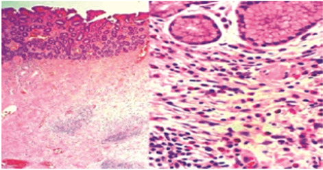
Microscopy evaluation of the lesion.
In our case, the stromal cells exhibit immunoreactivity for Vimentin +++, CD-34 +++ and actin-smooth muscle +; negative are investigations for S-100 protein, CD-117 (c-kit) and BRAF-V600E. Low growth fraction, evaluated with MIB-1 (Ki-67). The set of histomorphological, immunocytochemical and biomolecular data is compatible with a benign polyp fibroid inflammatory (Vanek’s Tumor)
Discussion
The etiology of IFP is unknown. An allergic hypothesis has been firstly proposed, on the base on eosinophilic infiltrate (1). Many have suggested etiologies possibly related to chemical, physical, or metabolic triggers (3). The role of H. Pylori infection has been also reported (4,5). Some cases of gastric IFP that morphologically changed and decreased in size after the cure of H. pylori infection were been described (4,6). In literature, we have been reported cases of IFPS associated with microorganisms such as Cytomegalovirus (7) and Actinomyces israelii (8). Jukic et al. have also shown for the first time the presence of androgen receptor-positive cells whose location corresponds with the distribution of Ki67 positive cells in IFP (9). Estrogen and androgen receptors in gastric cancer correlate to the tumor expression profile and estrogen is suggested to be a protective factor against gastric tumor in women before their menopausal period. As Vanek’s tumor seems to have a similar incidence in both genders and has been reported most frequently in the menopausal period, it could be speculated that its etiology may be related to a disturbance in testosterone levels, same as gastrointestinal malignancies are related to low testosterone levels (9). Therefore, it will be useful in the future to assess the link between IFP and the structure of sex hormones. The majority of IFPs are asymptomatic and discovered as occasional findings during endoscopy (performed for unrelated reasons) or laparotomy (10). The symptoms are dependent on size and localization of the IFP in the GI tract: abdominal pain, weight loss, bleeding, dyspepsia, obstruction, other GI symptoms, such as vomiting, diarrhea, bloody stools (larger polyps tend to erode and ulcerate superficially), tenesmus, and alterations in bowel habits. In the esophagus, it causes dysphagia (11). Abdominal pain is the main symptom in patients with lesions in the stomach. Acute presentations with intussusception or obstruction are also commonly described. Intussusception and obstruction are the most frequent initial symptoms when IFP is located in the small intestine. In some polyps, eosinophils are particularly abundant, but it is now clear that this feature is not associated with peripheral blood eosinophilia or high IgE levels and certainly does not reflect an allergic pathogenesis, regardless of GI location. There is no evidence to support a possible association between inflammatory fibroid polyps and eosinophilic enteritis. Surgical or endoscopic excision is the mainstay of treatment and IFPs are not thought to recur following complete resection. In our study endoscopic ultrasound (EUS) was performed, the fibroid polyp appeared as a hypo-echoic, homogenous mass within the second and third sonographic layer (10). We removed the gastric lesion by en bloc endoscopic mucosal resection (EMR), with the technique inject and cut (endoscopic hemostasis with injections of epinephrine diluted 1:20,000 in normal saline solution) without immediate post-procedure complications. An endoclip was placed at the base of the polyp to guarantee hemostasis. No local recurrence has been highlighted in subsequent endoscopy follow-up. Vanek’s tumor affects both sexes (with slight predominance of the condition in men) and all age groups, with a peak of incidence in the 5th to 7th decade of life. Recent studies have reported a disproportionately large number of gastric inflammatory fibroid polyps in female patients. Its occurrence in children is rare (12-14). IFPs are located in the stomach (about 70% of cases), where they account for up to 3%to 4% of all gastric polyps, mainly in the antrum, immediately proximal to, or overlying, the pyloric sphincter; but (other gastric sites are known), small bowel (about 20% of cases), only occasionally in the duodenum and jejunum, rarely in the large bowel and esophagus) (15).
Macroscopically IFPs are typically small, well circumscribed, submucosally based, pedunculated or sessile lesions that may show surface erosion/ulceration of the overlying mucosa, usually single (multiple synchronous and metachronous disease is rare). The majority of the lesions usually are between 2-5 cm in diameter, although in literature IFPs larger than 20 cm have been reported (16). There are no valid data regarding their growth and no clear evidence whether their growth stops after reaching a certain size or whether they continue to enlarge.
Microscopically the tumor lesion is submucosal and often shows an abrupt demarcation at the level of the muscularis propria, well marginated, but nonencapsulated. Mucosal involvement is common with gastric lesions. However, unlike small intestinal lesions, involvement the muscularis propria is unusual in gastric polyps. Extension of the tumor into the mucosa causes separation of gastric glands, which results in a disordered and atrophic appearance. Classical Vanek’s tumor (“gastric”) is composed of a loose mixture of spindle-shaped, and stellate to epithelioid stromal cells, citologically bland (CD34 positive) (17), inflammatory cells, and small, thin-valled blood vessels in an edematous or myxoid background; centered in the submucosa and appears “pale” at low magnification. The nuclei of tumoral cells are smooth-contoured, oval, or spindle-shaped, with finely granular chromatin and small nucleoli. The cytoplasm is scanty/elongate and amphophilic. Mitotic figures are rare but may occasionally be present in deeper portions of the lesion. Atypical mitoses are never present. The tumor lesion presents apparent concentric formations composed of mesenchymal proliferating spindle-shaped cells (CD 34 positive) (18), with long slender cytoplasmic processes arranged in an “onion skin-like” pattern around vessels and mucosal glands. The inflammatory reaction is predominantly composed of lymphocytes and eosinophilic granulocytes, but plasma cells and histiocytes were also evident. These lesions show many spindle cells but only little collagen. Some authors have shown that there is another morphological subtype (“intestinal” or atypical inflammatory pseudotumor-like Vanek’s tumor) which, in contrast, is pauci-cellular and collagen-rich, lacks concentric formations and CD34 negative. These last tumors tend to be larger than those of the gastric type, but this is not supported by several studies. Vanek’s tumor has low mitotic activity and an absence of necrosis and metastasis (19). Kim and Kim suggested that some atipical features in Vanek’s tumor represented evolutional changes of the lesion (20). They demonstrated different histologic patterns in relation to the size of the lesion, distinguishing four stages; the first two stages (nodular e fibrovascular) showing concentric formations of immature and mature fibroblasts, marked inflammatory infiltration and early collagenization. The latter two stages (sclerotic and edematous) thought to represent variants of one “organized stage” are devoid of concentric formations of fibroblast-like cells. Although the histogenesis of IFPs remains controversial (dendritic cells or CD34 positive perivascular cells); different cell types have been proposed: neural, fibroblastic, myofibroblastic, histiocytic, fibrohistiocytic, vascular and dendritic cells. The absence of stem cell factor (SCF), suggests that IFPs are not related with interstitial cells Cajal and GI stromal tumor (GIST) (21). The Immunohistochemical and ultrastructural profile of the proliferating cells is that of modified “fibroblasts/myofibroblasts”, with variable actin but no S100 protein or epithelial markers. The stromal cells can demonstrate diffuse positivity for Vimentin, CD34, Fascin, CD35, Cyclin D1, Calponin. A smaller proportion are also positive for HHF-35 and Mac-387. Factor VIII and cytokeratin are usually negative (22).
The differential diagnosis of IFPs includes numerous lesions, which show spindle cell proliferation admixed with inflammatory cell infiltrate. Some authors believe that their consistent expression of cyclin D1 and fascin suggests that they have dendritic cell differentiation, but their key feature is consistent CD34 reactivity in small tumors, which sometimes is diminished as lesion attain larger sizes. This latter finding of course, like the molecular findings raises the differential diagnostic consideration of GISTs, but the morphology is different and inflammatory fibroid polyps lack CD117 (c-Kit) expression (22,23). In large examples, sarcomas are often considered, but the bland appearance of the proliferating cells and the inflammatory background argue against this interpretation. In some respects, these polyps resemble nodular/proliferative fasciitis, but fasciitis lacks CD34 and is typically strongly actin reactive (22,24). Solitary fibrous tumor is also CD34 reactive, but it seldom arises in the gastrointestinal (GI) tract and lacks an inflammatory backdrop. However up to 8% of IFPs have been described as concomitant lesions with adenoma or carcinoma (25).
These polyps were believed to be reactive in the past, but they are now known to harbor mutations. Recent data show that the spindle cells express platelet-derived growth factor receptor alpha (PDGFRA), and the majority of IFPs harbor activating PDGFRA mutations, in exon 12 and 18, and, less frequently, in exon 14, suggesting a possible neoplastic factor in the etiology of this lesion (26,27) and relationship between a subset of PDGFRA mutated gastrointestinal stromal tumors (GISTs) and IFPs. However, in contrast to GISTs, inflammatory fibroid polyps are always benign. These are benign lesions that seldom recur after excision. Furthermore, because activating V600E BRAF mutation was recently suggested to represent a possible alternative early molecular event in some KIT and PDGFRA wild-type GISTs, we also performed molecular genetics analysis of a part of exon 15 of BRAF to clarify its potential role in development of Vanek’s tumor (17).
Conclusions
The results of this study suggest that the two morphological patterns of Vanek’s tumor, both “classical” (CD34 e CF positive) and “inflammatory pseudotumor-like” (CD34 e CF negative) IFPs, may be driven by activating PDGFRA mutations and more probably represent only variants of one type of tumor than two different lesions. This study remarked the possible oncogenic role of BRAF mutations in PDGFRA wild-type Vanek’s tumors. BRAF mutations were not shown growth PDGFRA wild-type Vanek’s tumor. Endoscopical mucosal resection (EMR) is a safe and efficacy solution for the resection of gastrointestinal polyps. In our case report EMR permitted a complete resection of the inflammatory fibroid polyp (Vanek’s tumor) as demonstrated by histological examination. The efficacy of this endoscopic procedure was confirmed by no local recurrence in subsequent endoscopy follow-up.
Ethical Statement:
The authors are accountable for all aspects of the work in ensuring that questions related to the accuracy or integrity of any part of the work are appropriately investigated and resolved. All procedures performed in studies involving human participants were in accordance with the ethical standards of the institutional and/or national research committee(s) and with the Helsinki Declaration (as revised in 2013). Written informed consent was obtained for publication of this case report and accompanying images.
Conflicts of Interest:
Each author declares that he or she has no commercial associations (e.g. consultancies, stock ownership, equity interest, patent/licensing arrangement etc.) that might pose a conflict of interest in connection with the submitted article.
Authors’ contributions.
CB and FF conceived, designed and coordinated the research; CB and CG carried out the esophagogastroduodenoscopy (EGD) exam and the surgical resection; FF carried out the histomorphological, immunocytochemical and biomolecular analysis for the histogenetic identification of the lesion; CG, GP, ADA, AF and FF contributed to the acquisition and interpretation of the data, wrote the paper and prepared figures. All Authors revised the manuscript and gave their contribution to improve the paper. All authors read and approved the final manuscript.
References
- Vanek J. Gastric submucosal granuloma with eosinophilic infiltration. Am J Pathol. 1949;25(3):397–411. [PMC free article] [PubMed] [Google Scholar]
- Helwig EB, Ranier A. Inflammatory fibroid polyps of the stomach. Surg Gynecol Obstet. 1953;96:335–67. [PubMed] [Google Scholar]
- Barussaud M, Regenet N, Briennon X, et al. Clinical Spectrum and Surgical Approach of Adult Intussusceptions: A Multicentric Study. Int J Colorectal Dis. 2006;21(8):834–9. doi: 10.1007/s00384-005-0789-3. [DOI] [PubMed] [Google Scholar]
- Nishiyama Y, Koyama S, Andoh A, et al. Gastric inflammatory fibroid polyp treated with Helicobacter pylori eradication therapy. Intern Med. 2003;42:263–7. doi: 10.2169/internalmedicine.42.263. [DOI] [PubMed] [Google Scholar]
- Shalom A, Wasserman I, Segal M, Orda R. Inflammatory fibroid polyp and Helicobacter pylori. Aetiology or coincidence? Eur J Surg. 2000;166:54–7. doi: 10.1080/110241500750009717. [DOI] [PubMed] [Google Scholar]
- Hirasaki S, Matsubara M, Ikeda F, Taniguchi H, Suzuki S. Gastric Inflammatory Fibroid Polyp Treated with Helicobacter pylori Eradication Therapy. Intern Med. 2007;46(12):855–8. doi: 10.2169/internalmedicine.46.6445. [DOI] [PubMed] [Google Scholar]
- Agaimy A, Mudter J, Märkl B, Chetty R. Cytomegalovirus infection presenting as isolated inflammatory polyps of the gastrointestinal tract. Pathology. 2011;43(5):440–6. doi: 10.1097/PAT.0b013e3283485e51. [DOI] [PubMed] [Google Scholar]
- Liu TC, Lin MT, Montgomery EA, Singhi AD. Inflammatory fibroid polyps of the gastrointestinal tract: spectrum of clinical, morphologyc and immunohistochemistry features. Am J Surg Pathol. 2013;37:586592. doi: 10.1097/PAS.0b013e31827ae11e. [DOI] [PubMed] [Google Scholar]
- Jukic Z, Ferencic Z, Radulovic P, Mijic A, Fucic A. Estrogen and androgen receptors in inflammatory fibroid polyp (Vanek’s tumor): case report. Anticancer Res. 2014;34(12):7203–6. [PubMed] [Google Scholar]
- Paikos D, Moschos J, Tzilves D, et al. Inflammatory fibroid polyp or Vanek’s tumour. Dig Surg. 2007;24(3):231–3. doi: 10.1159/000103326. [DOI] [PubMed] [Google Scholar]
- Wysocki AP, Taylor G, Windsor JA. Inflammatory fibroid polyps of the duodenum: a review of the literature. Dig Surg. 2007;24(3):162–8. doi: 10.1159/000102099. [DOI] [PubMed] [Google Scholar]
- Chongsrisawat V, Yimyeam P, Wisedopas N, Viravaidya D, Poovorawan Y. Unusual manifestations of gastric inflammatory fibroid polyp in a child. World J Gastroenterol. 2004;10:460–2. doi: 10.3748/wjg.v10.i3.460. [DOI] [PMC free article] [PubMed] [Google Scholar]
- Siminas S, Qasem E, Shukla R, Turnock R. Inflammatory Fibroid Polyp: A Rare Benign Tumor of the Alimentary Tract in Children Presenting as Intussusception—Case Report and Review of Literature. European J Pediatr Surg Rep. 2014;2(1):16–9. doi: 10.1055/s-0033-1354746. [DOI] [PMC free article] [PubMed] [Google Scholar]
- Righetti L, Parolini F, Cengia P, et al. Inflammatory fibroid polyps in children: A new case report and a systematic review of the pediatric literature. World J Clin Pediatr. 2015;4(4):160–6. doi: 10.5409/wjcp.v4.i4.160. [DOI] [PMC free article] [PubMed] [Google Scholar]
- Blackshaw AJ, Levison DA. Eosinophilic infiltrates of the gastrointestinal tract. J Clin Pathol. 1986;39:1–7. doi: 10.1136/jcp.39.1.1. [DOI] [PMC free article] [PubMed] [Google Scholar]
- Neishaboori H, Maleki I, Emadian O. Jejunal intussusception caused by a huge Vanek’s tumor: a case report. Gastroentero. Hepatol Bed Bench. 2013;6:210–3. [PMC free article] [PubMed] [Google Scholar]
- Daum O, Hatlova J, Mandys V, et al. Comparison of morphological, immunohistochemical, and molecular genetic features of inflammatory fibroid polyps (Vanek’s tumor) Virchows Arch. 2010;456:491–7. doi: 10.1007/s00428-010-0914-8. [DOI] [PubMed] [Google Scholar]
- Hasegawa T, Yang P, Kagawa N, Hirose T, Sano T. CD34 expression by inflammatory fibroid polyps of the stomach. Mod Pathol. 1997;10:451–6. [PubMed] [Google Scholar]
- De Petris G, Leung ST. Pseudoneoplasms of the gastrointestinal tract. Arch Pathol Lab Med. 2010;134:378–91. doi: 10.5858/134.3.378. [DOI] [PubMed] [Google Scholar]
- Kim YI, Kim WH. Inflammatory fibroid polyps of the gastrointestinal tract. Evolution of histologic patterns. Am J Clin Pathol. 1988;89:721–7. doi: 10.1093/ajcp/89.6.721. [DOI] [PubMed] [Google Scholar]
- Pantanowitz L, Antonioli DA, Pinkus GS, Shahsafaei A, Odze RD. Inflammatory fibroid polyps of the gastrointestinal tract: evidence for a dendritic cell origin. Am J Surg Pathol. 2004;28(1):107–14. doi: 10.1097/00000478-200401000-00013. [DOI] [PubMed] [Google Scholar]
- Montgomery AE, Voltaggio L. Biopsy Interpretation of the Gastrointestinal Tract Mucosa. Neoplastic. (3rd Edition) 2018;2 [Google Scholar]
- Kolodziejczyk P, Yao T, Tsuneyoshi M. Inflammatory fibroid polyp of the stomach. A special reference to an immunohistochemical profile of 42 cases. Am J Surg Pathol. 1993;17(11):1159–68. doi: 10.1097/00000478-199311000-00009. [DOI] [PubMed] [Google Scholar]
- Montgomery EA, Meis JM. Nodular fasciitis. Its morphologic spectrum and immunohistochemical profile. Am J Surg Pathol. 1991;15:942–8. [PubMed] [Google Scholar]
- Mucientes P, Mucientes F, Klaassen R. Inflammatory fibroid polyp associated with early gastric carcinoma: a case report. Ann Diagn Pathol. 2012;16(2):148–51. doi: 10.1016/j.anndiagpath.2011.01.001. [DOI] [PubMed] [Google Scholar]
- Schildhaus HU, Cavlar T, Binot E, Büttner R, Wardelmann E, Merkelbach-Bruse S. Inflammatory fibroid polyps harbour mutations in the platelet-derived growth factor receptor alpha (PDGFRA) gene. J Pathol. 2008;216(2):176–82. doi: 10.1002/path.2393. [DOI] [PubMed] [Google Scholar]
- Huss S, Wardelmann E, Goltz D, et al. Activating PDGFRA mutations in inflammatory fibroid polyps occur in exons 12, 14 and 18 and are associated with tumour localization. Histopathology. 2012;61:59–68. doi: 10.1111/j.1365-2559.2012.04203.x. [DOI] [PubMed] [Google Scholar]