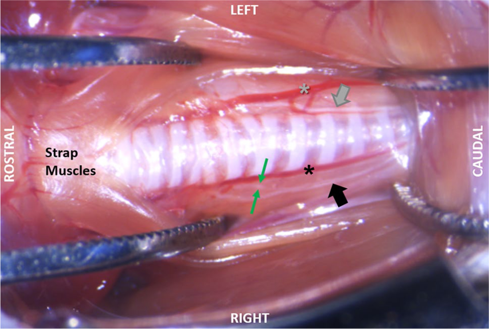
Fig. 9.

Post-mortem dissection demonstrating RLN branching. The left side shows that with minimal retraction of the soft tissues, as is used in our surgical approach, RLN branching is not visible. Instead, only the RLN trunk (gray arrow) can be seen running between the inferior thyroid artery (gray asterisk) and trachea. As shown on the right side, RLN branching is visible only during extreme lateral retraction of the midline strap muscles and fascia. In this specimen, the right RLN trunk (black arrow) has been pulled away from the inferior thyroid artery (black asterisk) to expose a single RLN branch (between the green arrows) near the 6th tracheal ring
