Abstract
Carbonic anhydrase (CA) plays a vital role in photosynthetic tissues of higher plants, whereas its non-photosynthetic role in the symbiotic root nodule was rarely characterized. In this study, 13 CA genes were identified in the model legume Lotus japonicus by comparison with Arabidopsis CA genes. Using qPCR and promoter-reporter fusion methods, three previously identified nodule-enhanced CA genes (LjαCA2, LjαCA6, and LjβCA1) have been further characterized, which exhibit different spatiotemporal expression patterns during nodule development. LjαCA2 was expressed in the central infection zone of the mature nodule, including both infected and uninfected cells. LjαCA6 was restricted to the vascular bundle of the root and nodule. As for LjβCA1, it was expressed in most cell types of nodule primordia but only in peripheral cortical cells and uninfected cells of the mature nodule. Using CRISPR/Cas9 technology, the knockout of LjβCA1 or both LjαCA2 and its homolog, LjαCA1, did not result in abnormal symbiotic phenotype compared with the wild-type plants, suggesting that LjβCA1 or LjαCA1/2 are not essential for the nitrogen fixation under normal symbiotic conditions. Nevertheless, the nodule-enhanced expression patterns and the diverse distributions in different types of cells imply their potential functions during root nodule symbiosis, such as CO2 fixation, N assimilation, and pH regulation, which await further investigations.
Keywords: carbonic anhydrase, Lotus japonicus, root nodule, symbiotic nitrogen fixation
1. Introduction
Carbonic anhydrases (CAs) are ubiquitous in living organisms including prokaryotes, plants, and animals [1]. CAs are among the most efficient enzymes, which mostly contain a zinc ligand and catalyze the reversible hydration of carbon dioxide: CO2 + H2O ⇌ HCO3− + H+ [2]. Higher plants contain three different types of CAs, namely αCA, βCA, and γCA. Each type of CA has multiple functional isoforms, which are widely expressed in photosynthetic or non-photosynthetic tissues and located in a variety of cellular compartments such as cytoplasm, plasma membrane, chloroplast, and mitochondria [3,4,5]. Various isoforms and diversified intracellular localizations of CAs correspond to the multiple biological functions of these enzymes in plants.
Three CA families have independent evolutionary history and likely have developed different biological functions. Although large numbers of αCA genes have been identified in plants, little information has been reported for their functions, intracellular locations, and expression patterns [3]. In contrast, γCAs are expressed in almost all tissues and play conserved roles in mitochondrial complex I, which indicates their housekeeping functions in maintaining mitochondrial function among different plant species [3,6]. βCAs are the most intensively studied CA family in plants. They were proposed to participate in facilitating the diffusion of CO2 across the chloroplast membranes and supply RuBisCO with CO2 in C3 plants [7]. They are also involved in the CO2 concentrating mechanism (CCM) in C4 plants and supply phosphoenolpyruvate carboxylase (PEPC) with HCO3− [8]. Additionally, βCAs were found to regulate CO2-controlled stomatal development and movement in Arabidopsis and rice [9,10,11]. Recently, several non-photosynthetic functions of βCAs have been identified, including the regulation of intracellular pH and cell differentiation in the tapetal cells [12] and the activation of plant basal immunity [13]. Although well proposed, non-photosynthetic functions of βCAs still require further investigations, such as amino acid biosynthesis, lipid biosynthesis, and CO2 fixation in the dark [3].
Legume root nodules exhibit relatively high CA activity, implying the non-photosynthetic role of CA in symbiotic nitrogen fixation (SNF) [14]. MsCA1, the first CA gene cloned from non-photosynthetic tissue in plants, was expressed in all cell types of nodule primordia but exclusively in the peripheral cortical cells of mature nodules [15]. Notably, the expression level of MsCA1 showed an inverse relationship with ambient O2 concentration, implying its potential role in the gas exchange of root nodules [16]. GmCA1 and LjCA1, two homologous genes of MsCA1 identified in Glycine max and Lotus japonicus respectively, showed similar expression patterns as MsCA1 in the nitrogen-fixing nodule, implying their evolutionarily conserved functions inside symbiotic root nodules [17,18,19]. Additionally, two α type CAs (LjCAA1 and LjCAA2) in Lotus japonicus and another α type CA (MlCAA1) in Mesorhizobium loti R7A were identified and proposed to play essential roles in root nodule symbiosis [20,21,22]. Nonetheless, the presence of multiple CA isoforms and the absence of corresponding genetic mutant materials greatly hindered the investigation of the biological function of CAs during SNF.
In this study, we firstly performed the genome-wide identification of candidate carbonic anhydrase genes in Lotus japonicus. Considering the specialized function of γCAs in mitochondrial complex I, here, we mainly focused on the characterization of αCAs and βCAs in non-photosynthetic root nodules. Three nodule-enhanced CA genes (LjβCA1, LjαCA2, and LjαCA6) were characterized and exhibited diverse expression patterns in different cell types of root nodules. To elucidate the biological functions of these candidate CA genes, CRISPR/Cas9-mediated gene knockout experiments were performed. Three LjβCA1 mutants and two LjαCA1/2 mutants were obtained and used for phenotypic comparisons. These results would contribute to the functional characterization of multiple CA isoforms in maintaining an efficient SNF during legume–rhizobia symbiosis.
2. Results
2.1. Identification and Phylogenetic Analysis of Carbonic Anhydrase Genes in Arabidopsis and Lotus
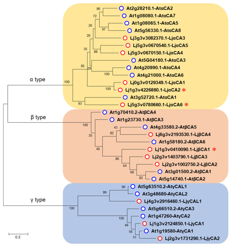
To identify the CA-encoding genes in the Lotus japonicus genome, we firstly performed a protein BLAST search using the Arabidopsis CA proteins as queries. Then, these protein sequences were examined by the NCBI Conserved Domain Database (CDD) to ensure that they contain the typical carbonic anhydrase domain. After removing the fragmentary and redundant sequences, a total of 13 putative CA genes were finally identified. To distinguish these candidates, these CA genes were subsequently labeled according to their sub-clade in CA phylogenetic tree and their chromosomal locations (Table 1, Figure 1). Notably, LjαCA1 was not mapped to any chromosome in the Lotus V3.0 genome, and it has no introns or putative 5′ or 3′ untranslated regions (Figure S1A). Therefore, LjαCA1 might be a pseudogene that was not regarded as the target for further analysis, although it shows above 95% homology with LjαCA2 in protein-coding sequence (Figure S1B). Additionally, three previously identified Lotus CA genes were renamed in this study, including LjCAA1 (renamed as LjαCA2), LjCAA2 (LjαCA6), and LjCA1 (LjβCA1) (Table 1).
Table 1.
Features of the 13 CA proteins (LjCAs) identified in Lotus japonicus.
| Type | Gene | Chr. | Transcript ID | Former Name | NO. of AA | MW (kDa) | pI | GRAVY |
|---|---|---|---|---|---|---|---|---|
| α type | LjαCA1 | Lj0g3v0129349.1 | 269 | 30.47 | 8.79 | −0.454 | ||
| LjαCA2 | Chr 1 | Lj1g3v4226880.1 | LjCAA1 [20] | 269 | 30.29 | 9.05 | −0.453 | |
| LjαCA3 | Chr 3 | Lj3g3v3082370.1 | 218 | 24.65 | 5.92 | −0.456 | ||
| LjαCA4 | Chr 5 | Lj5g3v0670150.1 | 280 | 32.01 | 9.66 | −0.619 | ||
| LjαCA5 | Chr 5 | Lj5g3v0670540.1 | 266 | 30.61 | 6.95 | −0.639 | ||
| LjαCA6 | Chr 5 | Lj5g3v0780660.1 | LjCAA2 [20] | 274 | 30.74 | 6.63 | −0.374 | |
| β type | LjβCA1 | Chr 1 | Lj1g3v0410090.1 | LjCA1 [17] | 263 | 29.87 | 6.00 | −0.303 |
| LjβCA2 | Chr 2 | Lj2g3v1002750.2 | 324 | 34.90 | 6.54 | −0.058 | ||
| LjβCA3 | Chr 2 | Lj2g3v1403790.1 | 256 | 27.91 | 5.49 | −0.129 | ||
| LjβCA4 | Chr 6 | Lj6g3v2193530.1 | 263 | 29.05 | 6.44 | −0.186 | ||
| γ type | LjγCA1 | Chr 1 | Lj1g3v2124850.1 | 273 | 29.58 | 6.23 | −0.095 | |
| LjγCA2 | Chr 2 | Lj2g3v1731290.1 | 271 | 29.46 | 6.07 | −0.101 | ||
| LjγCAL1 | Chr 4 | Lj4g3v2916460.1 | 186 | 20.20 | 9.44 | 0.176 |
Chr, chromosome; NO. of AA, number of amino acids; pI, isoelectric point; MW, molecular weight; GRAVY, grand average of hydropathicity.
Figure 1.
Phylogenetic tree of carbonic anhydrase genes in Lotus and Arabidopsis. The protein sequences were obtained from TAIR (http://www.arabidopsis.org/) (accessed on 19 September 2018) and Kazusa DNA Research Institute (http://www.kazusa.or.jp/lotus/) (accessed on 19 September 2018). Sequence alignment was performed using Clustal W, and the phylogenetic tree was generated by MEGA 7.0 with 1000 bootstrap replications. Blue circles indicate Arabidopsis CA proteins. Red circles indicate Lotus CA proteins. Red stars indicate three LjCAs with enhanced expression in root nodules. The scale bar indicates an evolutionary distance of 0.2 amino acid substitutions per position.
To identify the evolutionary relationships among these CA genes, a neighbor-joining phylogenic tree was constructed according to protein sequences of Arabidopsis and Lotus enzymes. Similar to Arabidopsis, the Lotus genome encodes three types of CAs, including six α type CAs (LjαCA1–6), four β type CAs (LjβCA1–4), and three γ type CAs (LjγCA1–2, LjγCAL1) (Figure 1, Table 1). Physicochemical property analysis shows that the lengths of Lotus carbonic anhydrase proteins (LjCAs) range from 186 to 324 amino acids, the molecular weights of the LjCAs range from 20.2 to 34.9 kDa, and the isoelectric points range from 5.49 to 9.66. Notably, both basic and acidic proteins are present in α and γ type CAs, whereas all these four β type CAs are acidic. Additionally, the grand average of hydropathicity (GRAVY) values range from −0.058 to −0.639 except for LjγCAL1, indicating that most of the LjCAs are hydrophilic (Table 1).
2.2. Expression Profiles of LjCAs across Different Tissues and Different Developmental Stages of Root Nodule
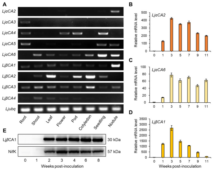
To identify the CA genes related to SNF, semi-quantitative RT-PCR was performed to investigate the expression profiles of LjCAs across different tissues, including root, shoot, leaf, flower, pod, cotyledon, seedling, and nodule. The results show that LjαCA2, LjαCA6, and LjβCA1 are highly expressed in root nodules, indicating their potential roles in maintaining nodule function. LjaCA6 demonstrates a higher expression level in seedlings, and it is also expressed in other tissues, including shoot and cotyledon. Hence, the biological function of LjaCA6 may not be restricted to root nodules (Figure 2A). For other CA genes, they tend to express in non-symbiotic tissues, such as LjαCA3 in root and LjβCA2 in leaf, pod, and cotyledon. Additionally, LjβCA3 is expressed in most tissues with a similar level, implying its potential housekeeping function (Figure 2A). The public expression profile data of LjCAs were also retrieved from Lotus japonicus Expression Atlas (https://lotus.au.dk/expat/) (accessed on 3 July 2021). As shown in Table S1, LjαCA2, LjαCA6, and LjβCA1 are three dominant CA genes with enhanced expression in root nodule consistent with the RT-PCR results (Figure 2A).
Figure 2.
Expression patterns of carbonic anhydrase genes in Lotus japonicus. (A) RT-PCR analyses of α and β type CA genes in different tissues of Lotus japonicus. A total of 33 cycles were used for amplifying α type CA genes, while 29 cycles were used for amplifying the ubiquitin gene and β type CA genes. The ubiquitin gene was used as an internal control. Root, uninoculated root; Nodule, mature nodule at 3 wpi (weeks post-inoculation). (B–D) Expression profiles of LjαCA2, LjαCA6, and LjβCA1 during nodule development. qRT-PCR was used to quantify the transcript abundance of LjαCA2, LjαCA6, and LjβCA1 in the uninoculated root (0 wpi) and in developing nodule (1 to 11 wpi). Relative mRNA levels of three genes in 1 to 11 wpi with respect to 0 wpi were calculated using ubiquitin as a a reference gene. Values are means ± SD of three technical replications. Similar results were observed in three independent experiments. For (A–D), cDNA from 5 ng total RNA was used as the template for a 10 μL PCR reaction. (E) Representative immunoblot of LjβCA1 and NifK in uninfected root (0 wpi) and in developing nodule (1 to 8 wpi). The primary antibodies were polyclonal antibodies against LjβCA1 and NifK.
To further investigate the expression patterns of three nodule-enhanced CA genes in detail, quantitative RT-PCR was performed to detect the transcript levels of LjαCA2, LjαCA6, and LjβCA1 at different stages of nodule development. The transcript levels of these three genes are increased after rhizobial inoculation or during nodule maturation (Figure 2B–D). Among them, LjαCA2 and LjαCA6 exhibit remarkably high expression levels in mature nodules (3, 5, 7 wpi) and maintain the high expression levels in senescent nodules (9, 11 wpi, Figure 2B,C). However, the expression of LjβCA1 is dramatically up-regulated at 3 wpi, reaching above 2,500-fold compared with that in uninoculated roots (0 wpi). After that, the expression level of LjβCA1 gradually declines along with the development of root nodules (Figure 2D). Additionally, we also performed immunoblot for detecting LjβCA1 and NifK proteins at different nodule developmental stages. The LjβCA1 protein accumulates at 2 wpi; then, it maintains a high level at the later stages of nodule development (2 to 8 wpi). In contrast, the protein level of NifK peaks at 3 wpi and decreases after nodule maturation (Figure 2E). In summary, the nodule-enhanced expression patterns of LjαCA2, LjαCA6, and LjβCA1 imply their potential functions in nodule maturation, nitrogen fixation, or even nodule senescence.
2.3. Spatiotemporal Expression Patterns of Nodule-Enhanced LjCAs in Root Nodule
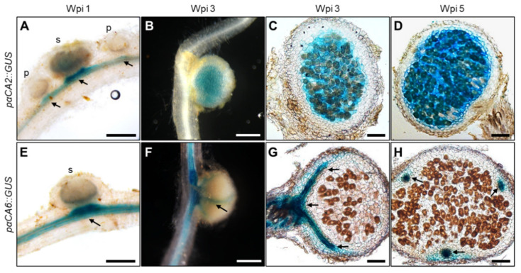
We next performed promoter–reporter fusion experiments for further investigating the cell-specific expression patterns of LjαCA2, LjαCA6, and LjβCA1. Around 3 kb promoter regions of three CA genes were amplified and then fused to the GUS reporter gene. Stably transformed plants were generated and used for GUS staining experiments. As shown in Figure 3, the GUS staining signal of pαCA2::GUS transgenic lines is located in the vascular bundle of the root near the nodule at 1 wpi (week post-inoculation). No signal is detectable inside nodule primordia, while a weak signal is visible in small nodules (Figure 3A). During nodule maturation, LjαCA2 is mainly expressed in the central nitrogen fixation zone, both in the infected and uninfected cells at 3 wpi (Figure 3B,C) and 5 wpi (Figure 3D). For LjαCA6, GUS staining signal is detectable in the root vascular bundle at 1 wpi (Figure 3E). In mature nodules, the expression of LjαCA6 is limited to the nodule vascular bundle at 3 and 5 wpi, and no signal is detectable in the central nitrogen-fixing zone (Figure 3F–H).
Figure 3.
Histochemical analysis of GUS expressions driven by LjαCA2 and LjαCA6 promoters in developing root nodules. Expression patterns of GUS reporter were analyzed for pαCA2::GUS (A–D) and pαCA6::GUS (E–H). Nodules at different developmental stages were investigated, including young nodules at 1 wpi (A,E) and mature nodules at 3 wpi (B,C,F,G) and 5 wpi (D,H). pαCA2::GUS and pαCA6::GUS constructs were introduced into wild-type plants using stable transformation. T2 generation plants were used for GUS staining experiments. Images are representative of at least eight independent transgenic plants. Black arrows indicate the vascular bundle. p, primordia; s, small nodule. Scale bars, 200 μm (A,E); 1 mm (B,F); 100 μm (C,D,G,H).
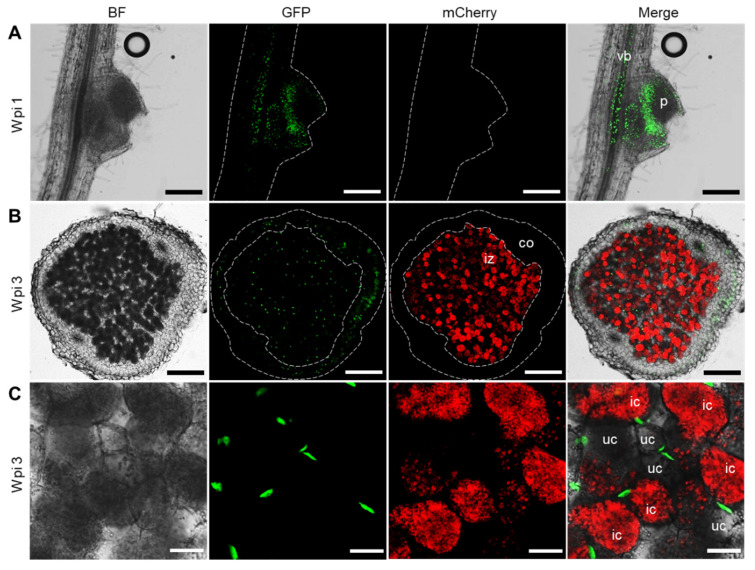
In contrast, we have not been able to detect any GUS staining signal in all the pβCA1::GUS transgenic lines. This is inconsistent with our qPCR and Western blot results, which shows an enhanced expression pattern of LjβCA1 in symbiotic root nodules (Figure 2A,D,E). Alternatively, a tYFP-NLS reporter system was used to analyze the expression of LjβCA1. This reporter consists of triple YFP protein fused to nuclear localization signal peptide (NLS), which shows an accumulated fluorescent signal in nuclei. Stably transformed plants containing the pβCA1::tYFP-NLS construct were inoculated with Mesorhizobium loti MAFF303099 expressing mCherry fluorescent protein for investigating the LjβCA1 expression pattern inside nodules. Both vascular bundle and nodule primordia show YFP fluorescent signals at 1 wpi (Figure 4A). In mature nodules, the fluorescent signals are detectable in both the inner nitrogen fixation zone and nodule cortical cell layers at 3 wpi (Figure 4B). More specifically, YFP fluorescent signals are only visible in the nuclei of uninfected cells but not in the infected cells, which are filled with mCherry-labeled rhizobia (Figure 4C). In summary, LjαCA2, LjαCA6, and LjβCA1 exhibit quite divergent expression patterns at different types of cells, although all of them are highly expressed inside the root nodules.
Figure 4.
Fluorescence observation of tYFP-NLS driven by LjβCA1 promoter in developing root nodule. Stable transgenic plants carrying the pβCA1::tYFP-NLS construct were inoculated with mCherry-labeled M. loti MAFF303099. Confocal microscopic images were captured at different developmental stages of root nodules, including young nodules at 1 wpi (A) and mature nodules at 3 wpi (B,C). Nuclei accumulating a green signal in the GFP channel show the fluorescence of tYFP-NLS reporter. The red signal in the mCherry channel shows the fluorescence of mCherry-labeled rhizobia. BF, bright field. Merged images of the BF, GFP, and mCherry channels were shown. vb, vascular bundle; p, primordia; iz, infected zone; co, cortical cell layers; ic, infected cell; uc, uninfected cell. Scale bars, 200 μm (A,B); 25 μm (C).
2.4. Construction of LjCAs Mutants and Symbiotic Phenotypic Analysis
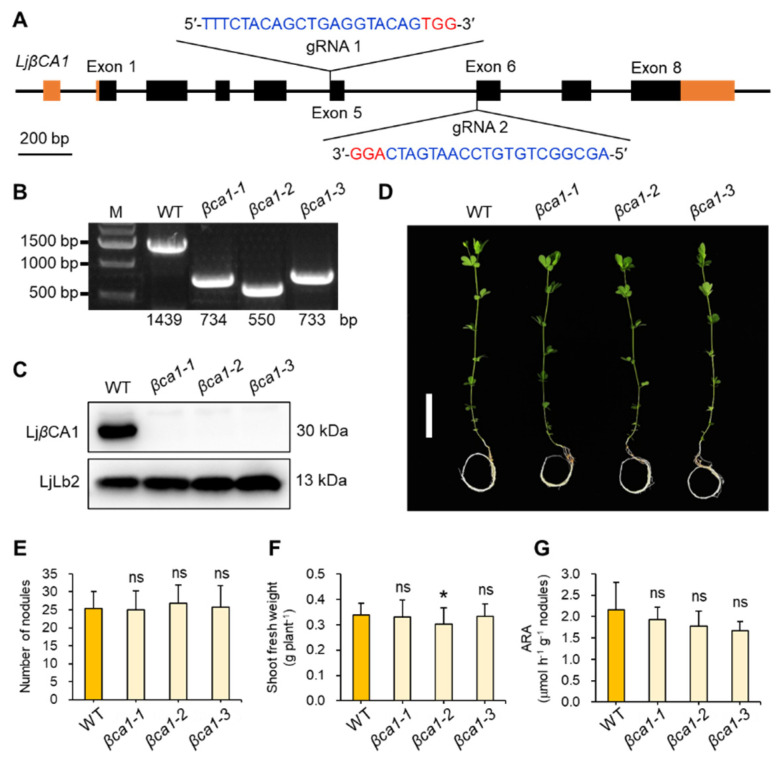
Since LjβCA1 is the only β type CA highly expressed in root nodules, we firstly constructed the corresponding mutants of LjβCA1 using CRISPR/Cas9 technology. As shown in Figure 5A, two gRNAs were designed against exon 5 and exon 6 of LjβCA1, respectively. Three independent mutant lines (βca1-1, βca1-2, βca1-3) were identified. All three mutant lines exhibit fragment deletion or insertion in the LjβCA1 genomic loci, which were identified by PCR-based genotyping (Figure 5B and Figure S2). To confirm the absence of LjβCA1 protein in three mutant lines, immunoblot analysis was performed using LjβCA1 antibody. The total protein was extracted from mature nodules at 4 wpi of WT and Ljβca1 mutants. As shown in Figure 5C, LjβCA1 protein is accumulated in wild-type mature nodules, but it is absent in the nodules of three mutant lines. In contrast, the leghemoglobin LjLb2 protein is accumulated to a similar level in mutant nodules compared with that in wild-type nodules. These results indicate that no functional LjβCA1 protein is properly translated in three independent βca1 mutant lines.
Figure 5.
Construction and symbiotic phenotype analyses of LjβCA1 mutants. (A) Gene structure and gRNA design of LjβCA1. Black boxes indicate the exons and orange boxes indicate the 5′ or 3′ untranslated regions (UTRs). Two gRNAs used for the LjβCA1 knockout experiment were located in exon 5 and exon 6, respectively. The PAM sequence is marked in red. The 20 bp gRNA sequence is marked in blue. (B) Genotyping information of three LjβCA1 mutants, including βca1-1, βca1-2, and βca1-3. (C) Western blot analysis of three LjβCA1 mutants using LjβCA1 and LjLb2 antibody. (D) The symbiotic phenotype of LjβCA1 mutants at 5 wpi. Plants were grown in nitrogen-deficient conditions after inoculation with M. loti MAFF303099. Three CRISPR/Cas9-derived independent LjβCA1 mutant lines (βca1-1, βca1-2, and βca1-3) were compared to the WT plants. Scale bar, 5 cm. SNF parameters include (E) root nodule number, (F) shoot fresh weight, and (G) ARA per nodule fresh weight of WT and mutant plants. Values are means ± SD of 30 plants per genotype. Student’s t-test was used for statistical analysis in (E–G) by comparing respective mutant lines to WT plants. ns, not significant; *, p < 0.05. Phenotyping analysis has been performed three times, and similar results were obtained.
To further investigate the biological function of LjβCA1, symbiotic phenotype analysis was performed under low nitrogen conditions (0.5 mM KNO3). After 5 weeks post-inoculation, three βca1 mutants grew similarly to wild-type plants (Figure 5D). Both nodule number and shoot fresh weight showed no significant differences between WT and βca1 mutants, except that βca1-2 showed a slight reduction compared with WT in shoot fresh weight (Figure 5E,F). Next, the acetylene reduction activity (ARA) per nodule fresh weight was analyzed to evaluate the nitrogen fixing activity. Three βca1 mutants showed slightly reduced ARA values, but there were no significant differences when compared with WT (Figure 5G). In conclusion, the absence of LjβCA1 does not result in obvious defects in SNF.
Considering the active expression of LjαCA2 in the mature nodules, it was regarded as another target for gene knockout. Two common gRNAs were designed for targeting both LjαCA1 and LjαCA2 due to the high sequence homology between these two genes, although LjαCA1 is a possible pseudogene (Figures S1 and S3). Two LjαCA1/2 double mutant lines were obtained with one or two base pairs deletion in the gRNA1 targeting sequence of each gene (Figure S3). Subsequently, the symbiotic phenotype was analyzed at 4 wpi. Two double mutants (αca12-1 and αca12-2) grew similarly to the wild-type plants (Figure S4A). Moreover, both nodule number and shoot biomass showed no significant difference in αca12-1 and were slightly higher in αca12-2 by comparison with that in wild-type plant (Figure S4B,C). Overall, knockout of both LjαCA1 and LjαCA2 does not influence the plant growth under normal symbiotic conditions.
3. Discussion
In this study, three nodule-enhanced carbonic anhydrase genes were functionally characterized in detail, which exhibited quite different expression patterns inside the root nodules of Lotus japonicus. Among them, LjβCA1 and its orthologs in alfalfa (MsCA1) and soybean (GmCA1) were identified previously. Using RNA in situ hybridization and immunolocalization, the mRNA and protein of LjβCA1, MsCA1, and GmCA1 were found to be located in most cells of nodule primordia and specific cell layers surrounding the infection zone of mature nodules [15,16,17,18,19]. In this study, the tYFP-NLS reporter system was used to confirm the promoter activity of LjβCA1. The tYFP-NLS expression was detected in nodule primordia and peripheral cortical cells of the mature nodules, which is consistent with the previous findings (Figure 4A,B) [15,16,17,18,19]. Interestingly, the YFP fluorescent signals were observed in uninfected cells inside the nodule infection zone, but no signal was observed in the infected cells (Figure 4C). By comparing the Western blot and qPCR results, the protein level of LjβCA1 does not decrease after nodule maturation, which is inconsistent with its reduced mRNA level after 3 wpi (Figure 2D,E). These results indicate that the LjβCA1 protein was relatively stable in the cortical and uninfected cells. However, the biological function of LjβCA1 in these cells was still unknown.
Previous work found that another two αCA genes (LjCAA1 and LjCAA2) were highly up-regulated in the root nodule of Lotus japonicus, which were renamed as LjαCA2 and LjαCA6 respectively [20] (Table 1). In this study, similar results were obtained for the expression pattern of LjαCA2, which is expressed in the central infection zone of mature nodules [20] (Figure 3A–D). However, different results were obtained for LjαCA6. The previous study has revealed that LjαCA6 mRNA was detectable in the inner cortical cell, vascular bundle, and central tissue of nodule by using RNA in situ hybridization method [20]. Here, the promoter activity of LjαCA6 was limited to the vascular bundle of the root and nodule using the promoter-GUS fusion system (Figure 3E–H). Two different methods obtained distinct results, which can be explained by the fact that ~3 kb of the promoter of LjαCA6 might be not sufficient to support its native expression pattern. Notably, there is another annotated gene (Lj5g3v0780650.1) located around 2186 bp up-stream of LjαCA6 translation starting site. Nevertheless, further work is needed to solve this discrepancy.
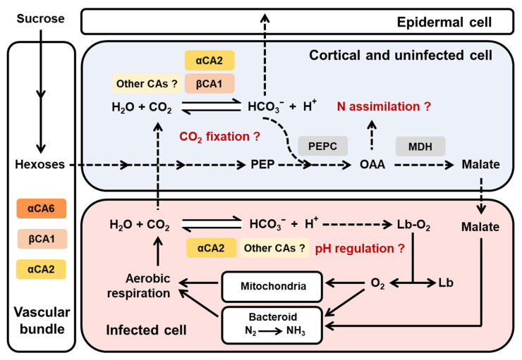
Numerous studies have revealed the CO2 fixation capability of root nodules in many legume species, such as soybean, alfalfa, pea, and Lotus [23,24,25,26]. Specific genes coding for phosphoenolpyruvate carboxylase (PEPC) and malate dehydrogenase (MDH) exhibit enhanced expression in root nodules [18,27,28]. The CA-PEPC-MDH pathway was regarded as the key component of dark CO2 fixation in legume root nodules [29]. As shown in Figure 6, CA catalyzes the hydration of carbon dioxide, providing bicarbonate for PEPC. Oxaloacetate (OAA), the refixed organic acid molecules, can be used as the C skeleton for N assimilation and a C resource for supporting rhizobial respiration. In this model, both LjβCA1 and LjαCA2 are expressed in the nodule cortical and uninfected cells, which are the cell types responsible for the carbon metabolism and gas exchange barrier [16,30]. Thus, these two CAs may function in nodule CO2 recycling or facilitating excess CO2 out of the nodule (Figure 6). Additionally, LjβCA1 and its orthologs (MsCA1 and GmCA1) lack the signal peptide sequence present in AtβCA1, which is a chloroplastic carbonic anhydrase (Figure S5A). The LjβCA1-GFP fusion protein was localized in the cytoplasm in Nicotiana benthamiana leaves, whereas GFP itself was localized in the cytoplasm and nucleus (Figure S5B,C). Thus, LjβCA1 or other CA isoforms are probably involved in CO2 metabolism coupled with PEPC enzyme in the cytoplasm of the cortical and uninfected cells (Figure 6).
Figure 6.
Proposed working model of carbonic anhydrases in root nodule symbiosis. Three carbonic anhydrase genes (LjαCA2, LjαCA6, and LjβCA1) exhibit different expression patterns inside root nodules. LjαCA2 is expressed in both infected and uninfected cells inside the central nitrogen fixation zone. LjαCA6 is only expressed in the vascular bundle. LjβCA1 is expressed in the uninfected cells of the central nitrogen fixation zone and also the cortical cells around the root nodule. Other CAs have not been characterized in this work but may collaboratively play important roles in maintaining an efficient SNF by contributing to pH regulation, CO2 fixation, and N assimilation in various types of cells within nodules. The solid arrows indicate the verified processes, and the dashed arrows indicate the hypothetical processes awaiting further investigations. PEP, phosphoenolpyruvate; PEPC, phosphoenolpyruvate carboxylase; OAA, oxaloacetate; MDH, malate dehydrogenase; Lb, leghemoglobin.
Another hypothetical function of CA enzymes inside symbiotic root nodules could be comparable to human red blood cells (RBCs), which contain carbonic anhydrase as the second most abundant protein next to hemoglobin [31]. CO2 produced by aerobic respiration can be quickly transformed into HCO3− and H+ through the catalysis mediated by α-type CAs in RBCs. Once the H+ is combined with oxyhemoglobin, the conformation of hemoglobin changes and oxygen is released to maintain normal respiration of aerobic tissue, which is known as the Bohr effect. Consequently, α-type CAs can trigger the oxygen release from oxyhemoglobin through pH regulation [31,32,33]. In the infected cells of root nodules, a large amount of leghemoglobin associates with oxygen and creates the micro-aerobic environment, which is crucial for efficient nitrogen fixation. Leghemoglobin needs to transfer a low concentration but high-flux rate of oxygen to mitochondria and bacteroid for sustaining normal cell respiration (Figure 6). However, the mechanism in regulating the release of oxygen from oxy-leghemoglobin is still unclear [34,35,36]. Thus, we speculate that a similar regulatory mechanism might be working in the nodule infected cells as that in RBCs. In this study, LjαCA2 showed strong induction in the infected cells of mature root nodules. LjαCA2-mediated CO2 rehydration may lead to the change of local pH, which influences the affinity between leghemoglobin and oxygen (Figure 6). Indeed, it has been reported that the pH value near the symbiosome membrane is significantly lower than that in other areas of the infected cells [37].
Several functions of CAs during SNF have been proposed, but none of them have been experimentally confirmed. In this study, knockout of LjβCA1 or both LjαCA1 and LjαCA2 does not influence the overall plant growth under normal symbiotic conditions (Figure 5 and Figure S4). Here, explanations are given for illustrating these unexpected results. Firstly, the multiple CA isoforms may function redundantly to support efficient SNF. Indeed, these LjCAs exhibit partially overlapped expression patterns inside root nodules (Figure 3, Figure 4, and Figure 6). In addition, the function of LjCAs with low expression levels in nodules can not be ignored, such as LjαCA5 and LjβCA3 (Figure 2A). The other explanation is that LjCAs may help root nodules adapt to a variety of environmental stresses, such as drought, flooding, alkaline, or salinity stresses. In Arabidopsis, stress conditions can change the expression and activity of the CA enzymes. Numerous findings supported the contributions of CAs in plants adaptation to various stresses [5,38]. Therefore, the phenotype of LjCAs mutants would be more obvious under specific stress conditions but is not shown under the normal symbiotic conditions. Future work will focus on the construction of multi-gene knockout mutants of LjCAs to decipher the functional redundancy of carbonic anhydrases inside root nodules. The symbiotic phenotype analysis of LjCAs mutants under different stress conditions could be systemically performed. Finally, the characterization of subcellular localizations of LjCAs in different cell types of root nodules would also provide valuable information regarding how an efficient SNF is systemically coordinated in mature nodules.
4. Materials and Methods
4.1. Gene Identification and Phylogenetic Analysis
The protein sequences of AtCAs were obtained from the TAIR database (http://www.arabidopsis.org) (accessed on 19 September 2018). To identify the CA-encoding genes in the Lotus japonicus genome, we performed a BLASTP search on the Kazusa DNA Research Institute website (http://www.kazusa.or.jp/lotus/) (accessed on 19 September 2018), using the known AtCAs as queries with the parameters id% > 50% and E-value < 10−15. Then, the protein sequences were analyzed in the NCBI conserved domain database with default parameters (https://www.ncbi.nlm.nih.gov/Structure/cdd/wrpsb.cgi) (accessed on 30 May 2020). The protein biochemical properties such as the theoretical isoelectric point (pI), the molecular weight (MW), and the grand average of hydropathicity (GRAVY) were calculated using the ProtParam program (https://web.expasy.org/protparam/) (accessed on 4 March 2021). Multiple sequence alignment of the AtCAs and LjCAs was performed using Clustal W. Subsequently, a phylogenetic tree was constructed using MEGA 7.0 with the neighbor-joining method (1000 bootstrap replications) [39].
4.2. Plant Growth and Transformation
Ecotype MG-20 of Lotus japonicus was used in all experiments [40]. MG-20 seeds were firstly treated with 98% sulfuric acid for 10 min, subsequently washed three times with sterile water, then surface-sterilized in 2% sodium hypochlorite for 5 min, and washed three times again. The sterilized seeds were kept at 4 °C for at least 24 h. Then, seeds were germinated on one-half strength Murashige–Skoog medium in the dark for 2 days at 24 °C, which was followed by illumination for another 3–4 days. The stable transformation was performed as previously described [41]. For inoculation assay, five-day-old seedlings were planted in pots containing sterile perlite:vermiculite (1:3) supplemented with B&D medium containing 0.5 mM KNO3 [42] and grown in a greenhouse at 24 °C under 16 h/8 h day/night cycle. Ten-day-old seedlings were inoculated with Mesorhizobium loti MAFF303099, wild-type, or mCherry-labeled strain [43,44]. For subcellular localization experiments, Nicotiana benthamiana seedlings were grown in pots filled with perlite:vermiculite:nutrient soil mixture (1:1:1). The one-month-old tobacco plants were used for infiltration with Agrobacterium tumefaciens EHA105.
4.3. GUS Staining and tYFP-NLS Observation
Promoter fragments (~3 kb) of LjαCA2, LjαCA6, and LjβCA1 were PCR amplified using MG-20 genomic DNA as a template and then cloned into the promoterless DX2181G vector. For LjβCA1, another construct (pC1300-tYFP-NLS) was used to indicate the promoter activity. Related primers are included in Table S2. Stable transgenic plants were used for GUS staining and fluorescence observation. At least eight independent transgenic lines were tested. For GUS staining, plant tissues were immersed in the staining buffer containing 0.5 mg/mL of 5-bromo-4-chloro-3-indolyl-β-D-glucuronic acid cyclohexylammonium salt (Sangon Biotech, Shanghai, China), 1 mM potassium ferricyanide, 1 mM potassium ferrocyanide, 100 mM potassium phosphate (pH 7.0), 10 mM EDTA, 0.1% (w/v) sodium lauroyl sarcosinate, and 0.1% (v/v) Triton X-100. The reaction was performed at room temperature in a vacuum overnight. If necessary, root nodules were sectioned to 50–80 μm thick slices using a vibratome (Leica VT 1000S, Nussloch, Germany). Images were captured with a fluorescence stereo microscope (Nikon SNZ18, Tokyo, Japan) for the whole nodule and a light microscope (Leica DM2500, Nussloch, Germany) for nodule sections. For fluorescence observation, nodules were sectioned to 80–100 μm slices first and then observed and photographed with a laser-scanning confocal microscope (Leica TCS SP8, Nussloch, Germany). Fluorescence was detected with excitation at 488 nm and emission at 500–550 nm for tYFP-NLS and GFP, excitation at 580 nm and emission at 560–630 nm for mCherry, and excitation at 488 nm and emission at 650–750 nm for chloroplast autofluorescence.
4.4. Construction of LjCAs Knockout Mutants
Using CRISPR/Cas9 technology, LjCAs mutants were obtained through stable transformation in a MG-20 background. The web tool CRISPR-P 2.0 was used for designing high-score guide RNAs with low off-target effect (http://cbi.hzau.edu.cn/crispr/) (accessed on 27 March 2016). Two guide RNAs were designed and cloned into the final CRISPR vector as described before [45]. PCR-sequencing-based genotyping was performed at T1 generation. Cas9-free and homozygous mutants were identified and allowed to self-cross to produce T2 seeds. The symbiotic phenotype was analyzed using at least T2 generation plants. The detailed procedure for CRISPR-Cas9 mediated genome editing in Lotus japonicus was described previously [46]. The genotypes of obtained LjCAs mutants are described in Figures S2 and S3. Related primers were included in Table S2.
4.5. RNA Extraction and qRT-PCR
Total RNA was isolated using TransZol Plant reagent (TransGen Biotech, Beijing, China). 1 µg total RNA was used to synthesize the first-strand cDNA according to the instructions of the HiScript II Q RT SuperMix (Vazyme Biotech, Nanjing, China). Real-Time qRT-PCR was performed on Bio-Rad CFX96 Real-Time PCR Detection System based on the instruction of the TransStart® Tip Green qPCR SuperMix (TransGen Biotech, Beijing, China). The ubiquitin gene (GenBank accession no. AW720576) served as a reference gene. All reactions were performed with three technical replications. Primers used in this study were included in Table S2.
4.6. Western Blot Analysis
Root and nodule tissues (~200 mg) were homogenized in liquid nitrogen. Total protein was precipitated with methanol–chloroform and dissolved in 50 mM Tris-HCl, pH 6.8, 2% (w/v) SDS [47]. Protein concentration was quantified by BCA protein assay (Sangon Biotech, Shanghai, China). Similar amounts of total protein (~30 µg) were separated on 10% SDS-PAGE gel and transferred to the PVDF membrane (Merck Millipore, Darmstadt, Germany). Immunoblot analysis was performed using a primary antibody raised against LjLb2, LjβCA1, and NifK. To generate these antibodies, full-length coding sequences of LjβCA1, LjLb2, and MlNifK were amplified and cloned into the pET-28a vector. The sequence-verified constructs were transformed into E. coli BL21 (DE3)-RIL strain. The recombinant proteins with an N-terminal 6xHis tag were expressed under 28°C with 0.5 mM IPTG and then purified by Ni-NTA affinity chromatography (Genscript, Nanjing, China). The purified recombinant proteins were used as antigens for obtaining the primary antibodies in rabbit (PHYTOAB, San Jose, CA, USA). HRP-conjugated goat anti-rabbit secondary antibody was used. Signals were detected with Western ECL Substrate (Bio-Rad, Hercules, CA, USA) under a ChemiScope western blot processor (ChemiScope 6300, Clinx Science Instruments, Shanghai, China).
4.7. Nitrogenase Activity Assay
Nitrogenase activity was measured using the acetylene reduction assay [48]. Nodulated roots were put into glass bottles sealed with rubber stoppers. Each bottle contained four nodulated roots, and 5 biological replicates of each genotype were analyzed. 2 mL of acetylene was injected into each bottle after the same volume of air was pumped out. Subsequently, all bottles were incubated for 2 h at 28°C. For each bottle, a 100 μL gas sample was used to measure the ethylene production using a GC-4000A gas chromatograph (East & West Analytical Instruments, Beijing, China).
Acknowledgments
We thank the Bai Chuan fellowship of College of Life Science and Technology, Huazhong Agricultural University, for funding support. We also thank the Core Facilities at College of Life Science and Technology, Huazhong Agricultural University, for assistance with laser scanning confocal microscopy.
Abbreviations
| ARA | Acetylene reduction activity |
| BCA | Bicinchoninic acid |
| CA | Carbonic anhydrase |
| CCM | CO2 concentrating mechanism |
| CRISPR | Clustered regularly interspaced short palindromic repeats |
| EDTA | Ethylenediaminetetraacetic acid |
| GRAVY | Grand average of hydropathicity |
| GUS | β-Glucuronidase |
| Lb | Leghemoglobin |
| MDH | Malate dehydrogenase |
| MW | Molecular weight |
| NLS | Nuclear localization signal |
| OAA | Oxaloacetate |
| PEP | Phosphoenolpyruvate |
| PEPC | Phosphoenolpyruvate carboxylase |
| pI | Isoelectric point |
| PVDF | Polyvinylidene difluoride |
| RBCs | Red blood cells |
| SDS | Sodium dodecyl sulfate |
| SNF | Symbiotic nitrogen fixation |
| tYFP | Triple yellow fluorescent protein |
Supplementary Materials
The following are available online at https://www.mdpi.com/article/10.3390/ijms22157766/s1.
Author Contributions
L.W. and D.D. designed the research; L.W., J.L., Y.Z., T.T. and B.Z. performed the experiments and analyzed the data; L.W. wrote the manuscript; D.D. revised the manuscript. All authors have read and agreed to the published version of the manuscript.
Funding
This research was funded by the China Postdoctoral Science Foundation (Grant No. 2020M680103), the National Natural Science Foundation of China (Grant No. 32000192, 31870220), and the Natural Science Foundation of Hubei Province (2020CFA008).
Conflicts of Interest
The authors have declared no conflict of interests.
Footnotes
Publisher’s Note: MDPI stays neutral with regard to jurisdictional claims in published maps and institutional affiliations.
References
- 1.Hewett-Emmett D., Tashian R.E. Functional diversity, conservation, and convergence in the evolution of the α-, β-, and γ-carbonic anhydrase gene families. Mol. Phylogenet. Evol. 1996;5:50–77. doi: 10.1006/mpev.1996.0006. [DOI] [PubMed] [Google Scholar]
- 2.Chegwidden W.R., Carter N.D. Introduction to the carbonic anhydrases. In: Chegwidden W.R., Carter N.D., Edwards Y.H., editors. The Carbonic Anhydrases: New Horizons. Birkhäuser Verlag; Basel, Switzerland: 2000. pp. 13–28. [Google Scholar]
- 3.DiMario R.J., Clayton H., Mukherjee A., Ludwig M., Moroney J.V. Plant carbonic anhydrases—Structures, locations, evolution and physiological roles. Mol. Plant. 2017;10:30–46. doi: 10.1016/j.molp.2016.09.001. [DOI] [PMC free article] [PubMed] [Google Scholar]
- 4.Fabre N., Reiter I.M., Becuwe-Linka N., Genty B., Rumeau D. Characterization and expression analysis of genes encoding α and β carbonic anhydrases in Arabidopsis. Plant Cell Environ. 2007;30:617–629. doi: 10.1111/j.1365-3040.2007.01651.x. [DOI] [PubMed] [Google Scholar]
- 5.Floryszak-Wieczorek J., Arasimowicz-Jelonek M. The multifunctional face of plant carbonic anhydrase. Plant Physiol. Biochem. 2017;112:362–368. doi: 10.1016/j.plaphy.2017.01.007. [DOI] [PubMed] [Google Scholar]
- 6.Fromm S., Senkler J., Zabaleta E., Peterhansel C., Braun H.P. The carbonic anhydrase domain of plant mitochondrial complex I. Physiol. Plant. 2016;157:289–296. doi: 10.1111/ppl.12424. [DOI] [PubMed] [Google Scholar]
- 7.Ignatova L., Rudenko N., Zhurikova E., Borisova-Mubarakshina M., Ivanov B. Carbonic anhydrases in photosynthesizing cells of C3 higher plants. Metabolites. 2019;9:73. doi: 10.3390/metabo9040073. [DOI] [PMC free article] [PubMed] [Google Scholar]
- 8.Studer A.J., Gandin A., Kolbe A.R., Wang L., Cousins A.B., Brutnell T.P. A limited role for carbonic anhydrase in C4 photosynthesis as revealed by a ca1ca2 double mutant in maize. Plant Physiol. 2014;165:608–617. doi: 10.1104/pp.114.237602. [DOI] [PMC free article] [PubMed] [Google Scholar]
- 9.Hu H., Boisson-Dernier A., Israelsson-Nordstrom M., Bohmer M., Xue S., Ries A., Godoski J., Kuhn J.M., Schroeder J.I. Carbonic anhydrases are upstream regulators of CO2-controlled stomatal movements in guard cells. Nat. Cell Biol. 2010;12:87–93. doi: 10.1038/ncb2009. [DOI] [PMC free article] [PubMed] [Google Scholar]
- 10.Engineer C.B., Ghassemian M., Anderson J.C., Peck S.C., Hu H., Schroeder J.I. Carbonic anhydrases, EPF2 and a novel protease mediate CO2 control of stomatal development. Nature. 2014;513:246–250. doi: 10.1038/nature13452. [DOI] [PMC free article] [PubMed] [Google Scholar]
- 11.Chen T., Wu H., Wu J., Fan X., Li X., Lin Y. Absence of OsβCA1 causes CO2 deficit and affects leaf photosynthesis and stomatal response to CO2 in rice. Plant J. 2017;90:344–357. doi: 10.1111/tpj.13497. [DOI] [PubMed] [Google Scholar]
- 12.Huang J., Li Z., Biener G., Xiong E., Malik S., Eaton N., Zhao C.Z., Raicu V., Kong H., Zhao D. Carbonic anhydrases function in anther cell differentiation downstream of the receptor-like kinase EMS1. Plant Cell. 2017;29:1335–1356. doi: 10.1105/tpc.16.00484. [DOI] [PMC free article] [PubMed] [Google Scholar]
- 13.Hu Z., Ma Q., Foyer C.H., Lei C., Choi H.W., Zheng C., Li J., Zuo J., Mao Z., Mei Y., et al. High CO2- and pathogen-driven expression of the carbonic anhydrase βCA3 confers basal immunity in tomato. New Phytol. 2021;229:2827–2843. doi: 10.1111/nph.17087. [DOI] [PubMed] [Google Scholar]
- 14.Atkins C.A. Occurrence and some properties of carbonic anhydrases from legume root nodules. Phytochemistry. 1974;13:93–98. doi: 10.1016/S0031-9422(00)91273-1. [DOI] [Google Scholar]
- 15.Coba de la Pena T., Frugier F., McKhann H.I., Bauer P., Brown S., Kondorosi A., Crespi M. A carbonic anhydrase gene is induced in the nodule primordium and its cell-specific expression is controlled by the presence of Rhizobium during development. Plant J. 1997;11:407–420. doi: 10.1046/j.1365-313X.1997.11030407.x. [DOI] [PubMed] [Google Scholar]
- 16.Gálvez S., Hirsch A.M., Wycoff K.L., Hunt S., Layzell D.B., Kondorosi A., Crespi M. Oxygen regulation of a nodule-located carbonic anhydrase in alfalfa. Plant Physiol. 2000;124:1059–1068. doi: 10.1104/pp.124.3.1059. [DOI] [PMC free article] [PubMed] [Google Scholar]
- 17.Flemetakis E., Dimou M., Cotzur D., Aivalakis G., Efrose R.C., Kenoutis C., Udvardi M., Katinakis P. A Lotus japonicus β-type carbonic anhydrase gene expression pattern suggests distinct physiological roles during nodule development. Biochim. Biophys. Acta. 2003;1628:186–194. doi: 10.1016/S0167-4781(03)00142-8. [DOI] [PubMed] [Google Scholar]
- 18.Kavroulakis N., Flemetakis E., Aivalakis G., Katinakis P. Carbon metabolism in developing soybean root nodules: The role of carbonic anhydrase. Mol. Plant Microbe Interact. 2000;13:14–22. doi: 10.1094/MPMI.2000.13.1.14. [DOI] [PubMed] [Google Scholar]
- 19.Kavroulakis N., Flemetakis E., Aivalakis G., Dahiya P., Brewin N.J., Fasseas K., Hatzopoulos P., Katinakis P. Tissue distribution and subcellular localization of carbonic anhydrase in mature soybean root nodules indicates a role in CO2 diffusion. Plant Physiol. Biochem. 2003;41:479–484. doi: 10.1016/S0981-9428(03)00056-1. [DOI] [Google Scholar]
- 20.Tsikou D., Stedel C., Kouri E.D., Udvardi M.K., Wang T.L., Katinakis P., Labrou N.E., Flemetakis E. Characterization of two novel nodule-enhanced α-type carbonic anhydrases from Lotus japonicus. Biochim. Biophys. Acta. 2011;1814:496–504. doi: 10.1016/j.bbapap.2011.01.007. [DOI] [PubMed] [Google Scholar]
- 21.Kalloniati C., Tsikou D., Lampiri V., Fotelli M.N., Rennenberg H., Chatzipavlidis I., Fasseas C., Katinakis P., Flemetakis E. Characterization of a Mesorhizobium loti α-type carbonic anhydrase and its role in symbiotic nitrogen fixation. J. Bacteriol. 2009;191:2593–2600. doi: 10.1128/JB.01456-08. [DOI] [PMC free article] [PubMed] [Google Scholar]
- 22.Vullo D., Flemetakis E., Scozzafava A., Capasso C., Supuran C.T. Anion inhibition studies of two α-carbonic anhydrases from Lotus japonicus, LjCAA1 and LjCAA2. J. Inorg. Biochem. 2014;136:67–72. doi: 10.1016/j.jinorgbio.2014.03.014. [DOI] [PubMed] [Google Scholar]
- 23.Fischinger S.A., Hristozkova M., Mainassara Z.A., Schulze J. Elevated CO2 concentration around alfalfa nodules increases N2 fixation. J. Exp. Bot. 2010;61:121–130. doi: 10.1093/jxb/erp287. [DOI] [PMC free article] [PubMed] [Google Scholar]
- 24.Fischinger S.A., Schulze J. The importance of nodule CO2 fixation for the efficiency of symbiotic nitrogen fixation in pea at vegetative growth and during pod formation. J. Exp. Bot. 2010;61:2281–2291. doi: 10.1093/jxb/erq055. [DOI] [PMC free article] [PubMed] [Google Scholar]
- 25.Fotelli M.N., Tsikou D., Kolliopoulou A., Aivalakis G., Katinakis P., Udvardi M.K., Rennenberg H., Flemetakis E. Nodulation enhances dark CO2 fixation and recycling in the model legume Lotus japonicus. J. Exp. Bot. 2011;62:2959–2971. doi: 10.1093/jxb/err009. [DOI] [PubMed] [Google Scholar]
- 26.King B.J., Layzell D.B., Canvin D.T. The role of dark carbon dioxide fixation in root nodules of soybean. Plant Physiol. 1986;81:200–205. doi: 10.1104/pp.81.1.200. [DOI] [PMC free article] [PubMed] [Google Scholar]
- 27.Nomura M., Mai H.T., Fujii M., Hata S., Izui K., Tajima S. Phosphoenolpyruvate carboxylase plays a crucial role in limiting nitrogen fixation in Lotus japonicus nodules. Plant Cell Physiol. 2006;47:613–621. doi: 10.1093/pcp/pcj028. [DOI] [PubMed] [Google Scholar]
- 28.Miller S.S., Driscoll B.T., Gregerson R.G., Gantt J.S., Vance C.P. Alfalfa malate dehydrogenase (MDH): Molecular cloning and characterization of five different forms reveals a unique nodule-enhanced MDH. Plant J. 1998;15:173–184. doi: 10.1046/j.1365-313X.1998.00192.x. [DOI] [PubMed] [Google Scholar]
- 29.Schwember A.R., Schulze J., Del Pozo A., Cabeza R.A. Regulation of symbiotic nitrogen fixation in legume root nodules. Plants. 2019;8:333. doi: 10.3390/plants8090333. [DOI] [PMC free article] [PubMed] [Google Scholar]
- 30.White J., Prell J., James E.K., Poole P. Nutrient sharing between symbionts. Plant Physiol. 2007;144:604–614. doi: 10.1104/pp.107.097741. [DOI] [PMC free article] [PubMed] [Google Scholar]
- 31.Gilmour K.M. Transport and exchange of respiratory gases in the blood, carbonic anhydrase in gas transport and exchange. In: Farrell A.P., editor. Encyclopedia of Fish Physiology: From Genome to Environment. Academic Press; San Diego, CA, USA: 2011. pp. 899–908. [DOI] [Google Scholar]
- 32.Knight K. Carbonic anhydrase short-circuit could release root haemoglobin oxygen. J. Exp. Biol. 2011;214:2319–2328. doi: 10.1242/jeb.061291. [DOI] [PubMed] [Google Scholar]
- 33.Okonjo K.O. Bohr effect of human hemoglobin: Separation of tertiary and quaternary contributions based on the Wyman equation. Biophys. Chem. 2017;228:87–97. doi: 10.1016/j.bpc.2017.07.006. [DOI] [PubMed] [Google Scholar]
- 34.Appleby C.A. Leghemoglobin and rhizobium respiration. Annu. Rev. Physiol. 1984;35:443–478. doi: 10.1146/annurev.pp.35.060184.002303. [DOI] [Google Scholar]
- 35.Ott T., van Dongen J.T., Gunther C., Krusell L., Desbrosses G., Vigeolas H., Bock V., Czechowski T., Geigenberger P., Udvardi M.K. Symbiotic leghemoglobins are crucial for nitrogen fixation in legume root nodules but not for general plant growth and development. Curr. Biol. 2005;15:531–535. doi: 10.1016/j.cub.2005.01.042. [DOI] [PubMed] [Google Scholar]
- 36.Wang L., Rubio M.C., Xin X., Zhang B., Fan Q., Wang Q., Ning G., Becana M., Duanmu D. CRISPR/Cas9 knockout of leghemoglobin genes in Lotus japonicus uncovers their synergistic roles in symbiotic nitrogen fixation. New Phytol. 2019;224:818–832. doi: 10.1111/nph.16077. [DOI] [PubMed] [Google Scholar]
- 37.Pierre O., Engler G., Hopkins J., Brau F., Boncompagni E., Herouart D. Peribacteroid space acidification: A marker of mature bacteroid functioning in Medicago truncatula nodules. Plant Cell Environ. 2013;36:2059–2070. doi: 10.1111/pce.12116. [DOI] [PubMed] [Google Scholar]
- 38.Polishchuk O.V. Stress-related changes in the expression and activity of plant carbonic anhydrases. Planta. 2021;253:58. doi: 10.1007/s00425-020-03553-5. [DOI] [PubMed] [Google Scholar]
- 39.Kumar S., Stecher G., Tamura K. MEGA7: Molecular evolutionary genetics analysis version 7.0 for bigger datasets. Mol. Biol. Evol. 2016;33:1870–1874. doi: 10.1093/molbev/msw054. [DOI] [PMC free article] [PubMed] [Google Scholar]
- 40.Kawaguchi M. Lotus japonicus Miyakojima MG-20: An early-flowering accession suitable for indoor handling. J. Plant Res. 2000;113:507–509. doi: 10.1007/PL00013961. [DOI] [Google Scholar]
- 41.Lombari P., Ercolano E., Alaoui H., Chiurazzi M. Agrobacterium-mediated in vitro transformation. In: Márquez A.J., editor. Lotus japonicus Handbook. Springer; Dordrecht, The Netherlands: 2005. pp. 251–259. [Google Scholar]
- 42.Broughton W., Dilworth M. Control of leghaemoglobin synthesis in snake beans. Biochem. J. 1971;125:1075–1080. doi: 10.1042/bj1251075. [DOI] [PMC free article] [PubMed] [Google Scholar]
- 43.Saeki K., Kouchi H. The Lotus symbiont, Mesorhizobium loti: Molecular genetic techniques and application. J. Plant Res. 2000;113:457–465. doi: 10.1007/PL00013956. [DOI] [Google Scholar]
- 44.Kaneko T., Nakamura Y., Sato S., Asamizu E., Kato T., Sasamoto S., Watanabe A., Idesawa K., Ishikawa A., Kawashima K. Complete genome structure of the nitrogen-fixing symbiotic bacterium Mesorhizobium loti. DNA Res. 2000;7:331–338. doi: 10.1093/dnares/7.6.331. [DOI] [PubMed] [Google Scholar]
- 45.Wang L.X., Wang L.L., Tan Q., Fang Q.L., Zhu H., Hong Z.L., Zhang Z.M., Duanmu D.Q. Efficient inactivation of symbiotic nitrogen fixation related genes in Lotus japonicus using CRISPR-Cas9. Front. Plant Sci. 2016;7:1333. doi: 10.3389/fpls.2016.01333. [DOI] [PMC free article] [PubMed] [Google Scholar]
- 46.Wang L.L., Wang L.X., Zhou Y., Duanmu D.Q. Use of CRISPR/Cas9 for symbiotic nitrogen fixation research in legumes. Prog. Mol. Biol. Transl. Sci. 2017;149:187–213. doi: 10.1016/bs.pmbts.2017.03.010. [DOI] [PubMed] [Google Scholar]
- 47.Duanmu D., Casero D., Dent R.M., Gallaher S., Yang W., Rockwell N.C., Martin S.S., Pellegrini M., Niyogi K.K., Merchant S.S., et al. Retrograde bilin signaling enables Chlamydomonas greening and phototrophic survival. Proc. Natl. Acad. Sci. USA. 2013;110:3621–3626. doi: 10.1073/pnas.1222375110. [DOI] [PMC free article] [PubMed] [Google Scholar]
- 48.Hardy R.W., Holsten R., Jackson E., Burns R. The acetylene-ethylene assay for N2 fixation: Laboratory and field evaluation. Plant Physiol. 1968;43:1185–1207. doi: 10.1104/pp.43.8.1185. [DOI] [PMC free article] [PubMed] [Google Scholar]
Associated Data
This section collects any data citations, data availability statements, or supplementary materials included in this article.


