Abstract
Indoor air environment contains a complex mixture of biological contaminants such as bacteria, fungi, viruses, algae, insects, and their by-products such as endotoxins, mycotoxins, volatile organic compounds, etc. Biological contaminants have been categorized according to whether they are allergenic, infectious, capable of inducing toxic or inflammatory responses in human beings. At present, there is a lack of awareness about biological contamination in the indoor environment and their potential sources for the spreading of various infections. Therefore, this review article examines the association of biological contaminants with human health, and it will also provide in-depth knowledge of various biological contaminants present in different places such as residential areas, hospitals, offices, schools, etc. Moreover, qualitative and quantitative data of bio-contaminants in various indoor environments such as schools, hospitals, residential houses, etc. have also been derived from the recent literature survey.
Keywords: Bio-contaminants, Bacteria, Fungi, Endotoxin, Viruses, Health effects
Introduction
Biological contaminants (bio-contaminants) are pollutants of biological origin. Major components in the air include microbes (bacteria, viruses, unicellular organisms), fungi, algae, mites, insect debris/animal epithelia, and their by-products (WHO 1988; Moldoveanu 2015; EPA 2020). These are originated by pets (dogs, cats, birds, etc.), plants (pollen, odors, allergens), building materials, and their effects can be aggravated by the heating modes (temperature, humidity), the degree of ventilation, carpets, and tissues (mites). Chirca (2019) reported that bio-contaminants are present everywhere in indoor air, even in highly controlled environments (e.g., the operating rooms of modern hospitals). People spent most of the time (90% or above) in indoor areas, e.g., houses, schools, university rooms, colonial buildings like shops, cars, planes, and workplaces (Moldoveanu 2015). Indoor air never remains free from microorganisms/spores, and even, clean rooms contain around 25 spores/m3 (Chirca 2019). Air conditioners, fans, coolers, humidifiers, etc. are major sources of propagation and spread of microorganisms in indoor buildings. Ventilation items, fans, Air conditioners are generally colonized by fungi (e.g., Aspergillus, Penicillium, Phialophora, and Geotrichium), bacteria and yeasts (Dubey and Maheshwari 2014). The outdoor and indoor environments are interlinked to each other thus, microorganisms might be spread randomly. Roshan et al. (2019) reported that the indoor to outdoor ratio (I/O) of bio-contaminants is the crucial in estimation of their sources. The (I/O) < 1 of the sampling sites supports the fact that, the indoor bio-contaminants may be originated from the outdoor air (Roshan et al. 2019).
Although air is not a suitable medium for multiplication and survival of microorganisms, fungal spore concentration in outdoor air (e.g., contaminated farm building) can reach up to 3 × 10⁹ spores/m3 (National academic press 2020). The presence of only 10–30% fungal spore concentration indoor than outdoor air shows a lack of additional contamination sources (WHO 2009). Abiotic factors such as temperature, moisture, relative humidity, insulation, air circulation equipment, and maintenance of ducts regulate the indoor air and survival of biological contaminants. Concentration and type of bacteria in aerosols may be less and different in outdoor air than indoors, where ultraviolet light is fatal to airborne microorganisms (Bragoszewska et al. 2018). Although there is no actual safety data available, the figure of l03 microorganisms/m3 is generally considered the maximum safety level (Macher et al. 1995).
Quality and quantity of airborne bioaerosol is an important concern, as types of microorganisms in the air are mainly responsible for mortality and morbidity among susceptible hosts (Sharma et al. 2011a, b). Hospitals are an important indoor environment responsible for the spread of airborne pathogenic microorganisms. They also act as reservoirs of pathogens, which later on transfer to other individuals, e.g., patients, workers, doctors, and visitors through coughing, sneezing, talking, and other human activities (Nevalainen 2002). In another study, it has been reported that exposure to biological and non-biological contaminants in the hospital environment may cause several adverse health complications in patients, staff, and visitors (Ghandizadeh and Godini 2018). Major airborne transmitted pathogens in hospitals are Mycobacterium tuberculosis, Staphylococcus aureus, Influenza virus, Aspergillus flavus, A. fumigates, Candida albicans (Kausar et al. 2016). Fungal genera e.g., Aspergillus sp., Penicillium sp., Alternaria sp., Talaromyces, Trichoderma, etc. were present predominantly in the hematological hospital environment (Cho et al. 2018). However, Penicillium, Aspergillus and Cladosporium may present at higher concentrations in intensive care units (Gonçalves et al. 2018).
University libraries are very crucial for propagation of common airborne microorganisms such as Staphylococcus, Micrococcus, Streptococcus, Neisseria, Bacillus, Cladosporium, Aspergillus, Penicillium and Alternaria (Kausar et al. 2016; Hayleeyesus and Manaye 2014). Similarly, indoor air of residential houses shows the presence of Brevibacillus brevis, Arthrobacter FB24, Trichoderma reesei and Aspergillus clavatus (Joshi and Srivastava 2013).
The bio-contaminants primarily cause lower and upper airway diseases by inducing immediate hypersensitivity (IgE) reactions, other types of immunologic responses, or infection (Seltzer 1994). They can injure other organ systems, such as contact or irritant dermatitis, mycotoxin-induced flulike symptoms, diarrhea, and cancer (generally from ingestion) (Seltzer 1994). These contaminants may act as potential irritants and even toxins.
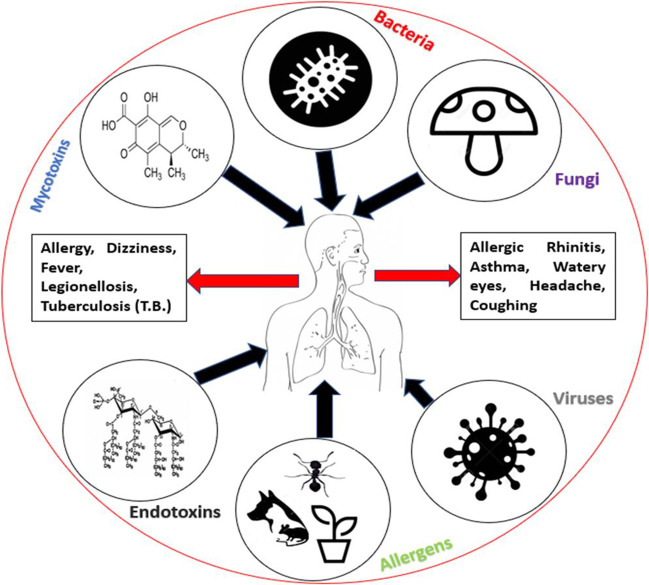
Bio-aerosols are airborne suspended particles, large molecules, volatile organic compounds, that are living or released from living organisms. The size of bioaerosol is generally 0.1 to 100 µm in diameter (Seltzer 1994). However, smaller bio-aerosols remain in the air for a longer time, whereas larger are deposited on surfaces. Table 1 represents sources, causal agents and the harmful effects of major bio-aerosols (ACGIH 1989). Moreover, the basic framework of the present review article is described in Fig. 1.
Table1.
Major bio-contaminants of indoor air, their sources, lifestyle and health effects
| Causative organism | Health effect | Lifestyle of organism | Sources in indoor area |
|---|---|---|---|
| Bacteria | |||
| M. tuberculosis | Tuberculosis (T.B.) | Obligate parasite | Human |
| Legionella spp. | Pneumonia | Facultative parasite | Hot water sources |
| Y. pestis | Plague | Facultative parasite | Infected fleas |
| B. anthracis | Anthrax | Facultative parasite | Animal handlers, veterinarians |
| Fungi | |||
| Alternaria spp. | Asthma, rhinitis | Saprophytes | Outdoor air, damp surfaces |
| Histoplasma spp. | Histoplasmosis | Facultative parasite | Bird droppings |
| Aspergillus (aflatoxin) | Cancer | Damp surfaces | |
| Penicillium spp. | Penicilliosis | Facultative parasite | Mold contaminated building |
| Viruses | |||
| Influenza virus | Respiratory infection | Obligate parasite | Human |
| Variola virus | Chicken pox | Obligate parasite | Human |
| Coronavirus (SARS-CoV-2) | Coronavirus disease | Obligate parasite | Human |
| Algae | |||
| Chlorococcus spp. | Asthma, rhinitis | Autotrophic | Outdoor air |
Fig. 1.
Framework of the study
However, evaluation of the indoor environment for the investigation of biological contaminants is a challenging task. Major challenges include the selection of appropriate sampling tests, to know where and when to sample and interpretation of results with harmful effects. If a threat is identified, the source of contamination must be investigated to reduce the risk of contamination. Another important aspect of the evaluation process is correlating the sampling data with health effects. The purpose of this review article is to represent an overview of airborne indoor bio-contaminant: their sources, physical properties, possible health consequences and evaluation methods associated with them.
Components of biological contaminants
Bacteria
Common bacteria associated with buildings are mostly saprophytic and they are a part of the normal microflora found in nose, mouth, ear, etc. and originating from outdoor air (Moldoveanu 2015). Few heterotrophic bacteria such as Mycobacteria, Legionella are found associated with biofilm formation, especially in water reservoirs of cooling systems and water pipes. Few species of bacteria (Actinobacteria and Bacillus) are associated with fungi in moist buildings (WHO 1988). Humans are responsible for the spread of various kinds of bacteria in the indoor environment. Bacteria found in the upper respiratory tract are often eliminated through talking, coughing or sneezing. Prominent bacterial species identified in the indoor areas were Corynebacterium spp., Streptococcus spp., Staphylococcus spp., and Micrococcus spp. (Moldoveanu 2015; Kausar et al. 2016).
Pets are an important source of bacteria and may be related to levels of endotoxins source in indoor environments (Ownby et al. 2013). Organic waste storage in the indoor areas increases the chances of bacterial contamination (Moldoveanu 2015).
Fungi
Fungi are unicellular or multicellular microorganisms having long, microscopic strands of hyphae to which are attached to various types of spore-forming structures (conidia). Group of fungi is extremely diverse, spore-bearing structures facilitate the process of aerosolization. Fungal spore concentration may range from 100 spores m3 to 100,000 spores m3 in the outdoor area (Moldoveanu 2015). Mold, mold spores and their by-products may be associated with the various respiratory problems (Americam Air and Water 2020). The concentration of fungi depends on the conditions of the area, for example, in damp houses with high water activity, the fungal spore count is very high (Nevalainen 2002). Aspergillus fumigates, Trichoderma, Fusarium, Yeast, Phialophora, and Stachybotrys, etc. are found in the high-water activity while Aspergillus versicolor & Penicillium grow well at relatively moderate to low water activity. Fungal spores are ubiquitous hence able to germinate in water activity between 0.80–0.98 (WHO 2009). Fungi need proteins, carbohydrates, and lipids for development. They can grow well on construction material, books, wood, paint, stored food, cooking oil, etc. (WHO 2009; Rautiainena et al. 2019). Fungal concentration and type depend upon the season, construction material, age of building and ventilation status of a building (WHO 1988). A review study on fungal bioaerosol shows that Aspergillus, Penicillium, Stachybotrys, Cladosporium, Alternaria, Trichoderma and Yeast are most common genera of fungi found in indoor and as well as outdoor air and may involve in causing severe health problems (WHO 2009; Sharma et al. 2011a, b).
Allergens
Allergens are substances that may be the natural cause of IgE-mediated allergic reactions in humans. They are high molecular weight protein or glycoproteins varying from 10 kDa to > 100 kDa. Allergens from Aspergillus, Penicillium, Cladosporium and Alternaria spp. induce type 1 hypersensitivity reaction, which causes various respiratory diseases (WHO 1988). Insects and their by-products are reported to act as potent allergens. A clinico-immunological study concluded that mosquito debris could be a potent inhalant allergen in indoor air (Kausar et al. 2007). Dermatophagoides pteronyssius (Der p I & Der p II) and Dermatophagoides farinae (Der f I) are major species of mites that produce allergens (Kausar 2018). Bakery, poultry, granary and sugar industry are major sources for allergens in the indoor environment. Penicillium, Aspergillus, Alternaria, Cladosporium, Curvularia, Epicoccum, Pullularia, Ganoderma, Trichoderma and Stemphylium are common fungi that act as allergens (Kausar 2018).
Mycotoxins
Mycotoxins are toxic secondary metabolites produced by the fungi, capable of causing disease and death in humans & animals (Bennett and Klich 2003). These interfere with RNA synthesis and cause DNA damage. Aflatoxin produced from Aspergillus flavus and A. parasiticus can cause cancer. Another mycotoxin produced by Stachybotrys chartarum can be related to acute pulmonary hemorrhage (Sharma et al. 2011a, b).
Volatile organic compounds (VOC) and fungi
Volatile organic compounds are low molecular weight chemicals with less solubility and high vapor pressure. VOC can also be produced as the metabolism of microorganisms and thus known as microbial volatile organic compounds (m VOC) (Rautiainena et al. 2019). The identification of mVOC in indoor air is an indicator of the presence of fungi even if quantitative measurements are negative. There are some mVOCs that function as indicators of biological contamination such as 2octen-1-ol. This compound is strongly emitted in the initial stages of growth of Aspergillus spp., Alternaria alternata and Penicillium spp., so it has been related as a non-specific indicator of fungal contamination (Jelen and Wasowicz 1998).
(1, 3)- β –D glucans
(1, 3)-β–D glucans are a group of β-D-glucose polysaccharides, a structural element of fungi and some bacteria that might be present in-house dust. The presence of (1,3)-β-D glucans in the indoor environment may induce respiratory infections (Seo et al. 2015). (Seo and co-workers investigated the concentration of (1,3)-β-D glucans in elementary schools before and after the rainy season concluded that their concentration and effects changes with time (Seo et al. 2015).
Viruses
Viruses are acellular entities that require a host for replication and multiplication that can grow on natural or man-made objects. Survival time of respiratory viruses and the risk of infection to cause disease is often induced by indoor air (Yu et al. 2004). Human activities like coughing, sneezing, talking, and laughing might spread millions of airborne viruses in the indoor environment through droplets (Diglisic et al. 1999). Hantavirus infection is very common in occupants in indoor environments due to the presence of rodents (Aitken and Jeffries 2001).
Endotoxins
Endotoxins are cell wall components gram negative-bacteria that made up of lipopolysaccharide (LPS) (Moldoveanu 2015, Dales et al. 2008). They are secreted by bacteria through small fragments in harsh conditions when the cell is dying. Pets, food waste storage, humidifiers and low ventilation are associated with a high level of endotoxin or gram-negative bacteria. Asthma and asthma-like symptoms are directly associated with the level of endotoxin in the indoor area (Michel et al. 1996).
Health effects induced due to biological contaminants
Biological contaminants may trigger allergic reactions, including hypersensitivity, allergic rhinitis, pneumonitis and some type of asthma. Infectious illnesses, such as measles, influenza, and chickenpox are transmitted through the air (Chirca 2019; Seltzer 1994; Rautiainena et al. 2019; Srikanth et al. 2008). Moulds and mildews release disease-causing toxins. Major symptoms of health problems caused by biological contaminants include sneezing, watery eyes, coughing, shortness of breath, dizziness, lethargy, fever, and digestive problems (EPA 2020; Stetzenbach et al. 2004). Various allergic reactions may occur after repeated exposure to a specific biological allergen (Chirca 2019; Seltzer 1994; Rautiainena et al. 2019; Srikanth et al. 2008). These reactions occur immediately due to re-exposure or multiple exposures over time. This is the reason behind the sudden high sensitivity to allergens in people with mild allergic reactions. Toxins produced by microorganisms grown in large building ventilation systems may be associated with diseases like humidifier fever. Microbes from home heating & cooling system and humidifiers can cause the same diseases. Susceptibility to disease causing bio-contaminants of indoor air is very high in children, older people and people with breathing problems, allergies, and lung diseases. Allergic reactions might be caused by pollens and molds in a significant portion of the population. Legionellosis, Tuberculosis, Staphylococcal infections, measles, and influenza are found to be transmitted by air (Seltzer 1994; Rautiainena et al. 2019). In this discussion health effects of indoor biological contaminants are divided into several categories based on reactions caused by specific organism (Table 2).
Table 2.
Major airborne microorganisms in different indoor spaces (SL- sampling location, SM-sampling method, ND- Not determined)
| SL | SM | Bacterial Concentration (cfu/m3) | Fungal Concentration (cfu/m3) | |||||||
|---|---|---|---|---|---|---|---|---|---|---|
| Ave | Min | Max | Major genus | Ave | Min | Max | Major genus | Reference | ||
| Library | Open plate | 1476 | 367 | 2595 |
Micrococcus Staphylococcus Streptococcus Bacillus, Neisseria |
1087 | 524 | 1992 | Cladosporium Alternaria Penicillium Aspergillus | Hayleeyesus and Manaye 2014 |
| Teaching hospital | Anderson sampler | ND | ND | ND | Bacillus Micrococcus Pseudomonas Staphylococcus | ND | ND | ND |
Kocuria Fusarium Aspergillus |
Sivagnanasundaram et al. 2019 |
| Maternity home | Open plate | ND | 259 | 2665 | Bacillus, Staphylococcus, Pseudomonas | ND | 79 | 826 |
Aspergillus Rhizopus Mucor |
Kumar et al. 2018 |
| Hospitals | Impactor | ND | 58 | 348 | Staphylococcus, Pseudomonas, Streptococcus, E. coli, Bacillus, Micrococcus | ND | ND | ND | ND | Kunwar et al. 2019; |
| Hospitals | Impactor | ND | ND | ND | ND | 954 | ND | ND | Aspergillus, Penicillium, Alternaria, Talaromyces | Cho et al. 2018 |
| Hospital | Anderson sampler | 720 | ND | ND | ND | 77 | ND | ND | - | Park et al. 2013 |
| Children medical Center | Anderson sampler | ND | ND | ND | ND | 40 | ND | ND |
Penicillium, Cladosporium, Aspergillus, Sterile mycelia |
Roshan et al. 2019 |
| Laboratory | Open plate | 320 | ND | ND | Staphylococcus, Bacillus | 460 | ND | ND |
Aspergillus, Penicillium, Alternaria |
Sheik et al. 2015 |
| College | Open plate | 290 | ND | ND | Staphylococcus, Micrococcus Bacillus | 340 | ND | ND |
Aspergillus, Alternaria, Penicillium |
Sheik et al. 2015 |
| College | Anderson sampler | ND | ND | ND | ND | ND | 12 | 8767 | Penicillium, Cladosporium, Alternaria, Aspergillus | Fang et al. 2019 |
| College | Anderson sampler | ND | 120 | 680 | Micrococcus, Bacillus, Staphylococcus, Pseudomonas, Streptococcus, | ND | 210 | 680 | Aspergillus, Penicillium, Fusarium, Mucor, Rhizopus | Bomala et al. 2016 |
| School | Anderson sampler | ND | 120 | 720 | Micrococcus, Bacillus, Staphylococcus, Streptococcus | ND | 220 | 420 | Aspergillus, Penicillium, Fusarium, Mucor, Rhizopus | Bomala et al. 2016 |
| Resident-ial house | Aerosol monitor | ND | 275 | 14,469 | Staphylococcus, Bacillus, Micrococcus, Serratia | ND | 315 | 1887 | Alternaria, Aspergillus | Sidra et al. 2015 |
| Office | Anderson sampler | ND | 14 | 494 | Micrococcus, Staphylococcus | ND | 0 | 176 | Penicillium,Aspergillus | Gołofit-Szymczak and Górny 2015 |
| Office | Anderson sampler | ND | 424 | 821 |
Staphylococcus Micrococcus |
ND | ND | ND | ND | Bragoszewska et al. 2018 |
| Offices | Open plate | ND | ND | ND |
Micrococcus, S. aureus, Streptococcus Bacillus |
ND | 734 | 4034 |
Aspergillus, Penicillium, Mucor, Cladosporium |
Moldoveanu 2015 |
Respiratory infections
Pathogens, viruses causing the common cold, flu and bacterial infections are causative agents for respiratory infections and the major cause behind respiratory problems is exposure to indoor molds (American Air and Water 2020; Fung and Hughson 2001; Curtis et al. 2004). Airborne fungi including Penicillium, Aspergillus, Acremonium, Paecilomyces, Mucor and Cladosporium can cause respiratory infection and diseases (Fung and Hughson 2001; Sharma et al. 2011a, b; Singh et al. 1994). Aspergillosis is the most common disease among them that can occur in immunocompromised hosts or as secondary infection (Maschmeyer et al. 2007). Long-lasting cold, watering eyes, prolonged muscle cramps and joint pain may be the symptoms of aspergillosis (Shah and Punjabi 2014). Soil-borne fungi such as Coccidioides, Histoplasma, and Blastomyces are usually carried by bats and birds that can cause wind-borne or animal-borne contamination. Aspergillus fumigatus and Aspergillus flavus are well growing indoor species of Aspergillus genus that can cause nosocomial infections (Verma et al. 2003), Allergic Bronchopulmonary Aspergillosis (ABPA) and sinusitis. The constant allergic response maintains the fungal colonization, and first-line therapy with steroids brings down the level of inflammation and may result in the elimination of the colonizing organism (Shah and Punjabi 2014). Although a lot of studies and consideration have been performed, however the actual mechanism behind the association of mold and respiratory infections of the indoor areas are still unknown. Some molds may cause an immunosuppressive effect on animals when they used as an experimental procedure (Srikanth et al. 2008).
Infectious diseases
Infectious diseases are caused by bacteria, viruses, fungi, etc. and can easily spread, from one person to another person. Infectious diseases may recognize by various symptoms in humans, including headache, rhinitis, fever, chills, coughs, shortness of breath, vomiting and diarrhea. The mode of action for the infectious diseases may occur by direct entry of pathogenic biological contaminants (mostly microorganisms in the form of bioaerosol) or entry of harmful toxins (e.g. neurotoxin, enterotoxin, etc.) produced by them. Some important infectious diseases with epidemiological issues are discussed here.
Bacterial diseases
Tuberculosis
Tuberculosis (T.B.) is among the top bacterial diseases responsible for mortalities all over the World. It is a common community-acquired disease caused due to inhalation of aerosolized bacilli in droplet nuclei of expectorated sputum-positive tuberculosis patients. The causal agent of T.B. is Mycobacterium tuberculosis. This is transmitted during coughing, sneezing and talking activities by infected persons to others in the form of bio-aerosols (Srikanth et al. 2008). Several outbreaks of multidrug-resistant (MDR) tuberculosis in the United Kingdom have highlighted the potential for transmission within the hospital environment (Srikanth et al. 2008).
Leprosy
Leprosy is an airborne infectious disease caused by slow-growing, intracellular acid-fast bacterium Mycobacterium laprae. Transmission of causative agent can be through respiratory droplets from the nasal mucosa of infected persons. It colonizes and usually infects at a lower temperature, symptoms may take 2–10 years to appear. Skin, nails, upper respiratory mucosa, superficial nerves, and lymph node are major affected organs (Anathanarayan and Panikar 2009). The reason behind giving more concern to Leprosy is its unculturable ability under laboratory conditions.
Legionellosis
Legionellosis is a hospital and community-acquired pneumonia caused by Legionella pneumophila in humans living in industrial or occupational works. The active aerosolizing processes (aeration of contaminated water) causes the occurrence of Legionella. The man-made water systems, biofilms in cooling towers and air conditioning systems are often inhabited by Legionella. However, the use of monochloramine for community residual drinking water disinfection may be helpful in preventing Legionnaires disease. Monochloramine is better than chlorine for reaching at distant points and penetrating biofilm in water sources but it requires higher pH for optimal disinfection (USEPA 2020; Kool et al. 1999).
Anthrax
Anthrax is a highly infectious airborne disease caused due to inhalation of the spores of Bacillus anthracis. Outbreaks of anthrax are often linked to bioterrorism that spreads through intentionally contaminated mail, apart from occupational exposures. Lungs, intestinal, skin, and injection are four major routes for spreading of disease (Vieira et al. 2017).
Fungal diseases
Mycosis
Mycosis is a common fungal infection in humans and animals. It can be divided into cutaneous, subcutaneous, systemic, or superficial depending upon host response to pathogen and various tissue infected (Fung and Hughson 2001; WHO 2009). Bird droppings and building dust may involve in systemic mycosis such as cryptococcosis, histoplasmosis and coccidioidomycosis in people associated with it (Fung and Hughson 2001; WHO 2009). Mycosis is a more severe infection in immunocompromised persons, e.g., suffering from AIDS, cancer chemotherapeutic patients and leukemia (WHO 1988; Fung and Hughson 2001; Prussin and Marr 2015).
Mycotoxicosis
Mycotoxicosis is caused by exposure to secondary metabolites produced by fungi. Mycotoxin shows their toxigenic effect by causing cytotoxicity, interfering with DNA/RNA synthesis, protein synthesis and cancer (Seltzer 1994; Fung and Hughson 2001). Only a few molds can produce mycotoxins depending upon seasons and environmental factors (dampness, relative humidity temperature, presence of oxygen and carbon dioxide) (Moldoveanu 2015; American Air and Water 2020). Mycotoxin isolated from few fungi acts as a potent carcinogens in occupants. Aflatoxin from Aspergillus flavus and A. parasiticus capable of causing liver cancer (Cirrhosis), ochratoxin A (Food containing mycotoxin) is a human carcinogen (Moldoveanu 2015; American Air and Water 2020). Various industries including livestock processing, peanut processing and other food production units are responsible for exposure to these carcinogens via ingestion and inhalation. Few studies have found an association between wood dust and various cancers including sinus cancer (Seltzer 1994; American Air and Water 2020; Fung and Hughson 2001).
Viral diseases
Different viruses including Influenza virus (H1N1), enteric viruses of intestinal origin, respiratory syncytial virus (RSV), hantavirus from rodent feces severe acute respiratory syndrome (SARS) virus, influenza virus, measles, mumps and rubella are easily transmitted through airborne route (Yu et al. 2004, Diglisic et al. 1999). SARS is a highly contagious respiratory illness, spread by a novel coronavirus that causes morbidity, mortality, and very severe atypical pneumonia.
(Srikanth et al. 2008). The transmission of SARS may be amplified by aerosol-generating procedures (such as endotracheal intubation, bronchoscopy, and treatment with aerosolized medication) used in hospitals (Yu et al. 2004). Recently, the whole World is suffering from coronavirus disease (COVID-19) caused by coronavirus (SARS-CoV-2). As per recent reports of WHO 7 million confirmed cases and more than 4 lakh deaths have occurred (WHO 2020). Chickenpox is one of the most common airborne diseases in children. This disease may occur in the adult with more virulence in specific areas.
Sources of biological contaminants in indoor air
Occurrence and concentration of microbiological agents may vary depending upon different conditions and activities such as by occupants, building systems, furnishings and food in different places (Kausar et al. 2007; Yassin and Almouqatea 2010; Sheik et al. 2015; Badea et al. 2015).
Humans
Humans are the most source for survival and transport of biological contaminants in the indoor area. A healthy person may have 1012 -1014 microbial cells in a different part of the body (Ananthanaryan and Paniker 2009). Inadequate ventilation in the closed indoor area including improper outdoor air, poor air distribution, improper air filtration and poor maintenance of ventilation system, may also risk in indoor air quality (Mandal and Brandl 2011). Human occupancy affects airborne microbes as well as community structure. The human-related bacteria are two times more in human-occupied area than the unoccupied outdoor area (Prussin and Marr 2015). Common bacterial species related to building are Micrococcus, Staphylococcus, Bacillus and Streptococcus (Charlson et al. 2011). Although most bacterial genera are detrimental for human and part of normal flora however, microflora of the male and female occupied area is variable. Corynebacterium spp., Roseburia spp., and Dermabacter genera are more abundant in male while Lactobacillus genera in females (Prussin and Marr 2015; Charlson et al. 2011).
Few fungal genera such as Candida, Rhodotorula, Trichosporon and Cryptococcus are related to human skin which spread in the form of bio-aerosols in occupied buildings. Pathogenic viruses are also associated with human beings but due to the knowledge gap pertaining to viral communities, their effect on the human community is unclear (Barberan et al. 2015).
Building systems
Construction material and other elements used in buildings may be the source of possible biological contamination. Biological contaminants may spread in a building through bio-aerosols formation in toilets, baths, sinks, drain traps, aquariums, etc. (WHO 1988). Construction materials on degradation convert into organic compounds that may very favourable for the growth of microorganisms. High relative humidity and high temperature also support the growth and proliferation of indoor microorganisms (WHO 1988). Some natural materials such as fabrics used for carpeting, furnishing, curtains, decorative articles, stuffed toys, cushions, and straw or reed baskets are also an important source of possible contamination in the indoor area (WHO 1988). Undesirable biological contamination in a building may be associated with problems such as sick building syndrome (SBS) and building-related illness (WHO 1988; Mandal and Brandl 2011).
Pets
Some reports suggested that exposure to bio-aerosols and dust originated from dogs is beneficial for the health of children and infants (Prussin and Marr 2015; Fujimura et al. 2010; Fujimura et al. 2014). Cats and dogs living in the indoor area have 56 and 24 bacterial genera, respectively. Arthrobacter spp., Bacteroides spp., Blautia spp., Neisseria spp., Moraxella spp., in dogs and Bifidobacterium spp., Sporosarcina spp., Jeotgalicoccus spp. Moraxella spp., Sporosarcina spp., Porphyromonas spp., and Prevotella spp. are predominant in cats (Prussin and Marr 2015).
Healthcare facilities
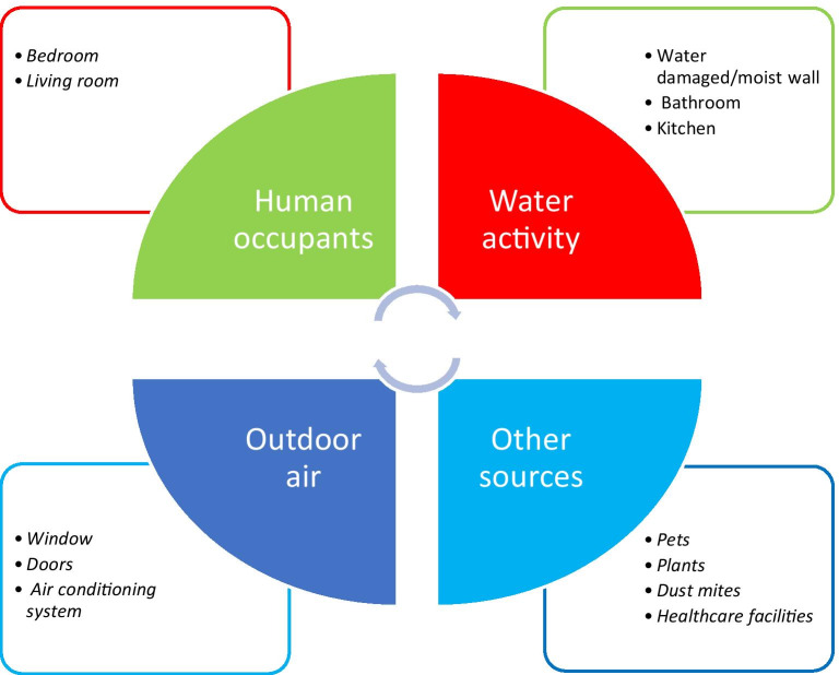
Hospital area is one of the major sources for spreading various microorganisms in the indoor space. Patients suffering from different diseases, ventilation facilities, sweeping, dressing beds, and other activities contribute airborne microorganisms through bio-aerosols formation (Knights et al. 2011). The different sources of bio-aerosols are presented in Fig. 2.
Fig. 2.
Sources of bioaerosols in indoor environment
Ambient conditions responsible for biological contamination
Temperature
Temperature is one of the most important factors for the growth and survival of biological contaminants in the indoor environment. The optimum temperature may vary from organism to organism even in the same genus. Extreme high and low temperature retards the growth of mesophilic bacteria (Seltzer 1994). Fungal species can grow on various temperatures eg. Yeast (Sporobolomyces), few mould spore formation and survival are better in cold environments and Aspergillus species can thrive at 57 °C with the optimum temperature around 40 °C (Seltzer 1994). Few species of bacteria (Legionella thermophila) and Actinomycetes are recorded at high temperature in the indoor environment. The rise in temperature causes high respiration, thus increasing CO2 and water activity, which promoting the growth of biological contaminants (Seltzer 1994).
Ventilation facility and sunlight
The presence of the number of ports of exit and entry in an indoor building is the source of microorganisms from the outdoor air. Inadequate ventilation leads to a lack of outdoor air, poor air distribution, thus resulting in poor indoor air quality (WHO 1988). Sizes of bioaerosol particles are also important as larger and dense particles settle quickly than larger particles. Ultraviolet light (UV) from sun cause dimerization in purine bases of bacterial DNA, which is bactericidal thus controlling factor for bacteria in outdoor air (WHO 1988). Some studies also suggested that indoor air quality may be significantly improved by proper use of ventilation facilities (Ghandizadeh and Godini 2018).
Available nutrients
The presence of dead organic matter (protein, carbohydrate, etc.) is a significant nutrient source that is the most limiting factor for bacteria, fungi and some viruses (Seltzer 1994).
Moisture
Bioaerosols are generated significantly through dehumidifiers, chillers, water sprays and stagnant water sources (WHO 1988). Installation of gravity-based drip pans and proper maintenance of non-stream humidifiers is suggested to reduce the process of bio-aerosolization (WHO 1988). High moisture level promotes the growth and survival of several species of microorganisms (WHO 1988; Stetzenbach et al. 2004; Lignell 2008). The concentration of microorganisms is relatively higher in moist damaged buildings than normal buildings (Lignell 2008). Control of indoor humidity level primarily through the regulation of ventilation rates and the careful use of dehumidifiers, chillers and humidifiers where necessary can reduce aerosol formation (WHO 1988; Stetzenbach et al. 2004). Humidity more than 65% may cause the incidence of upper respiratory problems which might increase and can have adverse effects on people suffering from asthma and allergies. Lower moisture levels (below 20%) may induce dryness or itching of the skin and aggravate certain skin conditions (WHO 1988).
Quantification of biological contaminants
Various sampling methods are needed to quantify and describe the microbial populations in the indoor environment. Evaluation methods may differ depending upon concentration and type of biological contaminants. Microbes (bacteria, fungi, spores, etc.) can be sampled from the air, surfaces, dust, and other materials. Since there is no technique that would cover all aspects of environmental sampling, the methods need to be selected according to the needs of the investigation. For recognition of any bio-contaminant in air first collection and then sample analysis followed by additional techniques are performed. These methods may be culture-based or non-culture based depending upon the viability of samples. Direct microscopy is also a useful method for the morphological identification of bacteria and fungi. The use of cello tape by sticking it on the surface and transferring it on slide followed by Carbela’s stain in case of most moulds is the cheaper and convenient non-culture method. Culture methods rely on the type of biological agents present in that indoor environment by providing them suitable medium and other requirements for growth in in-vitro. The study performed on the assessment of bio-contaminants proved that Diphtheroids (non-pathogenic gram-positive bacilli) & Staphylococcus were the most common bacteria and Cladosporium is most common fungi in the indoor area (Yassin and Almouqatea 2010). A similar study suggests that the concentration of microorganisms is quite low in college as compared to laboratories and toilets as per guidelines (Sheik et al. 2015). Fungal spore levels may vary distinctly in different seasons, indoor and outdoor as well (Dey et al. 2019).
Sampling methods
The sampling of air is performed for the collection of viable biological contaminants on a suitable medium. Sampling of dust, airborne microorganisms and other particulates are based on the same principle. Air sampling is mainly divided into two types based on sampler and protocol applied (a) Active sampling and (b) Passive sampling. Studies published about the comparison of active and passive methods to assess indoor quality concluded that active methods are more efficient than passive, but often quantitative data is imbalanced due to the overgrowth of viable organisms (Moldoveanu 2015, Napoli et al. 2012). Sampling duration is very crucial for the evaluation of airborne microorganisms, however, a study published in 2006 found that airborne culturable mold concentrations remain similar in duration in the range of 1–10 min (Godish and Godish 2007).
Passive sampling
Passive monitoring is based upon exposure of agar plates and agar coated glass slides in air and deposition of microorganisms along with airborne particles. Passive sampling of microorganisms is based on the Koch’s sedimentation method. Growth of viable biological particles was expressed in the form of colony-forming units (CFU) by the following formula:
where “n” is the number of colonies, “t” is exposure time, and “s” is the surface area of the Petri plate.
However, the Koch’s sedimentation method is less appropriate than the impaction method by the sampler. The sedimentation method is cheap and simple, provides an approximate enumeration of fungi and bacteria in the indoor environment (Moldoveanu 2015; Srikanth et al. 2008).
Active sampling
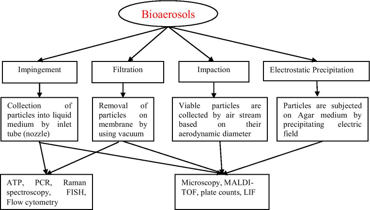
Active sampling methods rely on the collection of airborne microorganisms on the culture medium by any external force. Active sampling methods require specific setup of instruments for different air samples. Srikanth and co-workers reviewed the different types of active samplers used for the evaluation of bioaerosols in indoor air (Srikanth et al. 2008). Almost samplers used for quantitative measurement of airborne microorganisms are based on creating a vacuum inside. Active sampling methods for quantitative measurement of airborne microorganisms are based on solid impaction surfaces, an impingement in liquids, filtration, electric precipitation, thermal precipitation and centrifugation (Sutton 2004; Kang and Frank 1989). Active sampling methods are based on four principles: Impingement, impaction, filtration, and precipitation. Inlet efficiency (shape, size, density and collection efficiency of the airborne particle) is an important physical parameter of a sampler (Srikanth et al. 2008). All active methods for bioaerosol sampling and further methods used for identification are described in Fig. 3 (Mandal and Brandl 2011).
Fig. 3.
Bioaerosols sampling methods and identification by using different techniques
Impaction methods
Impaction methods collect airborne microorganisms with air current on the agar surface and sometimes on a dry surface. Anderson multistage sieve sampler is a typical example of an impaction method consisting of air-jet directed over a plate for the collision of the particle on a plate (Anderson 1958). Airborne microorganism’s particles along with suspended particles are settled down by the force of the vacuum. Impaction methods are more accurate and don’t require any manipulation procedure after a collection of samples (Moldoveanu 2015; Sutton 2004; Kang and Frank 1989).
Filtration methods
Filters are used routinely for the collection of microorganisms due to high-cost efficiency and easy procedure. Pore size and constructive material of filters may vary according to requirement e.g., cellulose, a gelatine membrane filter and glass fibre are the most commonly used material. Having a large number of pores, it can filter air easily; however, the disadvantage of filtration is least effective with vegetative cells (Sutton 2004; Kang and Frank 1989).
Impingement methods
Impingement liquid medium (salt solution with antifoam agents, proteins) is used for the collection of airborne microorganisms. Entrapping of air through liquid makes dispersal of particles in it, followed by quantitative measurement through serial dilution. All Glass Impinger-30 (AGI-30) sampler has high-velocity which is a typical example of this category. These methods are inexpensive and highly useful for the collection of viable cells (Sutton 2004; Kang and Frank 1989).
Control of biological contaminants
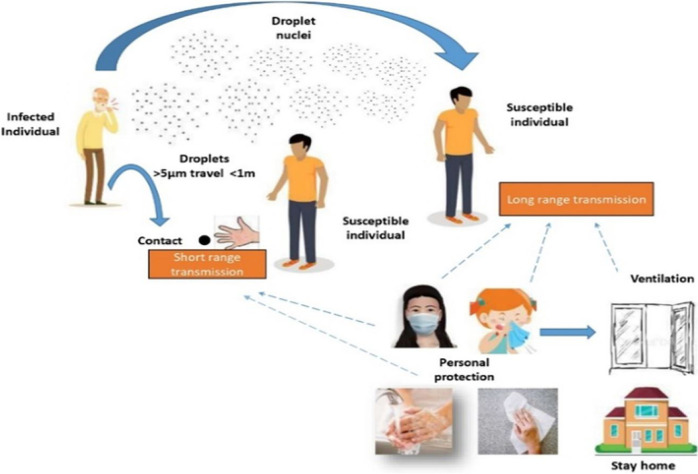
Control of biological contamination in indoor air is predominantly based on the avoidance of conditions that provide a substrate for the growth of viable particles, and secondarily on the containment and removal of such growth (WHO 1988). By the use of air filters and improving ventilation performance may be a viable solution to control bioaerosols (Vlad et al. 2013). Most control strategies applied in indoor areas are based on controlling the dew point, which reduces the growth of various microorganisms. Contaminated water on aerosolization may result in the formation and spread of harmful bioaerosols in indoor and outdoor areas (WHO 1988). Airborne transmission of infective disease inside a building depends upon occupant density e.g. higher the density higher the risk. Presence of vectors such as pets, insects, may be responsible for the transmission of non-viable aerosols causing allergy (WHO 1988). Possible transmission and control strategies of airborne diseases are described in Fig. 4.
Fig. 4.
Possible transmission pathways and personal prevention from airborne infectious diseases
Design and construction of a building
Major factors responsible for biological contamination in the indoor area such as regional climatic factors, behavioral factors etc. are originated from improper construction and design of buildings (WHO 1988; Dales et al. 2008). The use of concrete as a building material is the most efficient way to minimize the relative humidity of a building, which is the most prominent factor for microbial growth.
Humidity control
The use of air conditioners and humidifiers helps in controlling biological contamination by reducing relative humidity in a building. In humid weather, water vapor pressure is very low in the outdoor area; thus, humidification is required. Humidification through corrosion-proof injection of the stream with 65 °C temperature is the most preferable in a building.
Social and behavior factors control
There are many incidents related to biological contamination but mostly are avoided due to a lack of data or resources in indoor areas. Qualitative and quantitative profile of microorganisms of indoor space is strongly influenced by the climatic conditions of that area. Equator countries are likely to more chances of disease than temperate or polar regions (WHO 1988). Behavioral factors are often useful and least expensive for maintaining air quality and reducing adverse health effects on us (WHO 1988).
Bio-fumigation
Fumes from herbal plants and their products have been used in cleaning the air from ancient times. In Hindu culture, bio-fumigation is considered as holy act known as ‘Agnihotra’ in which cow dung, fine cow butter, meditational plant leaves, wood, etc., are burned and its fumes clean the air. Scientists performed agnihotra in different sites and recorded microflora before and after it, there was a huge reduction in microbial quantity. Bio-fumigation controls microorganisms by lowering relative humidity (Pachori et al. 2013). Studies also performed Vedic methods for purification in Haridwar. Lad and co-workers prepared Dhoop from herbal products for cleaning the air and its efficiency was also checked its efficiency against indoor microorganisms (Lad and Palekar 2016).
Conclusion
A variety of bio-contaminants are present in indoor as well as outdoor air. Human beings exposed to indoor bio-contaminants may suffer from various airborne health problems including respiratory infections, allergy, infectious diseases, and indirect effects. Due to the omnipotent presence of microbes in nature, they are present in every type of environment. Millions of microbial cells are inhaled with the breathing of air. Only a limited number of microbes are culturable in the laboratory due to the lack of all nutrients and conditions. Humans and other activities act as a source for high microbial load in different indoor sites. Qualitative and quantitative variation in bio-contaminants is associated with factors such as temperature, available nutrients, ventilation facility, moisture etc. The health effects of bio-contaminants may be minimized by following programmed hygiene and maintenance procedures. However, there is an urgent need to establish standard protocols for inspection of indoor air quality and hopefully, it will clarify the indoor environment and its effects on human health.
Acknowledgements
This work was financial supported by Science and Engineering Research Board, Department of Science and Technology (SERB-DST), New Delhi, India.
Abbreviations
- AC
Air conditioner
- RSV
Respiratory Syncytial Virus
- SARS
Severe acute respiratory syndrome
- WHO
World health organization
- COVID-19
Coronavirus disease
Authors contributions
P K wrote and designed the manuscript. A K and A B S drafted and edited the manuscript. R S designed, analyzed, wrote and supervised the manuscript.
Data availability
Not Applicable.
Declarations
Ethics approval and consent to participate
Not Applicable.
Consent for publication
Not Applicable.
Conflict of interest
The authors declare that they have no conflict of interest.
Footnotes
Publisher’s note
Springer Nature remains neutral with regard to jurisdictional claims in published maps and institutional affiliations.
References
- Aitken C, Jeffries DJ. Nosocomial spread of viral disease. Clin Microbiol Rev. 2001;14:528–546. doi: 10.1128/CMR.14.3.528-546.2001. [DOI] [PMC free article] [PubMed] [Google Scholar]
- American Air and Water (2020) Mold, mold spores and indoor air quality. American Air and Water. Available at https://www.americanairandwater.com/mold Accessed on: 8 June 2020
- American Conference of Governmental Industrial Hygienist (1989) Guidelines for the assessment of Bioaerosols in indoor environment. American Conference of Governmental Industrial Hygienist, Cincinnati
- Ananthanaryan R, Paniker CKJ (2009) Text of Microbiology, 8th Edition, Universities press
- Anderson AA. A new sampler for collection, sizing and enumeration of viable airborne particles. J Bact. 1958;76:471–484. doi: 10.1128/jb.76.5.471-484.1958. [DOI] [PMC free article] [PubMed] [Google Scholar]
- Badea S, Chirita AL, Androne C, Olaru BG (2015) Indoor air quality assessment through microbiological methods. Scientific Papers. Series Journal of Young Scientist 3:12–17
- Barberan A, Dunn RR, Reich BJ, Pacifici K, Laber EB, Menninger HL, et al. The ecology of microscopic life in household dust. Proc R Soc B. 2015;282:20151139. doi: 10.1098/rspb.2015.1139. [DOI] [PMC free article] [PubMed] [Google Scholar]
- Bennett JW, Klich M. Mycotoxins. Clin Microbiol Rev. 2003;16(3):497–516. doi: 10.1128/CMR.16.3.497-516.2003. [DOI] [PMC free article] [PubMed] [Google Scholar]
- Bomala K, Saramanda G, Reddy B, et al. Microbiological indoor and outdoor air quality of selected places in Visakhapatnam City India. Int J Curr Res. 2016;08(04):29059–29062. [Google Scholar]
- Bragoszewska E, Mainka A, Pastuszka JS, Czyk KL, Desta YG. Assessment of bacterial aerosol in a preschool primary school and high school in Poland. Atmosphere. 2018;9:87. doi: 10.3390/atmos9030087. [DOI] [Google Scholar]
- Charlson ES, Bittinger K, Haas AR, Fitzgerald AS, Frank I, Yadav A, et al. Topographical continuity of bacterial populations in the healthy human respiratory tract. Am J Resp Crit Care. 2011;184:957–963. doi: 10.1164/rccm.201104-0655OC. [DOI] [PMC free article] [PubMed] [Google Scholar]
- Chirca I (2019) The hospital environment and its microbial burden: challenges and solutions. Future Microbiol 14(12):1007–1010. 10.2217/fmb-2019-0140 [DOI] [PubMed]
- Cho SY, Myong JP, Kim WB, et al. Profiles of Environmental Mold: Indoor and Outdoor Air Sampling in a Hematology Hospital in Seoul, South Korea. Int J Environ Res Public Health. 2018;15:2560. doi: 10.3390/ijerph15112560. [DOI] [PMC free article] [PubMed] [Google Scholar]
- Curtis L, Lieberman A, Stark M, et al. Adverse Health Effects of Indoor Molds. J Nutr Environ Med. 2004;14(3):261–274. doi: 10.1080/13590840400010318. [DOI] [Google Scholar]
- Dales R, Liu L, Wheeler AJ, Gilbert NL (2008) Quality of indoor residential air and health. CMAJ 179(2):147–152. 10.1503/cmaj.070359 [DOI] [PMC free article] [PubMed]
- Dey D, Ghoshal K, Bhattacharya SG. Aerial fungal spectrum of Kolkata, India, along with their allergenic impact on the public health: a quantitative and qualitative evaluation. Aerobiologia. 2019;35:15–25. doi: 10.1007/s10453-018-9534-6. [DOI] [Google Scholar]
- Diglisic G, Rossi CA, Doti A, Walshe DK. Seroprevalence study of Hantavirus infection in the community-based population. Md Med J. 1999;48:303–306. [PubMed] [Google Scholar]
- Dubey RC, Maheshwari DK (2014) Textbook of Microbiology, 5th edition. S. Chand & Co., Limited, Ram Nagar, New Delhi 110055 (India)
- Fang Z, Zhang J, Guo W, et al. Assemblages of Culturable Airborne Fungi in a Typical Urban. Tourism-driven Center of Southeast China Aerosol and Air Quality Research. 2019;19:820–831. doi: 10.4209/aaqr.2018.02.0042. [DOI] [Google Scholar]
- Fujimura KE, Johnson CC, Ownby DR, Cox MJ, Brodie EL, Havstad SL, et al. Man’s best friend? The effect of pet ownership on house dust microbial communities. J Allergy Clin Immunol. 2010;126:410–412. doi: 10.1016/j.jaci.2010.05.042. [DOI] [PMC free article] [PubMed] [Google Scholar]
- Fujimura KE, Demoor T, Rauch M, Faruqi AA, Jang S, Johnson CC, et al. House dust exposure mediates gut microbiome Lactobacillus enrichment and airway immune defense against allergens and virus infection. Proc Natl AcadSci U S A. 2014;111:805–810. doi: 10.1073/pnas.1310750111. [DOI] [PMC free article] [PubMed] [Google Scholar]
- Fung F, Hughson WG. Health effects of indoor fungal bioaerosol exposure. Appl Occup Environ Hyg. 2001;18(7):535–544. doi: 10.1080/10473220301451. [DOI] [PubMed] [Google Scholar]
- Ghanizadeh F, Godini H. A review of the chemical and biological pollutants in indoor air in hospitals and assessing their effects on the health of patients, staff and visitors. Rev Environ Health. 2018;33(3):231–245. doi: 10.1515/reveh-2018-0011. [DOI] [PubMed] [Google Scholar]
- Godish DR, Godish TJ. Relationship between sampling duration and concentration of culturable airborne mould and bacteria on selected culture media. J ApplMicrobiol. 2007;102:1479–1484. doi: 10.1111/j.1365-2672.2006.03200.x. [DOI] [PubMed] [Google Scholar]
- Gołofit-Szymczak M, Górny RL (2015) Bacterial and fungal aerosols in air-conditioned office buildings in Warsaw, Poland—The Winter Season. Int J Occup Saf Ergon 16(4):465–476 [DOI] [PubMed]
- Gonçalves CL, Mota FV, Ferreira FG. Airborne Fungi in an Intensive Care Unit. Braz J Biol. 2018;78(2):265–270. doi: 10.1590/1519-6984.06016. [DOI] [PubMed] [Google Scholar]
- Hayleeyesus SF, Manaye AM (2014) Microbiological quality of indoor air in university libraries. Asian Pac J Trop Biomed 4(Suppl 1):S312–7. 10.12980/APJTB.4.2014C807 [DOI] [PMC free article] [PubMed]
- Jelen HH, Wasowicz E. Volatile fungal metabolites and their relation to the spoilage of agricultural commodities. Food Rev Int. 1998;14(4):391–426. doi: 10.1080/87559129809541170. [DOI] [Google Scholar]
- Joshi M, Srivastava RK. Identification of indoor airborne microorganisms in residential rural houses of Uttarakhand, India. Int J CurrMicrobiol App Sci. 2013;2(6):146–152. [Google Scholar]
- KangFrank YJJF. Biological Aerosols: A Review of Airborne Contamination and its Measurement in Dairy Processing Plants. J Food Prot. 1989;52(7):512–524. doi: 10.4315/0362-028X-52.7.512. [DOI] [PubMed] [Google Scholar]
- Kausar MA. A review on Respiratory allergy caused by insects. Bioinformation. 2018;14(9):540–553. doi: 10.6026/97320630014540. [DOI] [PMC free article] [PubMed] [Google Scholar]
- Kausar MA, Vijayan VK, Bansal SK, Menon BK, Vermani M, Agarwal MK. Mosquitoes as sources of inhalant allergens: Clinico-immunologic and biochemical studies. J Allergy ClinImmunol. 2007;120:1219–1221. doi: 10.1016/j.jaci.2007.07.017. [DOI] [PubMed] [Google Scholar]
- Kausar MA, Arif JM, Alanazi SMM, Alshmmry AMA, Alzapni YAA, Alanazy FKB, Shahid SMA, Hossain A. Assessment of microbial load in indoor environment of University and hospitals of Hail. KSA Biochem Cell Arch. 2016;16(1):177–183. [Google Scholar]
- Knights D, Kuczynski J, Charlson ES, Zaneveld J, Mozer MC, Collman RG, et al. Bayesian community-wide culture-independent microbial source tracking. Nat Meth. 2011;8:761–763. doi: 10.1038/nmeth.1650. [DOI] [PMC free article] [PubMed] [Google Scholar]
- Kool JL, Bergmire-Sweat D, Butler JC, Brown EW, Peabody DJ, Massi DS, et al. Hospital characteristics associated with colonization of water systems by Legionella and risk of nosocomial legionnaires disease: a cohort study of 15 hospitals. Infect Control Hosp Epidemio. 1999;20:798–805. doi: 10.1086/501587. [DOI] [PubMed] [Google Scholar]
- Kumar H, Kaur A, Kaur B, et al. Assessment of microbial contamination in indoor air of private maternity homes in Moga, Punjab. J Clin Diagn Res. 2018;12(5):DC01–DC05. [Google Scholar]
- Kunwar A, et al. Bacteriological assessment of the indoor air of different hospitals of Kathmandu District. Int J Microbiol. 2019 doi: 10.1155/2019/5320807. [DOI] [PMC free article] [PubMed] [Google Scholar]
- Lad N, Palekar S. Preparation and evaluation of Herbal Dhoop for cleansing the air. Int J Herb Med. 2016;4(6):98–103. [Google Scholar]
- Lignell U (2008) Characterization of microorganisms in indoor environments. Publications of the National Public Health Institute KTL, Kuopio
- Macher JM, et al. Sampling airborne microorganisms and aeroallergens. In: Cohen BS, Hering SV, et al., editors. Air Sampling Instruments for Evaluation of Atmospheric Contaminants. 8. Cincinnati: ACGIH; 1995. pp. 589–617. [Google Scholar]
- Mandal J, Brandl H. Bioaerosols in indoor environment - A review with special reference to residential and occupational locations. Open Environ Biol Monit J. 2011;4:83–96. doi: 10.2174/1875040001104010083. [DOI] [Google Scholar]
- Maschmeyer G, Haas A, Cornely OA. Invasive Aspergillosis: Epidemiology, Diagnosis and Management in Immunocompromised Patients. Drugs. 2007;67(11):1567–1601. doi: 10.2165/00003495-200767110-00004. [DOI] [PubMed] [Google Scholar]
- Michel O, Kips J, Duchateau J, et al. Severity of asthma is related to endotoxin in house dust. Am J Respir Crit Care Med. 1996;154:1641–1646. doi: 10.1164/ajrccm.154.6.8970348. [DOI] [PubMed] [Google Scholar]
- Microbiomes of the Built environment: A research agenda for indoor microbiology, human health and buildings. The National Academics Press. 10.17226/23647 [PubMed]
- Moldoveanu AM (2015) Biological Contamination of Air in Indoor Spaces. Current Air Quality issues 489-514. 10.5772/59727
- Napoli, et al. Air sampling procedures to evaluate microbial, contamination: a comparison between active and passive methods in operating theatres. BMC Public Health. 2012;12:594. doi: 10.1186/1471-2458-12-594. [DOI] [PMC free article] [PubMed] [Google Scholar]
- Nevalainen A, Seuri M (2005) Of microbes and men. Indoor Air 15(Suppl 9):58–64. 10.1111/j.1600-0668.2005.00344.x [DOI] [PubMed]
- Ownby DR, Peterson EL, Wegienka G. Are Cats and Dogs the Major Source of Endotoxin in Homes? Indoor Air. 2013;23(3):219–226. doi: 10.1111/ina.12016. [DOI] [PMC free article] [PubMed] [Google Scholar]
- Pachori RR, Kulkarni NS, Sadar PS, Mahajan NN. Effect of Agnihotra Fumes on Aeromicroflora. The Bioscan. 2013;8(1):127–129. [Google Scholar]
- Park DU, Yeom JK, Lee WJ. Assessment of the levels of airborne bacteria, gram-negative bacteria, and fungi in hospital lobbies. Int J Environ Res Public Health. 2013;10:541–555. doi: 10.3390/ijerph10020541. [DOI] [PMC free article] [PubMed] [Google Scholar]
- Prussin AJ, Marr LC (2015) Sources of airborne microorganisms inthe built environment. Microbiome 3(1) [DOI] [PMC free article] [PubMed]
- Rautiainena P, Hyttinenb M, Ruokolainenb J, et al. Indoor air-related symptoms and volatile organic compounds in materials and air in the hospital environment. Int J Env Health Res. 2019;29(5):479–488. doi: 10.1080/09603123.2018.1550194. [DOI] [PubMed] [Google Scholar]
- Roshan SK, Sedighe NB, et al. Study on the relationship between the concentration and type of fungal bio-aerosols at indoor and outdoor air in the Children's Medical Center, Tehran, Iran. Environ Monit Assess. 2019;191(2):48. doi: 10.1007/s10661-018-7183-4. [DOI] [PubMed] [Google Scholar]
- Seltzer JM (1994) Biological contaminants. J Allergy Clin Immunol 94(2 Pt 2):318–26 [PubMed]
- Seo S, Ji YG, Yoo Y, Kwon MH, Choung JT (2015) Submicron fungal fragments as another indoor biocontaminant inelementary schools. Environ Sci Process Impacts 17(6):1164–1172 [DOI] [PubMed]
- Shah A, Punjabi C. Allergic aspergillosis of the respiratory tract European Respiratory. Review. 2014;23:8–29. doi: 10.1183/09059180.00007413. [DOI] [PMC free article] [PubMed] [Google Scholar]
- Sharma R, Gaur SN, Singh VP, Singh AB. Association between Indoor fungi in Delhi Homes and Sensitization in Children of Respiratory Allergy. Med Mycol. 2011;50(3):281–290. doi: 10.3109/13693786.2011.606850. [DOI] [PubMed] [Google Scholar]
- Sharma R, Deval R, Priyadarshia V, Gaur SN, Singh VP, Singh AB. Indoor fungal concentration in the homes of allergic/asthmatic children in Delhi India. Allergy Rhinol. 2011;2:21–32. doi: 10.2500/ar.2011.2.0005. [DOI] [PMC free article] [PubMed] [Google Scholar]
- Sheik GB, Rheam AIA, Shehri ZS, Otaibi OBM. Assessment of Bacteria and Fungi in air from College of Applied Medical Sciences (Male) at AD-Dawadmi Saudi Arabia. Int Res J Biol Sci. 2015;4(9):48–53. [Google Scholar]
- Sidra S, Ali Z, Sultan S, Ahmed S, Colbeck I, Nasir ZA (2015) Assessment of airborne microflora in the indoor micro-environments of residential houses of Lahore, Pakistan. Aerosol and AirQuality Research 15(6):2385–2396
- Singh A, Gangak SV, Singh AB. Airborne fungi in the hospitals of metropolitan Delhi. Aerobiologia. 1994;10:11–21. doi: 10.1007/BF02066742. [DOI] [Google Scholar]
- Sivagnanasundaram P, Amarasekara RWK, Madegedara RMD et al. Assessment of Airborne Bacterial and Fungal Communities in Selected Areas of Teaching Hospital, Kandy, Sri Lanka. Bio Med Res Int Volume 2019. 10.1155/2019/7393926 [DOI] [PMC free article] [PubMed]
- Srikanth P, Sudharsanam S, Steinberg R. Bio-aerosols in indoor environment: composition, health effects and analaysis. Indian J Med Microbiol. 2008;26(4):302–312. doi: 10.1016/S0255-0857(21)01805-3. [DOI] [PubMed] [Google Scholar]
- Stetzenbach LD, Buttner MP, Cruz P (2004) Detection and enumeration of airborne biocontaminants. Curr Opin Biotechnol 15(3):170–174. 10.1016/j.copbio.2004.04.009 [DOI] [PubMed]
- Sutton GHC (2004) Enumeration of total Airborne Bacteria, Yeast and Mold Contaminants and Identification of Escherichia coli O157:H7, Listeria Spp., Salmonella Spp., and Staphylococcus Spp. in a Beef and Pork Slaughter Facility
- Szymczak MZ, Górny RL. Bacterial and Fungal Aerosols in Air-Conditioned Office Buildings in Warsaw, Poland—The Winter Season. International Journal of Occupational Safety and Ergonomics (JOSE) 2010;16(4):465–476. doi: 10.1080/10803548.2010.11076861. [DOI] [PubMed] [Google Scholar]
- United States Environmental Protection agency (EPA) (2020) Chloramine in Drinking Water, Drinking water requirements for states and public watewr Systems. Available on: https://www.epa.gov/dwreginfo/chloramines-drinking-water Accessed on: 9 June 2020
- Verma KS, Jain V, Rathore AS. Role of Aspergillus spp. in causing possible Nosocomial Aspergillosis among Immunocompromised Cancer Patients. Indian J Asthma Immunol. 2003;17:77–83. [Google Scholar]
- Vieira A R, Salzer J S, Traxler R M et al., Enhancing Surveillance and Diagnostics in Anthrax-Endemic Countries, Emerging Infectious Diseases; Vol. 23, Supplement to December 2017 10.3201/eid2313.170431 [DOI] [PMC free article] [PubMed]
- Vlad DC, Popescu R, Filimon MN, Gurban C, Tutelka A, Nica DV, Dumitrascu V. Assessment of microbiological indoor air quality in public buildings: A case study (Timisoara, Romania) Afr J Microbiol Res. 2013;17(9):1957–1963. [Google Scholar]
- WHO (2009) WHO Guidelines for Indoor Air Quality: Dampness and Mould, World Health Organization, Copenhagen [PubMed]
- WHO (2020) Coronavirus disease (COVID-19) World Health Organization situation reports 142. Available on: https://www.who.int/docs/default-source/coronaviruse/situation-reports Accessed on-10 June 2020.
- World Health Organization. Regional Office for Europe: Indoor air quality: biological contaminants. Report on a WHO meeting, Rautavaara, 29 August -2 September 1988
- Yassin MF, Almouqatea S. Assessment of airborne bacteria and fungi in an indoor and outdoor environment. Int J Environ Sci Tech. 2010;7(3):535–544. doi: 10.1007/BF03326162. [DOI] [Google Scholar]
- Yu ITS, Li Y, Wong TW, Tam W, Chan AT, Lee JHW, et al. Evidence of Airborne Transmission of the Severe Acute Respiratory Syndrome Virus. N Engl J Med. 2004;350:1731–1739. doi: 10.1056/NEJMoa032867. [DOI] [PubMed] [Google Scholar]
Associated Data
This section collects any data citations, data availability statements, or supplementary materials included in this article.
Data Availability Statement
Not Applicable.


