Abstract
Introduction
Cranial radiotherapy (CRT) is the main treatment for non-small cell lung cancer (NSCLC) with brain metastasis (BM) and non-EGFR/ALK/ROS1-TKIs indication, and anlotinib can improve overall prognosis. However, the clinical effects of CRT combined with anlotinib for the treatment of NSCLC with BM remain unclear.
Methods
We retrospectively analyzed the clinical effects of anlotinib + CRT versus CRT alone in NSCLC patients with BM and non-EGFR/ALK/ROS1-TKIs indication from September 2016 to June 2020. The progression-free survival (PFS) and overall survival (OS) of anlotinib + CRT versus CRT alone were analyzed. After evaluation of the clinical characteristics to generate a baseline, the independent prognostic factors for intracranial PFS (iPFS) and OS were subjected to univariate and multivariate analysis. Finally, subgroup analysis for iPFS and OS was performed to assess treatment effects using randomized stratification factors and stratified Cox proportional hazards models.
Results
This study included data for 73 patients with BM at baseline. Of the 73 patients, 45 patients received CRT alone, and 28 patients received CRT + anlotinib. There was no significant difference in clinical features between the two groups (P > 0.05). Compared with the CRT group, the combined group had longer iPFS (median iPFS [miPFS]: 3.0 months vs 11.0 months, P = 0.048). However, there were no significant differences in OS, extracranial PFS, and systemic PFS. For clinical features, univariate and multivariate analysis showed that the plus anlotinib treatment was an independent advantage predictor of iPFS (hazard ratio [HR] 0.51; 95% confidence interval [CI] 0.27–0.95; P = 0.04), and age ≥57 years (HR 1.04, 95% CI 1.01–1.08, P = 0.014) and KPS score ≤80 (HR 1.04, 95% CI 1.01–1.08, P = 0.014) were independent disadvantage predictors of OS (P < 0.05). In addition, although this difference was not statistically significant (p > 0.05), the patients with the anlotinib + local CRT (LCRT) treatment had the longest iPFS (miPFS: 27.0 months) and OS (median OS [mOS]: 36 months). The miPFS and mOS values for the LCRT group were 11 months and 18 months, respectively, with shorter values for whole-brain RT (WBRT) + anlotinib group, WBRT + LCRT + anlotinib group, WBRT, and WBRT + LCRT.
Conclusion
Anlotinib can improve the intracranial lesion control and survival prognosis of NSCLC patients with CRT.
Keywords: radiotherapy, lung cancer, brain metastases, progression-free survival, overall survival
Background
Lung cancer is the most common of all malignant tumors worldwide.1–3 The proportion of non-small cell lung cancer (NSCLC) in all lung cancer is about 80%, and 30–43% of patients will have brain metastasis (BM) in the process of the disease.4 The prognosis of lung cancer patients with BM is poor, and the median overall survival (mOS) of patients that do not receive treatment is only 1–3 months.5
In recent years, significantly improved prognosis has been achieved for lung cancer patients by treatment using small molecular targeted tyrosine kinase inhibitors (TKIs) to target anaplastic lymphoma kinase (ALK), epidermal growth factor receptor (EGFR), and C-ros oncogene 1-receptor tyrosine kinase (ROS1).6 Patients with BM from NSCLC receiving these treatments also showed significantly improved survival.7 However, for advanced NSCLC patients with non-gene mutation or resistance to EGFR/ALK/ROS1-TKIs, alternative TKIs are not available, making it urgent to develop specific targeted drugs.
Anlotinib hydrochloride was independently developed in China as an orally administered, multi-target TKI.8 Anlotinib can inhibit tumor cell proliferation and tumor angiogenesis by inhibiting tumor-related kinases, such as VEGFR, FGFR, PDGFR α/β, c-kit, and RET.8–10 In ALTER1202, ALTER0302 and ALTER0303 trials, the overall survival (OS) and progression-free survival (PFS) of the anlotinib group were significantly better than that of the placebo (p < 0.05).5,10–12 The ALTER0303 clinical trial evaluated the efficacy of anlotinib for the treatment of BM. For these patients with BM at baseline, the mPFS for anlotinib treatment was 4.17 months, considerably higher than the 1.3 months for placebo treatment, and the mOS for anlotinib treatment was 8.57 months compared to 4.55 months for placebo treatment. The patients in the anlotinib group exhibited a longer time to brain progression (TTBP) than the placebo group, indicating that anlotinib delays the progression of intracranial lesions from advanced NSCLC patients with non-gene mutation or resistance to EGFR/ALK/ROS1-TKIs.12 Therefore, in May 2018, the China Food and Drug Administration officially approved anlotinib for third-line or higher treatment of advanced NSCLC patients with non-EGFR/ALK/ROS1-TKIs indication.
Clinical studies have confirmed that anlotinib can effectively treat some patients with advanced lung cancer, including patients with BM.12 However, for NSCLC patients with no specific gene mutation or EGFR/ALK/ROS1-TKIs resistance, cranial radiotherapy (CRT) is still considered the standard treatment regime, as this treatment can quickly relieve central nervous system symptoms and improve the survival time of patients.13 CRT can increase the permeability of the blood–brain barrier (BBB),14 which may increase anlotinib content in brain tissue, so the curative effect of CRT combined with anlotinib may be better than that of CRT alone for NSCLC patients with no specific gene mutation or EGFR/ALK/ROS1-TKIs resistance. In this study, we retrospectively analyzed the treatment effects of CRT combined with anlotinib compared with CRT alone for patients with lung cancer BM and multi-line chemotherapy failure or patients with EGFR/ALK/ROS1-TKIs resistance or patients with non-EGFR/ALK/ROS1 mutations or intolerable chemotherapy.
Methods
Patients
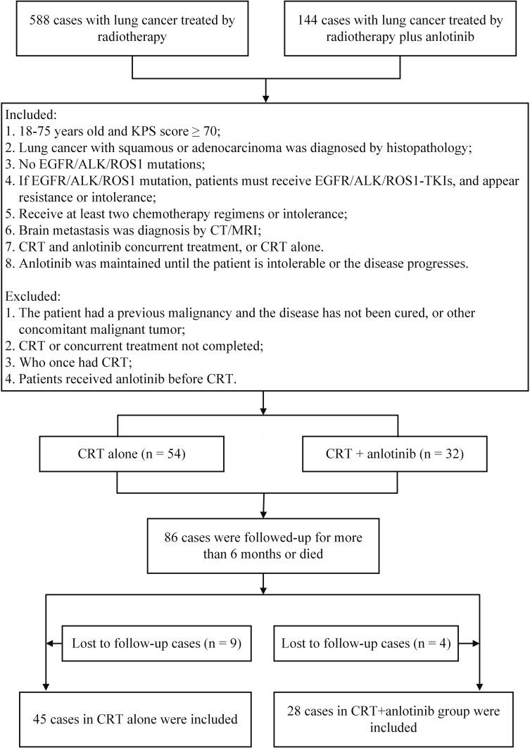
We reviewed the clinical records of patients diagnosed with NSCLC and BM between September 2016 and June 2020 at The First Affiliated Hospital of Bengbu Medical College (China). The clinical records of these patients included their clinical information, imaging data, tumor-related features, treatment process and clinical outcomes. Clinical information included gender, age, smoking and drinking history, previous disease history, and Karnofsky Performance Status (KPS) score. Tumor-related features included pathological type, EGFR/ALK/ROS1 mutation status, extracranial metastasis, number of BM, and treatment process (including CRT and drug therapy).12 The imaging data were evaluated by two radiologists performed single-blind evaluation of tumor volume. When the two had different opinions, a third radiologist reviewed them. The TNM staging criteria for patients were based on the Union for International Cancer Control/American Joint Committee on Cancer (UICC/AJCC) 8th Edition.15 The inclusion criteria were 1) 18–75 years old and KPS score ≥70; 2) NSCLC diagnosed by histopathology; 3) no EGFR/ALK/ROS1 mutations; 4) if EGFR/ALK/ROS1 mutation, patients must have received EGFR/ALK/ROS1-TKIs and exhibited resistance or intolerance; 5) received at least two chemotherapy regimens or intolerance, 6) BM diagnosed by computed tomography/magnetic resonance imaging (CT/MRI); 7) patients received CRT and anlotinib concurrent treatment, or CRT treatment alone; 8) anlotinib was maintained until the patient became intolerant or the disease progressed (Figure 1).16 The exclusion criteria were 1) diagnosis with a previous malignancy and the disease was not cured, or presence of other concomitant malignant disease; 2) CRT or concurrent treatment not completed; 3) previously received CRT treatment; 4) received anlotinib treatment before CRT. According to the treatment process, all collected patients were divided into two groups: CRT combined with anlotinib concurrent therapy group (CRT + anlotinib group) or CRT alone group.
Figure 1.
The flow diagram of included patients.
Treatment
Anlotinib treatment was performed according to the drug guidelines of 8–12 mg daily (recommended dose) for 14 days orally and then 7 days off.10 The CRT treatment (6 MV X-ray) was the first CRT treatment for all patients. Fifteen patients received intensity modulated radiotherapy (IMRT), 43 patients received conformal radiotherapy, and 15 patients received IMRT for local lesions and conformal radiotherapy for whole brain. CRT treatment included whole-brain radiotherapy (WBRT), WBRT plus local CRT (LCRT), or LCRT, as decided by the multidisciplinary team based on the number of BM, patient KPS score, pathological type, and other factors. BM with ≤3 lesions were mainly assigned to LCRT, and >3 lesions were mainly assigned to WBRT or WBRT + LCRT treatments. For CRT, the dose for WBRT was 30–40 Gy in 10–20 fractions. The dose for LCRT was 25–54 Gy in 5–27 fractions. The dose for WBRT + LCRT was 30–40 Gy for WBRT and 10–24 Gy for LCRT. Clinical follow-up was carried out every 3–6 months, and included imaging, physical, and routine laboratory tests. The therapeutic effect was evaluated according to the Response Evaluation Criteria in Solid Tumors (RECIST) 1.1.17
Outcomes
Overall response rate (ORR) was defined as the proportion of complete response (CR) and partial response (PR) cases relative to the total number of evaluable cases. OS was defined based on the initiation of CRT to the death time or last follow-up time.18,19 The intracranial PFS (iPFS) and extracranial PFS (ePFS) were defined from the initiation of CRT to intracranial/extracranial progression time or death time, or the last follow-up time for patients who showed no progress or died. Systematic PFS (sPFS) was defined from the initiation of CRT to death, or tumor progression, or the last follow-up time for patients who showed no progress or died.12 The last follow-up time was December 2020. The primary endpoints included iPFS and OS, and the secondary endpoints included ePFS and sPFS.
Statistical Analysis
Patient characteristics were expressed as categorical variables and analyzed by Pearson’s chi-square test or Fisher’s exact test. The age as a patient characteristic was calculated as mean ± standard deviation (S.D). Differences in PFS and OS between CRT + anlotinib group and CRT alone group were compared using Cox proportional hazards models. Subgroup analyses in PFS and OS were accomplished by randomized stratification factors and stratified Cox proportional hazards models. Statistical analyses were carried out using SPSS 25.0 (International Business Machines Corporation, Armonk, New York, USA). The figures were prepared using GraphPad Prism v8.3 (GraphPad Software Inc., San Diego, USA). A value of P < 0.05 with 2 sides was defined as statistical significance.
Results
Patient Characteristics
According to the included and excluded criteria, 86 NSCLC patients with CRT and non-EGFR/ALK/ROS1-TKIs indication were included in this retrospective study. Thirteen cases lacking sufficient follow-up data were excluded (Figure 1). Finally, 73 patients were included in the study, including 14 cases of squamous carcinoma (19.18%), and 59 cases of adenocarcinoma (80.82%). The median and average ages of all patients were 57 and 58.5 years (range 30–75 years), respectively. Of these, 39 patients (53.42%) were male and 52 patients (71.23%) were never smokers. The KPS scores of 48 patients (65.75%) were in the range of 90–100, and scores of 25 patients (34.25%) were in the range of 70–80. The left lung was the primary cancer site for 38 patients (52.05%), and the right lung was the primary cancer site for the other 35 patients (47.95%). There were 5 (6.85%), 16 (21.92%), 11 (15.07%), and 21 (28.77%) of patients classified as stage T1, T2, T3, and T4, respectively; and the classifications for the remaining 20 patients (27.40%) were not available. There were 4 (5.48%), 7 (9.59%), 31 (42.47%) and 17 (23.29%) patients classified as stage N0, N1, N2, and N3; the remaining 14 patients (19.18%) lacked classification data. A total of 39 patients (53.43%) had extracranial distant metastasis, and the presence of extracranial distant metastasis was not assessed for 7 patients (9.60%). Fourteen patients had the EGFR gene mutation; these patients exhibited resistance to treatment with EGFR/ALK/ROS1-TKIs and started CRT after BM diagnosis. There were 16, 12, 33, 7, and 4 patients that received zero-line, first-line, second-line, third-line and fourth-line treatments before CRT, with no significant differences in the baseline characteristics (P > 0.05). The patient baseline characteristics of the CRT alone and the anlotinib + CRT groups are listed in Table 1. Of the 73 patients, 28 cases received anlotinib plus CRT, and the other 45 cases received CRT alone.
Table 1.
Clinical Baseline Characteristics of Included Patients
| Characteristic | CRT Alone (n = 45) | CRT +Anlotinib (n = 28) | P value |
|---|---|---|---|
| Age (years) | |||
| Average (mean±SD) | 57.4±8.87 | 60.29±10.04 | 0.33 |
| Median | 56 | 63.5 | |
| Range | 41–73 | 30–75 | |
| Gender | |||
| Female | 21 (46.67%) | 13 (46.43%) | 0.98 |
| Male | 24 (53.33%) | 15 (53.57%) | |
| KPS score | |||
| 90–100 | 28 (62.22%) | 20 (71.43%) | 0.42 |
| 70–80 | 17 (37.78%) | 8 (28.57%) | |
| Smoking history | |||
| Yes | 11 (24.44%) | 10 (35.71%) | 0.3 |
| No | 34 (75.56) | 18 (64.29%) | |
| Primary site | |||
| Left | 22 (48.89%) | 16 (57.14%) | 0.49 |
| Right | 23 (51.11%) | 12 (42.86%) | |
| Pathological type | |||
| Adenocarcinoma | 38 (84.44%) | 21 (75%) | 0.32 |
| Squamous carcinoma | 7 (15.56%) | 7 (25%) | |
| T stage | |||
| T1 | 1 (2.22%) | 4 (14.29%) | 0.11 |
| T2 | 9 (20%) | 7 (25%) | |
| T3 | 7 (15.56%) | 4 (14.29%) | |
| T4 | 17 (37.78%) | 4 (14.29%) | |
| Tx | 11 (24.44%) | 9 (32.14%) | |
| N stage | |||
| N0 | 2 (4.44%) | 2 (7.14%) | 0.41 |
| N1 | 4 (8.89%) | 3 (10.71%) | |
| N2 | 23 (51.11%) | 8 (28.57%) | |
| N3 | 8 (17.78%) | 9 (32.14%) | |
| Nx | 8 (17.91%) | 6 (21.43%) | |
| Brain metastases | |||
| ≤3 | 10 (22.22%) | 8 (28.57%) | 0.54 |
| >3 | 35 (77.78%) | 20 (71.43%) | |
| CRT pattern | |||
| WBRT | 30 (66.67%) | 13 (46.43%) | 0.16 |
| WBRT + LCRT | 8 (17.78%) | 7 (25.0%) | |
| LCRT | 7 (15.55%) | 8 (28.57%) | |
| Extracranial distant metastasis | |||
| Yes | 24 (53.33%) | 15 (53.57%) | 0.84 |
| No | 16 (35.56%) | 11 (39.29%) | |
| Not available | 5 (11.11%) | 2 (7.14%) | |
| Treatment-line | |||
| Zero-Line | 8(17.78%) | 8(28.57%) | 0.084 |
| First-Line | 4(8.89%) | 8(28.57%) | |
| Second-Line | 25(55.56%) | 8(28.57%) | |
| Third-Line | 5(11.11%) | 2(7.14%) | |
| Fourth-Line | 2(4.44%) | 2(7.14%) |
Abbreviations: CRT, cranial radiotherapy; KPS, Karnofsky Performance Status; WBRT, whole brain radiotherapy; LCRT, local cranial radiotherapy.
Efficacy
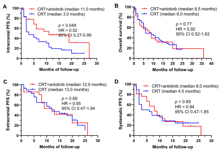
The median and mean follow-up time of all patients were 8.0 and 9.82 months, respectively. The ORR values of the CRT + anlotinib group and the CRT alone group were 89.29% and 80.0%, respectively. Of the 45 patients in the CRT alone group, six patients (13.33%) were alive with no evidence of disease progression, 32 patients (71.11%) died with intracranial progression, 24 patients (53.33%) died with extracranial progression, and four patients (8.89%) were alive with detected intracranial progression. Of the 28 patients in the CRT + anlotinib group, four patients (14.29%) were alive without evidence of disease progression, 15 patients (53.57%) were dead with intracranial progression, 13 patients (46.43%) were dead with extracranial progression, and three patients (10.71%) were alive with intracranial progression. For the whole group, the median iPFS (miPFS) and mOS were 6.0 and 8.0 months, respectively. The miPFS values were 11.0 months for the anlotinib + CRT group and 3.0 months for the CRT alone group (HR 0.52, 95% CI 0.27–0.99, P = 0.048) (Figure 2). This indicated that plus anlotinib treatment was closely associated with a significantly longer iPFS when combined with CRT. The mOS of the anlotinib + CRT group was longer than that of the CRT alone group (8.5 vs 6.0 months), although this difference was not statistically significant (HR 0.92, 95% CI 0.52–1.62, P = 0.77). The CRT + anlotinib group vs CRT alone group was 12.0 vs 13.0 months for median ePFS (mePFS, HR 0.95, 95% CI 0.47–1.94, P = 0.89) and 8.0 vs 4.5 months for median sPFS (msPFS, HR 0.85, 95% CI 0.47–1.85, P = 0.85). There was no significant difference for ePFS and sPFS in these two groups.
Figure 2.
The survival analysis of different treatment groups. (A) iPFS, (B) OS, (C) ePFS and (D) sPFS for patients with BM at baseline.
A value of P < 0.1 was considered a significant difference for univariate analysis. The analysis revealed that iPFS was significantly related to KPS score, pathological type, age, and plus anlotinib treatment (Table 2). OS was related to age, gender, KPS score, and pathological type.
Table 2.
Univariate Analysis Between Different Characteristics and iPFS or OS
| Variable | iPFS | OS | ||
|---|---|---|---|---|
| HR (95% CI) | P value | HR (95% CI) | P value | |
| Group | ||||
| CRT + Anlotinib vs CRT | 0.58 (0.32–1.03) | 0.06 | 1.08 (0.63–1.87) | 0.77 |
| Gender | ||||
| Female vs Male | 1.37 (0.81–2.32) | 0.24 | 1.65 (0.96–2.84) | 0.07 |
| Age | ||||
| ≥ 57 years vs < 57 years | 1.03 (1–1.06) | 0.05 | 1.04 (1.01–1.08) | 0.01 |
| KPS score | ||||
| 90–100 vs 70–80 | 0.53 (0.31–0.91) | 0.02 | 0.52 (0.30–0.90) | 0.02 |
| Smoking history | ||||
| Yes vs No | 1.63 (0.92–2.89) | 0.1 | 1.54 (0.87–2.71) | 0.14 |
| Pathological type | ||||
| Squamous vs adenocarcinoma | 0.56 (0.29–1.1) | 0.09 | 0.47 (0.24–0.90) | 0.02 |
| T stage | ||||
| Tx | 1 | – | ||
| T1 | 0.66 (0.22–2) | 0.47 | 0.50 (0.14–1.71) | 0.27 |
| T2 | 0.79 (0.38–1.66) | 0.53 | 1.01 (0.48–2.10) | 0.99 |
| T3 | 0.68 (0.3–1.53) | 0.35 | 0.76 (0.33–1.72) | 0.51 |
| T4 | 0.54 (0.27–1.1) | 0.1 | 0.65 (0.31–1.34) | 0.24 |
| N stage | ||||
| Nx | 1 | – | ||
| N0 | 0.27 (0.05–1.36) | 0.11 | 0.31 (0.07–1.37) | 0.12 |
| N1 | 0.88 (0.26–2.92) | 0.83 | 0.69 (0.24–2.01) | 0.5 |
| N2 | 0.9 (0.26–3.16) | 0.87 | 0.96 (0.32–2.9) | 0.94 |
| N3 | 0.86 (0.24–3.11) | 0.82 | 0.74 (0.23–2.36) | 0.66 |
| Primary site | ||||
| Left vs right | 1.28 (0.75–2.17) | 0.36 | 0.74 (0.43–1.27) | 0.27 |
| Number of brain metastases | ||||
| >3 vs ≤3 | 1.22 (0.58–2.57) | 0.61 | 1.83 (0.78–4.27) | 0.16 |
| Extracranial distant metastasis | ||||
| Yes vs No | 1.14 (0.64–2.02) | 0.66 | 1.1 (0.62–1.96) | 0.75 |
| Treatment-line | ||||
| Zero-Line | 1 | 1 | ||
| First-Line | 0.93 (0.26–3.33) | 0.91 | 0.76 (0.24–2.37) | 0.63 |
| Second-Line | 1.78 (0.49–6.46) | 0.38 | 0.91 (0.28–2.97) | 0.88 |
| Third-Line | 1.32 (0.4–4.37) | 0.65 | 0.91 (0.32–2.64) | 0.87 |
| Fourth-Line | 0.48 (0.1–2.38) | 0.37 | 0.61 (0.15–2.43) | 0.48 |
Abbreviations: OS, overall survival; iPFS, intracranial progression free survival; CRT, cranial radiotherapy; KPS, Karnofsky Performance Status; HR, hazard ratio; CI, confidence interval.
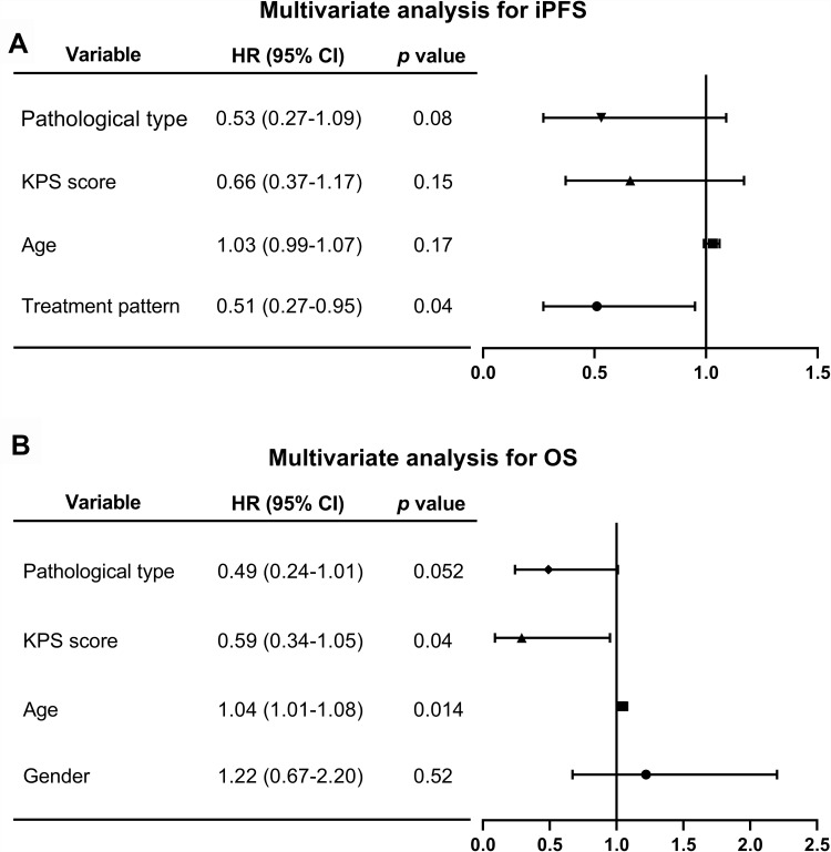
The characteristics identified as significant by univariate analysis were then subjected to adjusted Cox multivariate analyses to analyze the correlation between these characteristics and iPFS or OS. In multivariate analyses, only plus anlotinib significantly prolonged iPFS (HR 0.51, 95% CI 0.27–0.95, P = 0.04) (Figure 3A). Age ≥57 years (HR 1.04, 95% CI 1.01–1.08, P = 0.014) can significantly decreased OS and KPS score ≥90 (HR 0.59, 95% CI 0.34–1.05, P = 0.04) correlated with significantly prolonged OS (Figure 3B). No statistically significant differences were observed between gender, smoking history, T stage, N stage, primary site, extracranial distant metastasis, number of brain metastases, number of lines of therapy, and iPFS or OS in this study (P > 0.05).
Figure 3.
After univariate analysis, the significant variables were chosen for multivariate analysis for iPFS and OS. In multivariate analysis, (A) only the plus anlotinib treatment was positively correlated with prolonged iPFS (P < 0.05); (B) age < 57 years and KPS score ≥ 90 were positively correlated with prolonged OS (P < 0.05).
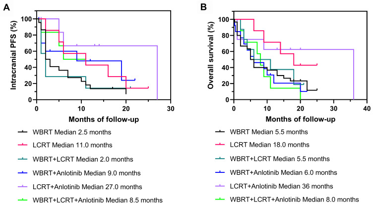
Subgroup analyses indicated that iPFS of LCRT + anlotinib group (miPFS 27.0 months) exhibited the strongest benefits of the groups (Figure 4A). Although there was no statistical significance of the effect of LCRT + anlotinib on OS, the mOS of LCRT + anlotinib group was 36 months, higher than the other groups. The second highest iPFS and OS values were for the LCRT group (miPFS 11.0 months; mOS 18.0 months) (Figure 4B). These results suggest that treatment that combined anlotinib with LCRT was better than LCRT alone, although the difference was not statistically significant. The iPFS of the LCRT group (miPFS 11.0 months) was longer than WBRT + LCRT + anlotinib group (miPFS 8.5 months) and WBRT group (miPFS 2.5 months), OS of LCRT group (mOS 18.0 months) was longer than the WBRT + LCRT + anlotinib group (mOS 8.0 months) and WBRT group (mOS 5.5 months). Overall, these results indicated greater importance of CRT pattern for prognosis than supplemental treatment with anlotinib (Figure 4).
Figure 4.
The subgroup analysis of different treatment patterns for BM patients at baseline. (A) iPFS and (B) OS of CRT alone group and CRT + anlotinib group for patients with different treatment patterns.
Discussion
The mechanism of BM from lung cancer is complex, but is closely related to angiogenesis. Angiogenesis in metastasizing lesions can develop through multiple signal pathways, and an important one is the VEGF pathway. Studies have found that the expression level of VEGF in tumor is negatively related to poor prognosis.8 As a new type of small molecule and multi-targeting TKI, anlotinib mainly acts through the anti-VEGF pathway for anti-tumor effect.8 In 2020 and 2021, oncologists suggested that anlotinib has intracranial activity and can control intracranial tumors.12,20,21 The ALTER0303 study also showed that anlotinib can prolong PFS in lung cancer patients with BM.12 Anlotinib normalizes the blood vessels in a metastatic tumor, adjusts the internal microenvironment of the tumor, restores the normal permeability of blood vessels, and acts synergistically with CRT to enhance radiosensitivity and reduce brain edema. However, there is no evidence shown that CRT can enhance anlotinib cross the BBB, and the detailed mechanism of anlotinib action in lung cancer patients with BM requires further study.13 Additionally, further clinical studies are required to determine whether the combination of anlotinib and CRT is better than CRT alone for patients with BM from lung cancer who failed to respond to multi-line chemotherapy or without EGFR/ALK/ROS1-TKIs indication.
In our study, CRT + anlotinib treatment was significantly superior to treatment with CRT alone (miPFS: 11.0 vs 3.0 months, P = 0.048). However, this treatment did not obviously improve OS and sPFS (P > 0.05), although mOS and msPFS of CRT + anlotinib group were longer than those of the CRT alone group. These results were consistent with those reported for the ALTER0303 study, where anlotinib affected PFS but did not significantly prolong OS in patients with BM. In our study, intracranial control was more effective than systemic control, which is likely related to CRT mainly acting to control intracranial lesions. Univariate analysis and multivariate analysis of clinic baseline characteristics and patient survival data showed that plus anlotinib was an independent prognostic factor to improve iPFS (P < 0.05), and younger age and higher KPS score were independent prognostic factors to improve OS (P < 0.05). This may be because the good physical condition of a patient can influence the effectiveness of treatment. The subgroup analysis of survival data showed that LCRT + anlotinib treatment of lung cancer patients improved and extended iPFS and OS compared to those of the other treatment groups, but there was no significant difference in the treatment pattern (P > 0.05). This suggests that the addition of anlotinib may be the most beneficial for LCRT patients, and this effect should be studied further.
Our study is limited in that it is a retrospective study, which does not allow randomization of patients and affects homogeneity, thus reducing the level of evidence. Another limit is that the number of cases analyzed was less than ideal. Therefore, future studies should utilize an expanded sample size, conduct prospective research, and eliminate heterogeneity to be able to draw more vigorous conclusions. Despite these limitations, our study can serve as strategic reference for the current clinical treatment of NSCLC patients with BM and non-EGFR/ALK/ROS1-TKIs indications.
Conclusion
In this study, we analyzed the efficacy of anlotinib (multi-target inhibitors) combined with CRT for patients with BM from advanced NSCLC with non-EGFR/ALK/ROS1-TKIs indications. The results indicated that the concurrent use of anlotinib has obvious clinical value to prolong the iPFS of patients with CRT. Our study has important reference significance for the clinical treatment of BM from NSCLC with non-EGFR/ALK/ROS1-TKIs indications.
Acknowledgments
The study was supported by the First Affiliated Hospital of Bengbu Medical College Science Fund for Distinguished Young Scholars (No. 2019BYYFYJQ04), the Natural Science Foundation of Bengbu Medical College (No. BYKY2019097ZD), the Bengbu Medical College Science Fund for “Excellent Young Teachers in 512 Talent Development Programme” (No. BY51201314), and the Bengbu – Bengbu Medical College Joint Research Project (No. BYLK201810).
Abbreviations
NSCLC, non-small cell lung cancer; BM, brain metastasis; mOS, median survival time; OS, survival time; TKIs, tyrosine kinase inhibitors; ALK, anaplastic lymphoma kinase; EGFR, epidermal growth factor receptor; ROS1, C-ros oncogene 1-receptor tyrosine kinase; CR, complete remission; mPFS, median progression-free survival; TTF, time to treatment failure; PFS, progression-free survival; BBB, blood–brain barrier; HR, hazard ratio; CI, confidence interval; ORR, objective response rate; TTBP, time to brain progression; CRT, cranial radiotherapy; KPS, Karnofsky Performance Status; UICC, Union for International Cancer Control; AJCC, American Joint Committee on Cancer; CT/MRI, computed tomography/magnetic resonance imaging; WBRT, whole-brain radiotherapy; RECIST, Response Evaluation Criteria in Solid Tumors; ORR, Overall response rate; PR, partial response; iPFS, intracranial progression-free survival; ePFS, extracranial progression-free survival; sPFS, Systematic progression-free survival; S.D, standard deviation; miPFS, median intracranial progression-free survival; mePFS, median extracranial progression-free survival; msPFS, median systematic progression-free survival.
Data Sharing Statement
The data used and/or analyzed in this study are available from the corresponding author upon reasonable request.
Ethics Approval and Consent to Participate
This study was a retrospective study, which utilized collected clinical data of patients, did not interfered with the treatment plan of patients, did not present physiological risks to patients, and maintained the privacy of patients. Some data were for patients who had died before this study. The ethics committee of the First Affiliated Hospital of Bengbu Medical College considered the risk-benefits and determined that informed consent was not required for this analysis. All processes conformed to the Declaration of Helsinki. The study design was reviewed and approved by the First Affiliated Hospital of Bengbu Medical College.
Disclosure
The authors report no conflicts of interest in this work.
References
- 1.Siegel RL, Miller KD, Jemal A. Cancer statistics, 2020. CA Cancer J Clin. 2020;70(1):7–30. doi: 10.3322/caac.21590 [DOI] [PubMed] [Google Scholar]
- 2.Zhang H, Wang A, Tan Y, et al. NCBP1 promotes the development of lung adenocarcinoma through up-regulation of CUL4B. J Cell Mol Med. 2019;23(10):6965–6977. doi: 10.1111/jcmm.14581 [DOI] [PMC free article] [PubMed] [Google Scholar]
- 3.He Z, Shi Z, Sun W, et al. Hemocompatibility of folic-acid-conjugated amphiphilic PEG-PLGA copolymer nanoparticles for co-delivery of cisplatin and paclitaxel: treatment effects for non-small-cell lung cancer. Tumour Biol. 2016;37(6):7809–7821. doi: 10.1007/s13277-015-4634-1 [DOI] [PubMed] [Google Scholar]
- 4.Sung H, Ferlay J, Siegel RL, et al. Global cancer statistics 2020: GLOBOCAN estimates of incidence and mortality worldwide for 36 cancers in 185 countries. CA Cancer J Clin. 2021;71(3):209–249. doi: 10.3322/caac.21660 [DOI] [PubMed] [Google Scholar]
- 5.Yang S, Zhang Z, Wang Q. Emerging therapies for small cell lung cancer. J Hematol Oncol. 2019;12(1):47. doi: 10.1186/s13045-019-0736-3 [DOI] [PMC free article] [PubMed] [Google Scholar]
- 6.Yang ZR, Liu MN, Yu JH, et al. Treatment of stage III non-small cell lung cancer in the era of immunotherapy: pathological complete response to neoadjuvant pembrolizumab and chemotherapy. Transl Lung Cancer Res. 2020;9(5):2059–2073. doi: 10.21037/tlcr-20-896 [DOI] [PMC free article] [PubMed] [Google Scholar]
- 7.Alcibar OL, Nadal E, Romero Palomar I, Navarro-Martin A. Systematic review of stereotactic body radiotherapy in stage III non-small cell lung cancer. Transl Lung Cancer Res. 2021;10(1):529–538. doi: 10.21037/tlcr-2020-nsclc-04 [DOI] [PMC free article] [PubMed] [Google Scholar]
- 8.Shen G, Zheng F, Ren D, et al. Anlotinib: a novel multi-targeting tyrosine kinase inhibitor in clinical development. J Hematol Oncol. 2018;11(1):120. doi: 10.1186/s13045-018-0664-7 [DOI] [PMC free article] [PubMed] [Google Scholar]
- 9.Si X, Zhang L, Wang H, et al. Quality of life results from a randomized, double-blinded, placebo-controlled, multi-center Phase III trial of anlotinib in patients with advanced non-small cell lung cancer. Lung Cancer. 2018;122:32–37. doi: 10.1016/j.lungcan.2018.05.013 [DOI] [PubMed] [Google Scholar]
- 10.Si X, Zhang L, Wang H, et al. Management of anlotinib-related adverse events in patients with advanced non-small cell lung cancer: experiences in ALTER-0303. Thorac Cancer. 2019;10(3):551–556. doi: 10.1111/1759-7714.12977 [DOI] [PMC free article] [PubMed] [Google Scholar]
- 11.Han B, Li K, Wang Q, et al. Effect of anlotinib as a third-line or further treatment on overall survival of patients with advanced non-small cell lung cancer: the ALTER 0303 phase 3 randomized clinical trial. JAMA Oncol. 2018;4(11):1569–1575. doi: 10.1001/jamaoncol.2018.3039 [DOI] [PMC free article] [PubMed] [Google Scholar]
- 12.Jiang S, Liang H, Liu Z, et al. The impact of anlotinib on brain metastases of non-small cell lung cancer: post hoc analysis of a phase III randomized control trial (ALTER0303). Oncologist. 2020;25(5):870–874. doi: 10.1634/theoncologist.2019-0838 [DOI] [PMC free article] [PubMed] [Google Scholar]
- 13.Brun L, Dupic G, Chassin V, et al. Hypofractionated stereotactic radiotherapy for large brain metastases: optimizing the dosimetric parameters. Cancer Radiother. 2021;25(1):1–7. doi: 10.1016/j.canrad.2020.04.011 [DOI] [PubMed] [Google Scholar]
- 14.Kahrom A, Grimley R, Jeffree RL. A case of delayed cyst formation post brain AVM stereotactic radiosurgery for arteriovenous malformation: case report. J Clin Neurosci. 2021;87:17–19. doi: 10.1016/j.jocn.2021.01.051 [DOI] [PubMed] [Google Scholar]
- 15.Lancia A, Merizzoli E, Filippi AR. The 8(th) UICC/AJCC TNM edition for non-small cell lung cancer staging: getting off to a flying start? Ann Transl Med. 2019;7(Suppl6):S205. doi: 10.21037/atm.2019.07.02 [DOI] [PMC free article] [PubMed] [Google Scholar]
- 16.Duan Y, Qian J, Chen K, Zhang Z. Necessity of adjuvant postoperative radiotherapy for diffuse pigmented villonodular synovitis of the knee: a case report and literature review. Medicine. 2018;97(3):e9637. doi: 10.1097/md.0000000000009637 [DOI] [PMC free article] [PubMed] [Google Scholar]
- 17.He Z, Zhang H, Cui Z, et al. A case of chemorefractory metastatic type AB thymoma sensitive to helical tomotherapy. Ann Transl Med. 2019;7(12):274. doi: 10.21037/atm.2019.05.34 [DOI] [PMC free article] [PubMed] [Google Scholar]
- 18.Han B, Li K, Zhao Y, et al. Anlotinib as a third-line therapy in patients with refractory advanced non-small-cell lung cancer: a multicentre, randomised phase II trial (ALTER0302). Br J Cancer. 2018;118(5):654–661. doi: 10.1038/bjc.2017.478 [DOI] [PMC free article] [PubMed] [Google Scholar]
- 19.Li Y, Liu J, Li HX, et al. Radiomics signature facilitates organ-saving strategy in patients with esophageal squamous cell cancer receiving neoadjuvant chemoradiotherapy. Front Oncol. 2020;10:615167. doi: 10.3389/fonc.2020.615167 [DOI] [PMC free article] [PubMed] [Google Scholar]
- 20.Gao XH, Zheng J, Ma L, et al. Mitigation of acute radiation-induced brain injury in a mouse model using anlotinib. Ann Palliat Med. 2021;10(1):312–322. doi: 10.21037/apm-20-2284 [DOI] [PubMed] [Google Scholar]
- 21.Wang Y, Liang D, Chen J, et al. Targeted therapy with anlotinib for a patient with an oncogenic FGFR3-TACC3 fusion and recurrent glioblastoma. Oncologist. 2021;26(3):173–177. doi: 10.1002/onco.13530 [DOI] [PMC free article] [PubMed] [Google Scholar]


