Abstract
Background
Cellular transplantations have promising effects on treating spinal cord injury (SCI) patients. Mesenchymal stem cells (MSCs) and Schwann cells (SCs), which have safety alongside their complementary characteristics, are suggested to be the two of the best candidates in SCI treatment. In this study, we assessed the safety and possible outcomes of intrathecal co-transplantation of autologous bone marrow MSC and SC in patients with subacute traumatic complete SCI.
Methods
Eleven patients with complete SCI (American Spinal Injury Association Impairment Scale (AIS); grade A) were enrolled in this study during the subacute period of injury. The patients received an intrathecal autologous combination of MSC and SC and were followed up for 12 months. We assessed the neurological changes by the American Spinal Injury Association’s (ASIA) sensory-motor scale, functional recovery by spinal cord independence measure (SCIM-III), and subjective changes along with adverse events (AE) with our checklist. Furthermore, electromyography (EMG), nerve conduction velocity (NCV), magnetic resonance imaging (MRI), and urodynamic study (UDS) were conducted for all the patients at the baseline, 6 months, and 1 year after the intervention.
Results
Light touch AIS score alterations were approximately the same as the pinprick changes (11.6 ± 13.1 and 12 ± 13, respectively) in 50% of the cervical and 63% of the lumbar-thoracic patients, and both were more than the motor score alterations (9.5 ± 3.3 in 75% of the cervical and 14% of the lumbar-thoracic patients). SCIM III total scores (21.2 ± 13.3) and all its sub-scores (“respiration and sphincter management” (15 ± 9.9), “mobility” (9.5 ± 13.3), and “self-care” (6 ± 1.4)) had statistically significant changes after cell injection. Our findings support that the most remarkable positive, subjective improvements were in trunk movement, equilibrium in standing/sitting position, the sensation of the bladder and rectal filling, and the ability of voluntary voiding. Our safety evaluation revealed no systemic complications, and radiological images showed no neoplastic overgrowth, syringomyelia, or pseudo-meningocele.
Conclusion
The present study showed that autologous SC and bone marrow-derived MSC transplantation at the subacute stage of SCI could reveal statistically significant improvement in sensory and neurological functions among the patients. It appears that using this combination of cells is safe and effective for clinical application to spinal cord regeneration during the subacute period.
Keywords: Subacute complete spinal cord injury, Combination cell therapy, Schwann cells, Bone-marrow-derived mesenchymal stem cell
Introduction
Spinal cord injury (SCI) is a devastating condition that leads to physical, social, and vocational impairment due to the irreversible loss of neural function below the injury site [1]. Based on Lancet Neurology, the global burden of diseases (GBD), injuries, and risk factors between 1990 and 2016, the age-standardized incidence of SCI was 13 per 100,000. Furthermore, with global population growth, the absolute number of people living with the effects of SCI is expected to increase [2]. Most of the SCI patients suffer from a profound disability and its related complications, which impact the quality of life [3]. Therefore, functional improvement after SCI remains an important issue in recent decades. Regarding the lack of capacity for central nervous system regeneration, there is no definitive cure for these disorders. Advanced therapies like cell transplantation could be a promising option for treating SCI patients [4].
Numerous studies on animal models of SCI and human patients have demonstrated that cellular transplantations for SCI treatment might provide a source of neural cells and have neuroprotective and immunomodulatory effects after injury [5, 6]. Various cell types can be used due to their capacity for self-renewal and differentiation ability, but among them, mesenchymal stem cells (MSCs) and Schwann cells (SCs) have better safety alongside their complementary characteristics, so these cells are suggested to be one of the best candidates for transplantation in SCI subjects [7, 8].
Bone marrow MSC as a multipotent stromal cell has a potential effect to differentiate into osteoblast, adipocytes, chondrocytes, mature neurons, and glial cells [9]. Many studies have shown that MSCs could be considered for the SCI treatment. Zhu et al. conducted a phase I–II clinical trial on 28 chronic complete SCI patients to assess the safety and efficacy of umbilical cord blood mononuclear cell transplant. They concluded that transplantation could be safe and would lead to locomotor, bowel, and bladder recovery [10]. Also, Ghobrial and colleagues enrolled 12 patients with traumatic SCI in a phase II safety and efficacy study of intramedullary injections of human neural stem cells. They presented five total patients with 12 months of follow-up and observed that transplantation can be safely performed with improvement in overall mean functional outcomes measures [11]. Moreover, MSCs can produce various types of growth factors and neuroprotective cytokines which enable them to improve or restore damaged spinal cord function [12–14]. Despite the beneficial effects of MSC transplantation, according to the previous findings, their remyelinating ability is inadequate in SCI patients. The importance of remyelination in spinal cord repair after injury suggests that stem cells could be combined with remyelinating cells to improve the effectiveness of transplantation [15]. SCs which are normally located in the peripheral nerves could migrate and colonize in lesion sites to myelinate injured axons and are one of the suitable choices for transplantation in combination with MSCs [16–18].
Successful functional recovery in the patients suffering from SCI will most likely rely on effective treatment in the period corresponding with the natural history of neuro recovery. The clinical trials have been conducted to assess the possible outcome of combinational cell therapy for treating patients with chronic SCI. Like our previous study, they have indicated an insufficient recovery in patients with chronic disease [8, 15]. So, in this study, we aimed to assess the safety and possible outcomes of co-transplantation of autologous bone-marrow MSC and SC in the patients with subacute traumatic complete SCI (within 12 months post-injury) [19].
Patients and methods
Study design and selection criteria
This study was designed on the basis of the Declaration of Helsinki and approved by Ethics in Medical Research Committee, Shahid Beheshti University of Medical Sciences (code of ethics: 106, approved in October 2011). All the interventions were performed after obtaining informed consent from patients.
Our inclusion criteria for the study were as follows: (1) complete SCI (ASI A); (2) ≥ 3 and ≤ 12 months post-injury; (3) no improvement in sensory and motor scale after 3 months despite regular rehabilitation program; (4) absence of brain disease or psychological disorders; (5) no stenosis, tethering, syringomyelia, or compression in the magnetic resonance images (MRI) of the spinal cord taken at the beginning of the study; (6) absence of joint stiffness or pain, rashes, or any manifestation of rheumatologic disorders; and (7) aged between 18 and 60 years old.
Study exclusion criteria were (1) presence of any movement disorder not related to SCI; (2) a major complication such as urinary tract infection with sepsis, pneumonia, venous thromboembolism (deep vein thrombosis and pulmonary embolism), etc.; (3) fracture of upper or lower limbs leading to deformity and ankylosis; and (4) abnormal findings on baseline complete blood count.
Patients were selected from among those with spinal cord injury who referred to the neurosurgery clinic of Shohada Tajrish Hospital. Eleven patients (9 men and 2 women) with a mean age of 29.09 ± 9.41 years old met our inclusion and exclusion criteria and successfully enrolled in this study. Four cases of the patients had cervical and seven had thoracic lesions due to road traffic accidents and falls from the height (Table 1).
Table 1.
Demographic, clinical features, motor, and sensory level changes of the patients
| Patient number | Sex | Age (years) | Cause of injury | LOI | Interval between injection and trauma (months) | Motor level pre-treatment | Motor level 6 months after treatment | Motor level 12 months after treatment | Sensory level pre-treatment | Sensory level 6 months after treatment | Sensory level 12 months after treatment |
|---|---|---|---|---|---|---|---|---|---|---|---|
| 1 | Male | 18 | Accident | C4 | 3 | C7 | C7 | C8 | T4 | T4 | T4 |
| 2 | Female | 42 | Accident | T3 | 7 | T1 | T1 | T1 | T3 | T4 | T4 |
| 3 | Female | 38 | Accident | T10 | 3.5 | L1 | L1 | L1 | L1 | L1 | L1 |
| 4 | Male | 27 | Accident | C5 | 5 | C6 | C7 | C7 | C5 | C5 | C5 |
| 5 | Male | 21 | Accident | T12 | 9 | T1 | L3 | L3 | L5 | L5 | L5 |
| 6 | Male | 30 | Accident | T6 | 5.5 | T1 | T1 | T1 | T8 | T10 | T10 |
| 7 | Male | 17 | Falling | T5 | 3 | T1 | T1 | T1 | T5 | T9 | T9 |
| 8 | Male | 30 | Accident | C5 | 4 | C7 | C7 | C7 | T2 | T3 | T3 |
| 9 | Male | 19 | Accident | T2 | 7 | T1 | T1 | - | T3 | T3 | T12 |
| 10 | Male | 40 | Accident | T11 | 3 | T1 | T1 | - | T12 | T12 | - |
| 11 | Male | 38 | Accident | C5 | 8 | C7 | C8 | C8 | C4 | C4 | C4 |
LOI level of injury
Cell isolation and transplantation
All tests, including cell isolation and culture, were performed in Gandi Hospital’s cell therapy laboratory following Good Manufacturing Practice (GMP). Following the daycare procedure, the patients hospitalized, SCs, and MSCs were extracted from the patients in the operating room under sterile conditions, and they were discharged immediately after the procedure.
To collect SCs, as we previously reported [15], the sural nerve of the patient posterior to lateral malleolus was cut and sliced into 1- to 2-mm pieces, then was incubated with collagenase (1.4 U ml−1; Sigma, St. Louis, MO, USA) and Dispase (2.4 U ml−1; Sigma, USA). After washing the collagenase two times with DMEM/F12 and mesh filtering, the cells were treated with DMEM/F12, not including fetal bovine serum (FBS, Gibco, USA) for 5 days (37 °C, 5% CO2). After the fasting period, we gradually increased the concentration of FBS in culture progressively up to 10% during 1 week. The characterization of the isolated cells was approved by S-100 immunocytological staining, as described in our previous study [15].
To isolate bone marrow MSC, bone marrow blood (100–150 ml) was aspirated from the iliac bone. After the samples underwent a density gradient by Ficoll (1.077 g/l, Sigma, USA) at the ratio of 1:3, the mononuclear cell layer was recovered from the gradient interface after bone marrow blood was centrifuged (400g for 40 min). To separate the platelets and mononuclear cells, the cells were centrifuged three times with less gradient and time. To confirm that isolated cells were MSCs, we assessed the differentiation ability of these isolated cells to adipogenic and osteogenic cells. In addition, we assessed the cell surface markers (CD73, CD105, CD45, and CD34) through flow cytometry analysis to ensure the characteristic of the isolated cells which should be positive for CD73, CD90, and CD105 and negative for CD45.
MSCs and SCs were cultured and prepared separately then mixed before transplanting which was composed of MSCs at the final concentration of 5 × 10⁷ cells per ml and SCs at the final concentration of 5 × 10⁷ cells per ml. The cells were resuspended in 1 ml saline, and a total volume of 1 ml was injected into the SCI patient.
According to previous studies [12, 20, 21], transplantation was performed 3 weeks after cell harvesting, because 3 weeks is enough time to cultivate this number of cells. In this way, the cells were stored and cultured in the laboratory environment in the shortest possible time, and the cells have the highest quality at the time of transplantation. A physician transplanted the mixture of MSCs and SCs into the L4/L5 level in the operation room through lumbar puncture using spinal needle 24 G. We assured the entrance to the subarachnoid space by the existing CSF from the spinal needle. The mixture of cells (6 ml) was slowly injected. We kept the needle in place for 1 min to avoid leakage. The patients were discharged 1 h after the procedure.
Quality control of MSCs
We use clinical-grade material from reputable companies such as Gibco. All cell operations were performed in a sterile environment to prevent possible contamination. GMP clean rooms for cells were designed to ensure the quality, safety, and efficacy of those biological treatments, which makes it extremely important to have a good monitoring system.
The existence of mycoplasma infection was investigated before and after antibiotic treatment using the PCR-based method. A universal generic-specific primer capable of detecting all mycoplasma species was used to target the conserved region of 16S rDNA intragenic spacer regions including those representing 90–95% of mycoplasma cell culture contaminations. This allows for the detection of a wide variety of mycoplasma strains, including fastidious strains that are difficult to detect even by conventional growth-based methods. We used universal Mycoplasma primer: forward primer—GGCGAATGGGTG AGTAACACG; reverse primer—CGGATAACGC TTGCGACCTATG.
Mycoplasma test was performed on cells before and after antibiotic application, and no mycoplasma infection was confirmed.
Follow-up procedure
The patients were involved in a 12-month follow-up process after cell injection. We assessed neurological changes by the AIS score, functional recovery by the spinal cord independence measure (SCIM-III), and subjective changes along with adverse events (AE), presented in Table 2. A medical team including doctors, a physical therapist, and an occupational therapist assessed the patients for subjective changes, the severity, and the relevance of adverse events (AEs). All patients were monitored for adverse effects based on the Medical Dictionary for Regulatory Activities (MedDRA v. 18.1). The assessment of the changes in neuropathic pain and spasticity among the patients was based on subjective reports through exact history taking.
Table 2.
Adverse events of the patients
| Patient number | Adverse event | |||||||
|---|---|---|---|---|---|---|---|---|
| Fever | Numbness or tingling sensation | Facial flushing | Headache | General ache | Neuropathic pain | Spasticity | ||
| 1 | Before | − | − | − | − | − | − | − |
| After 6 m | − | + | − | − | − | + | + | |
| After 12 m | − | + | − | − | − | − | ↑ | |
| 2 | Before | − | − | − | − | − | − | − |
| After 6 m | − | − | − | − | + | + | + | |
| After 12 m | − | − | − | − | + | + | ↑ | |
| 3 | Before | − | + | − | − | − | − | − |
| After 6 m | − | ↑ | + | − | − | − | + | |
| After 12 m | − | ↑ | + | − | − | − | ↑ | |
| 4 | Before | − | + | − | − | − | − | + |
| After 6 m | − | + | − | − | − | − | + | |
| After 12 m | − | + | − | − | − | − | + | |
| 5 | Before | − | − | − | − | − | + | − |
| After 6 m | − | − | − | − | − | + | − | |
| After 12 m | − | − | − | − | − | ↓ | − | |
| 6 | Before | − | − | − | − | − | − | − |
| After 6 m | − | − | − | − | − | − | − | |
| After 12 m | − | − | − | − | − | − | − | |
| 7 | Before | − | − | + | − | − | + | + |
| After 6 m | − | − | + | − | − | + | ↑ | |
| After 12 m | − | − | + | − | − | + | ↑ | |
| 8 | Before | − | − | + | − | − | − | + |
| After 6 m | − | + | + | − | − | − | ↑ | |
| After 12 m | − | + | + | − | − | − | ↑ | |
| 9 | Before | − | − | − | − | − | − | + |
| After 6 m | − | − | − | − | − | − | + | |
| After 12 m | − | − | − | − | − | − | + | |
| 10 | Before | − | + | − | − | − | − | − |
| After 6 m | − | ↑ | − | − | − | − | − | |
| 11 | Before | − | + | − | − | − | − | + |
| After 6 m | − | + | − | − | − | − | + | |
| After 12 m | − | + | − | − | − | − | + | |
+, presence of sign; −, absence of sign; ↑, increase in severity; ↓, decrease in severity
Furthermore, electromyography (EMG), nerve conduction velocity (NCV), magnetic resonance imaging (MRI), and urodynamic study (UDS) were conducted for all the patients at the baseline, 6 months, and 1 year after the intervention. We performed EMG-NCV based on the previous trials [8, 20] and to differentiate voluntary muscle contraction from reflex or involuntary spontaneous limb movement.
We also ensured that all the participants received standard therapy for SCI injuries such as regular rehabilitation programs.
Statistical analysis
To study the significance of the changes in the clinical scales, the Wilcoxon rank test was used in SPSS 16.0 (IBM Crop., Armonk, USA). In all cases, the significance limit was placed on p < 0.05.
Result
Cell assessments
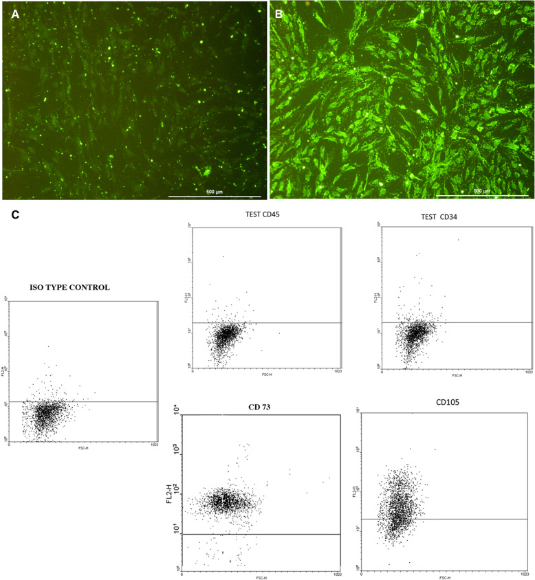
The extracted cells had the same characteristics as the cells we used in our previous study [15]. Cells isolated from the sural nerve were positive for S-100 marker, which indicated that these cells had the properties of SCs (Fig. 1A). MSCs were positive for CD73 and CD 105 and negative for CD45 and CD34 in flow cytometry analysis (Fig. 1B). In addition, the isolated cells could differentiate into adipogenic and osteogenic cells (data not shown).
Fig. 1.
Results of bone marrow MSC and SC laboratory assessments. A S-100 immunocytological staining negative control. B S-100 immunocytological staining test. C The cell surface marker (CD73, CD105, CD45, and CD34) analysis through flow cytometry
Adverse events
We observed some mild adverse events that based on medical team assessment; 38.46% were unlikely, 23.08% were possible, and 38.46% were probable AE. According to the MedDRA, the severity for each AE was mild. An increase in spasticity, numbness, or tingling sensation, and neuropathic pain was reported by 5, 4, and 2 out of 11 patients, respectively. Headache and facial flushing appeared in two patients after transplantation that was resolved spontaneously. Furthermore, none of the patients reported fever after injection (Table 2).
Other systemic complications such as anaphylactic shock, hypersensitivities, rush, or inflammation were not observed. Infectious complications associated with transplantation-like meningitis were not evident in the study. Since all AEs were mild, there was no need for medical treatment. However, the dose of the drug was increased for patients who already had a medical problem such as spasticity and had a mild increase after cell injection.
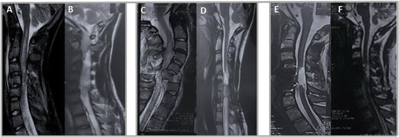
Although previous spinal instrumentation caused some worst effects on the visibility of images in the patients, MRI indicated no neoplastic overgrowth, syringomyelia, or pseudo-meningocele after transplantation (Fig. 2).
Fig. 2.
Pre- and post-injection MRIs of patients with numbers 1, 6, and 8. A Patient number 1, pre-injection. B Patient number 1, 6-month post-injection. C Patient number 6, pre-injection. D Patient number 6, 6-month post-injection. E Patient number 8, pre-injection. F Patient number 8, 6-month post-injection
AIS score evaluation
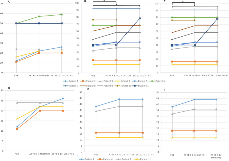
Sensory and/or motor improvement was evident in 9 patients according to the AIS assessment (in both score and motor and/or sensory level). Six patients experienced positive sensory changes in their AIS score (five patients also had changes in sensory level) and four patients had motor recovery (Table 1, Fig. 3). In our assessment, the cervical SCI patients showed more improvement rate in motor aspects (75% of the cervical and 14% of the lumbar-thoracic patients had motor improvement) and lumbar-thoracic SCI patients experienced more improvement rate in sensory AIS score (63% of the lumbar-thoracic and 50% of the cervical patients had sensory improvement) (Fig. 3). Among our patients, none of them showed AIS grade alteration from A to other grades.
Fig. 3.
A–C Motor, sensory light touch, and pin-prick AIS scores in total population. D–F Motor, sensory light touch, and pin-prick AIS scores in cervical patients (*p-value ≤ 0.05)
In terms of the intensity of the changes, light touch AIS score alterations were approximately the same as the pinprick changes (11.6 ± 13.1 and 12 ± 13, respectively), and both were more than the motor score alterations (9.5 ± 3.3). Light touch scores among all the patients improved statistically significant after 6 and 12 months in comparison with pre-transplantation scores (p-value = 0.042 and 0.027, respectively). Score differences between the 6th and the12th months were not significant (p-value = 0.317). For pinprick, the results were the same as the light touch changes (p-value = 0.041, 0.027, and 0.317, respectively). The difference between the motor score in the pre-transplantation evaluation and 6- and 12-month follow-up was not significant (p-value = 0.068 and 0.066, respectively) (Fig. 3, Table 3).
Table 3.
ASIA and SCIM III scores at different time points
| Score subject | Population | Time | Mean | SD | p-value | |
|---|---|---|---|---|---|---|
| ASIA | Motor score | Total study population | Before injection | 37.54 | 17.58 | – |
| After 6 months | 40.45 | 14.80 | 0.068 | |||
| After 12 months | 41.00 | 14.58 | 0.066 | |||
| Thoracic patients | Before injection | 50.00 | 00.00 | – | ||
| After 6 months | 51.00 | 2.64 | 0.317 | |||
| After 12 months | 51.28 | 3.40 | 0.317 | |||
| Cervical patients | Before injection | 15.75 | 5.90 | – | ||
| After 6 months | 22.00 | 1.63 | 0.109 | |||
| After 12 months | 23.00 | 2.58 | 0.109 | |||
| Light touch | Total study population | Before injection | 47.63 | 24.37 | – | |
| After 6 months | 50.54 | 24.46 | 0.042* | |||
| After 12 months | 51.80 | 25.74 | 0.027* | |||
| Thoracic patients | Before injection | 60.57 | 19.51 | – | ||
| After 6 months | 63.71 | 17.12 | 0.109 | |||
| After 12 months | 68.00 | 16.44 | 0.068 | |||
| Cervical patients | Before injection | 25.00 | 12.05 | – | ||
| After 6 months | 27.50 | 15.00 | 0.180 | |||
| After 12 months | 27.50 | 15.00 | 0.180 | |||
| Pin prick | Total study population | Before injection | 48.54 | 25.66 | – | |
| After 6 months | 51.63 | 25.71 | 0.041* | |||
| After 12 months | 53.00 | 27.00 | 0.027* | |||
| Thoracic patients | Before injection | 62.00 | 20.81 | – | ||
| After 6 months | 65.42 | 19.13 | 0.102 | |||
| After 12 months | 70.00 | 17.15 | 0.066 | |||
| Cervical patients | Before injection | 25.00 | 12.09 | – | ||
| After 6 months | 27.50 | 15.86 | 0.180 | |||
| After 12 months | 27.50 | 15.86 | 0.180 | |||
| SCIM III | Total score | Total study population | Before injection | 28.9 | 12.99 | – |
| After 6 months | 37.54 | 18.40 | 0.012* | |||
| After 12 months | 43.10 | 25.77 | 0.018* | |||
| Thoracic patients | Before injection | 37.14 | 6.28 | – | ||
| After 6 months | 49.28 | 7.08 | 0.018* | |||
| After 12 months | 60.50 | 14.18 | 0.028* | |||
| Cervical patients | Before injection | 14.50 | 7.00 | – | ||
| After 6 months | 17.00 | 12.00 | 0.317 | |||
| After 12 months | 17.00 | 12. 00 | 0.317 | |||
| Self-care | Total study population | Before injection | 8.00 | 5.53 | – | |
| After 6 months | 10.36 | 6.74 | 0.043* | |||
| After 12 months | 11.40 | 7.93 | 0.005** | |||
| Thoracic patients | Before injection | 11.71 | 2.49 | – | ||
| After 6 months | 14.71 | 2.98 | 0.068 | |||
| After 12 months | 17.16 | 2.48 | 0.042* | |||
| Cervical patients | Before injection | 1.50 | 1.00 | – | ||
| After 6 months | 2.75 | 3.50 | 0.317 | |||
| After 12 months | 2.75 | 3.50 | 0.317 | |||
| Respiration and sphincter management | Total study population | Before injection | 17.18 | 5.89 | – | |
| After 6 months | 21.72 | 8.22 | 0.016* | |||
| After 12 months | 26.20 | 13.63 | 0.027* | |||
| Thoracic patients | Before injection | 19.57 | 4.64 | – | ||
| After 6 months | 26.00 | 4.24 | 0.026* | |||
| After 12 months | 34.16 | 10.04 | 0.043* | |||
| Cervical patients | Before injection | 13.00 | 6.00 | – | ||
| After 6 months | 14.25 | 8.50 | 0.317 | |||
| After 12 months | 14.25 | 8.50 | 0.317 | |||
| Mobility | Total study population | Before injection | 3.72 | 3.58 | – | |
| After 6 months | 5.45 | 4.78 | 0.039* | |||
| After 12 months | 8.90 | 12.81 | 0.043* | |||
| Thoracic patients | Before injection | 5.85 | 2.60 | – | ||
| After 6 months | 8.57 | 2.63 | 0.039* | |||
| After 12 months | 14.83 | 13.77 | 0.043* | |||
| Cervical patients | Before injection | 0.00 | 0.00 | – | ||
| After 6 months | 0.00 | 0.00 | 1.00 | |||
| After 12 months | 0.00 | 0.00 | 1.00 | |||
Bold values indicate statistical significance (*p-value ≤ 0.05; **p-value ≤ 0.01)
SCIM III changes
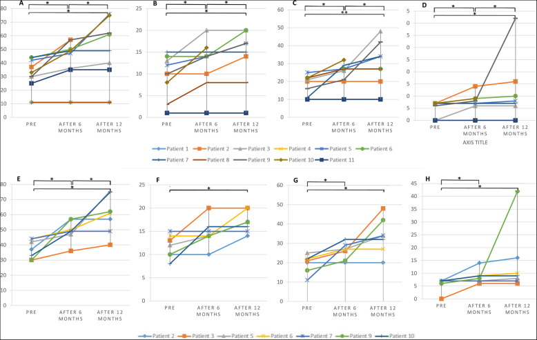
Regarding our SCIM III assessment, 8 out of 11 patients had some degrees of functional recovery, most of which were thoracic SCI patients. So, all the thoracic SCI patients experienced a positive change in SCIM III evaluation. Among the cervical SCI patients, only one patient (number 8) had improvement in the “sphincter management-bladder” item. The mean ± SD of the SCIM III changes was 21.2 ± 13.3, and among the SCIM III sub-scores, “respiration and sphincter management” (15 ± 9.9), “mobility” (9.5 ± 13.3), and “self-care” (6 ± 1.4) comprised the most of the score changes (Fig. 4).
Fig. 4.
A–D Total, self-care, respiration and sphincter management, and mobility SCIM III scores in total population. E–H Total, self-care, respiration and sphincter management, and mobility SCIM III scores in thoracic patients (*p-value ≤ 0.05; **p-value ≤ 0.01)
Statistical analysis revealed that the patients experienced statistically significant progressive changes after every 6 months in SCIM III total score and its sub-scales (Fig. 4). Differences between the pre- and post-transplantation groups and between each post-transplantation group (cervical and thoracolumbar) were statistically significant in total SCIM III score, respiration and sphincter management, mobility, and self-care (p-values of changes after 6 months were 0.012, 0.043, 0.016, and 0.039, and p-values of changes after 12 months were 0.018, 0.005, 0.027, and 0.043, respectively). In thoracolumbar patients, our statistical evaluation revealed that the differences between pre-transplantation and 6 and 12 months were statistically significant (except self-care changes after 6 months, which were not significant). The p-values for changes of SCIM III score in thoracolumbar SCI patients after 6 and 12 months were 0.018 and 0.028, respectively (Table 3).
Subjective outcomes
Our findings supported that the most remarkable positive, subjective improvement was in the trunk movement (in 8 patients) and equilibrium in standing/sitting positions (in 7 patients). Furthermore, three patients (patients with numbers 1, 3, and 5) experienced a reduction in the severity of constipation. We also observed that the two of our patients (numbers 1 and 9) claimed that they obtained the sensation of the filling bladder and rectum in the 6th and 12th months of their follow-up (patient number 1 acquired a sense of rectal filling in 12th months of follow-up). Furthermore, one patient had successful changes as the empowerment of voiding (patient number 5) (Table 4).
Table 4.
Subjective changes of the patients
| Patient number | Subjective changes | ||||||||||
|---|---|---|---|---|---|---|---|---|---|---|---|
| Trunk movement, 8 | Equilibrium in sitting position, 7 | Equilibrium in standing position, 7 | Constipation | Urination sensation | Voiding | Consistency | Feeling of defecation | Bowel sphincter management | Regularity of bowel movement | ||
| 1 | Before | − | − | + | + | − | − | − | − | − | − |
| After 6 m | − | − | ↓ | ↓ | + | − | − | − | − | + | |
| After 12 m | + | − | ↓ | ↓ | + | − | − | + | − | − | |
| 2 | Before | + | + | + | + | − | − | + | − | − | + |
| After 6 m | ↑ | ↑ | + | + | − | − | + | − | − | + | |
| After 12 m | ↑ | ↑ | + | + | − | − | + | − | − | + | |
| 3 | Before | + | + | + | + | − | − | − | − | − | − |
| After 6 m | ↑ | ↑ | ↓ | ↓ | − | − | − | − | − | − | |
| After 12 m | ↑ | ↑ | ↓ | ↓ | − | − | − | − | − | − | |
| 4 | Before | − | − | − | − | − | − | − | − | − | − |
| After 6 m | − | − | − | − | − | − | − | − | − | − | |
| After 12 m | − | − | − | − | − | − | − | − | − | − | |
| 5 | Before | + | + | + | + | − | − | − | + | + | − |
| After 6 m | ↑ | + | + | + | − | + | − | + | + | − | |
| After 12 m | ↑ | + | ↓ | ↓ | − | + | − | + | + | − | |
| 6 | Before | + | − | − | − | − | − | − | − | − | + |
| After 6 m | ↑ | + | − | − | − | − | − | − | − | + | |
| After 12 m | ↑ | ↑ | − | − | − | − | − | − | − | + | |
| 7 | Before | + | − | − | − | − | − | − | − | − | − |
| After 6 m | ↑ | + | − | − | − | − | − | − | − | − | |
| After 12 m | ↑ | ↑ | − | − | − | − | − | − | − | − | |
| 8 | Before | + | − | + | + | − | − | − | − | − | − |
| After 6 m | ↑ | + | + | + | − | − | − | − | − | − | |
| After 12 m | ↑ | ↑ | + | + | − | − | − | − | − | − | |
| 9 | Before | − | − | − | − | − | − | − | − | − | − |
| After 6 m | + | + | − | − | + | − | − | + | − | − | |
| After 12 m | ↑ | ↑ | − | − | + | − | − | + | − | − | |
| 10 | Before | + | − | + | + | − | − | − | − | − | + |
| After 6 m | + | + | + | + | − | − | − | − | − | + | |
| 11 | Before | − | − | − | − | − | − | − | − | − | − |
| After 6 m | − | − | − | − | − | − | − | − | − | − | |
| After 12 m | − | − | − | − | − | − | − | − | − | − | |
+, presence of sign; −, absence of sign; ↑, increase in severity; ↓, decrease in severity
UDS assessment of patient number 5 also supported this change. Pre-transplantation UDS of the patient revealed the presence of uninhibited contraction (no voiding), maximum detrusor pressure of 55cmH2O, maximum flow during cytometry less than 1 cc/s, and post-voiding residue of 314 cc. Post-transplantation UDS changed to maximum detrusor pressure of 15 cmH2O, maximum flow during cytometry of 3.3 cc/s, a post-voiding residue of 410 cc, and voided volume of 40 cc.
Discussion
The present study showed that autologous SC and bone marrow-derived MSC transplantation at the subacute stage of SCI could reveal statistically significant improvement in sensory and neurological function among the patients. Describing changes of patients with spinal cord injury includes many aspects consist of neurological, functional, and quality of life changes. The distinction of these improvements could be presented by AIS and SCIM III scores which indicated neurological and functional changes, respectively. Also, there is a difference between statistically significant and clinically significant. Although some changes in the AIS score are statistically significant, the transplantation did not change the AIS grade, and therefore, no clinically significant improvement could be considered. Furthermore, the changes in SCIM score which is statistically significant could be considered clinically significant because SCIM is a functional scale. In the present study, 75% of the cervical patients showed some degrees of motor improvements and 50% of them had sensory changes. But, thoracolumbar SCI patients experienced more improvement rate in sensory AIS score (63% of the patients) and less in the motor score (14%). Two patients obtained the sensation of the bladder and rectal filling and one patient claimed that he acquired the ability of voluntary voiding. There were no systemic nor serious complications such as fever, anaphylactic shock, hypersensitivities, rush, or inflammation after autologous transplantation. Also, radiological images showed no neoplastic overgrowth, syringomyelia, or psuedomeningocele, which documents the safety of using this cell combination therapy at the subacute stage.
The current clinical trial used the combination of autologous SC and bone marrow-derived MSC transplantation because of the multi-faceted inhibitory nature of the CNS lesion [22]. After spinal cord injury, multiple changes occur in damaged tissues that require various treatment strategies like neuroprotection, axonal regeneration promotion, and rehabilitation [23].
Since one of the issues following spinal cord injury is an inflammatory environment, previous treatment strategies have focused on reducing inflammatory responses. In the past, only high-dose methylprednisolone has been shown as the standard of care and has limited effectiveness for regenerating the spinal cord [24].
Cell transplantation is a targeted new promising therapeutic strategy for spinal cord regeneration based on a series of animal and clinical studies, and it has been previously reported that stem cells have a potential effect on the SCI treatment [20, 25, 26]. The safety and clinical application of SCs and MSCs separately have been reported in the treatment of SCI patients [7, 8, 27, 28]. MSCs are an appropriate source for cell therapy due to their ability of high growth rate, low immunogenicity, and favorable ethical profile [29]. Also, MSCs could enhance and support neurite outgrowth, axonal survival, and remyelination [30]. In spite of the beneficial effects of MSC transplantation, according to the previous findings, their remyelinating ability is inadequate in SCI patients [15]. So far, some studies have performed combination therapies for spinal cord regeneration; using two types of effective cells (SCs and MSCs) for spinal cord regeneration is one of these combination methods [23, 31, 32]. In our previous work, we reported the safety of SCs and MSCs combination therapy in the damaged chronic human spinal cord with no new deficit or adverse effect in the patients during short- and longer-term assessments [15]. Park et al. evaluated the therapeutic effects of autologous bone marrow cell transplantation in conjunction with administering granulocyte macrophage-colony stimulating factor (GM-CSF) in six complete SCI patients and reported neurologic improvements in AIS grades (from A to C) in five patients [33].
The delivery time of the stem cells after spinal cord injury is important for a better prognosis. This is because oligodendrocyte death and myelin loss after SCI are not a static phenomenon and demyelination continues and increases up to at least 450 days post-injury [33]. Generally, 12–18 months post-injury optimally correspond with the natural history of neuro recovery; this time is the golden time for supporting and improving neuro recovery and promoting conditions for the long-term maintenance of health and function [19]. In spite of promising results in chronic SCI patients after cell therapy [13, 34, 35], it seems that the sub-acute phase of stem cell transplantation after SCI may lead to better results [34]. The study by Yoon et al. reported improvement in AIS grade of 30.4% of the acute and subacute treated patients (AIS A to B or C), whereas no statistically significant changes were observed in the chronic patients. The majority of their study population had cervical injuries; nevertheless, they did not report the changes separately regarding the cervical and thoracic SCI patients [8]. Some differences between our results and theirs could be explained by the different composition levels of injury and cell combination therapy in our study [35].
Among the 11 patients, one reported improvement in urination management after cell therapy. Likely, Jiang et al.’s study [12] showed 80% of patients experienced recovery of urinary function. Nevertheless, Kishk et al. reported that no one in the treated or control groups reached the complete recovery of bladder control [20]. This controversy in the results could arise from different evaluation systems in each study. Despite the importance of urinary problems in SCI, there was a lack of comprehensive study for assessing cell transplantation in human subjects.
Neuropathic pain is one of the irritable complications that increases after cell transplantation, so is observed in some patients based on our study and other previous works [20, 36]. One of the possible reasons for neuropathic pain after cell therapy may be due to MSC differentiation into astrocytes which could lead to increased CGRP-positive sprouting and a consequent hypersensitive state [21, 37]. Because of the small sample size and lack of a control group, we could not conclude that the intrathecal injection of this combination cell therapy may increase neuropathic pain. So, clinical trials with larger volume sizes and control groups are needed for definitive comment on this topic.
In the present study, we used SC and MSC combination therapy in complete subacute SCI subjects to assess the safety and possible outcomes of this intervention. Unlike our previous findings [13], the results of this treatment in subacute patients were promising for the patients’ rehabilitation. Our data were in reasonably close agreement with the hopeful effects of combination cell therapy in sensory, motor, and functional status of subacute SCI patients alongside the positive subjective changes like empowerment in trunk movement, equilibrium in standing/sitting position, and sensation of the bladder and rectal filling. According to our limitations, we suggest a clinical trial with a large number of patients to unveil the effects of this kind of therapy on the suitable subgroups of SCI patients.
The nature of neural regeneration of SCI encountering many factors which mostly presents in the golden time for neurological improvements over the first 1–2 years post-injury. This fact is one of the important parameters affecting our results and limits our conclusion.
Conclusion
It seems that the combination of autologous SC and bone marrow-derived MSC transplantation at the subacute stage of SCI could significantly improve sensory and neurological function among the patients. Also, there were no systemic nor serious complications after autologous transplantation. This study is a phase 1/2 trial that aimed to provide the important information needed for the design of each respective trial phase. It is a preliminary study on the safety and feasibility of stem cells on spinal cord injury and to develop a preliminary sense of potential efficacy that its results can be used as a guide for configuring future studies because clinical efficacy could not be made without a randomized controlled clinical trial.
Acknowledgements
None to declare.
Abbreviations
- SCI
Spinal cord injury
- MSCs
Mesenchymal stem cells
- SCs
Schwann cells
- ASIA
American Spinal Injury Association Impairment Scale
- SCIM
Spinal cord independence measure
- AE
Adverse event
- EMG
Electromyography
- NCV
Nerve conduction velocity
- MRI
Magnetic resonance imaging
- UDS
Urodynamic study
- GBD
Global burden of diseases
- GMP
Good Manufacturing Practice
- FBS
Fetal bovine serum
- CSF
Cerebrospinal fluid
- PCR
Polymerase chain reaction
- CNS
Central nervous system
- GM-CSF
Granulocyte macrophage-colony stimulating factor
- DMEM/F12
Dulbecco’s modified Eagle medium/Nutrient Mixture F-12
Authors’ contributions
MHAP, SOY, MG, and MOY prepared the original draft. MHAP and MH conducted the formal analysis of the data. SOY, MG, and MSZ reviewed and finalized all drafts. MHAP, SOY, and FA conceptualized the study, and SOY, MEA, and ARZ provided overall supervision. All authors were involved in the investigation and provided guidance on the methodology. Also, they reviewed and revised the manuscript and approved the final revision of the manuscript.
Funding
None to declare.
Availability of data and materials
Researchers could submit their research proposals to the corresponding author to access datasets of this clinical trial.
Declarations
Ethics approval and consent to participate
The present study was conducted with the approval of the Ethics in Medical Research Committee of the Shahid Beheshti University of Medical Sciences. All procedures were performed after written informed consent was obtained from all patients.
Consent for publication
Consent for publication was obtained from all patients.
Competing interests
The authors declare that they have no competing interests.
Footnotes
Publisher’s Note
Springer Nature remains neutral with regard to jurisdictional claims in published maps and institutional affiliations.
Saeed Oraee-Yazdani and Mohammadhosein Akhlaghpasand contributed equally to this work.
Contributor Information
Saeed Oraee-Yazdani, Email: Saeed_o_yazdani@sbmu.ac.ir.
Alireza Zali, Email: dr_alirezazali@sbmu.ac.ir.
Masoud Soleimani, Email: ssoleim_m@modares.ac.ir.
References
- 1.Furlan JC, Noonan V, Singh A, Fehlings MG. Assessment of impairment in patients with acute traumatic spinal cord injury: a systematic review of the literature. J Neurotrauma. 2011;28(8):1445–1477. doi: 10.1089/neu.2009.1152. [DOI] [PMC free article] [PubMed] [Google Scholar]
- 2.James SL, Theadom A, Ellenbogen RG, Bannick MS, Montjoy-Venning W, Lucchesi LR, Abbasi N, Abdulkader R, Abraha HN, Adsuar JC, Afarideh M, Agrawal S, Ahmadi A, Ahmed MB, Aichour AN, Aichour I, Aichour MTE, Akinyemi RO, Akseer N, Alahdab F, Alebel A, Alghnam SA, Ali BA, Alsharif U, Altirkawi K, Andrei CL, Anjomshoa M, Ansari H, Ansha MG, Antonio CAT, Appiah SCY, Ariani F, Asefa NG, Asgedom SW, Atique S, Awasthi A, Ayala Quintanilla BP, Ayuk TB, Azzopardi PS, Badali H, Badawi A, Balalla S, Banstola A, Barker-Collo SL, Bärnighausen TW, Bedi N, Behzadifar M, Behzadifar M, Bekele BB, Belachew AB, Belay YA, Bennett DA, Bensenor IM, Berhane A, Beuran M, Bhalla A, Bhaumik S, Bhutta ZA, Biadgo B, Biffino M, Bijani A, Bililign N, Birungi C, Boufous S, Brazinova A, Brown AW, Car M, Cárdenas R, Carrero JJ, Carvalho F, Castañeda-Orjuela CA, Catalá-López F, Chaiah Y, Champs AP, Chang JC, Choi JYJ, Christopher DJ, Cooper C, Crowe CS, Dandona L, Dandona R, Daryani A, Davitoiu DV, Degefa MG, Demoz GT, Deribe K, Djalalinia S, Do HP, Doku DT, Drake TM, Dubey M, Dubljanin E, el-Khatib Z, Ofori-Asenso R, Eskandarieh S, Esteghamati A, Esteghamati S, Faro A, Farzadfar F, Farzaei MH, Fereshtehnejad SM, Fernandes E, Feyissa GT, Filip I, Fischer F, Fukumoto T, Ganji M, Gankpe FG, Gebre AK, Gebrehiwot TT, Gezae KE, Gopalkrishna G, Goulart AC, Haagsma JA, Haj-Mirzaian A, Haj-Mirzaian A, Hamadeh RR, Hamidi S, Haro JM, Hassankhani H, Hassen HY, Havmoeller R, Hawley C, Hay SI, Hegazy MI, Hendrie D, Henok A, Hibstu DT, Hoffman HJ, Hole MK, Homaie Rad E, Hosseini SM, Hostiuc S, Hu G, Hussen MA, Ilesanmi OS, Irvani SSN, Jakovljevic M, Jayaraman S, Jha RP, Jonas JB, Jones KM, Jorjoran Shushtari Z, Jozwiak JJ, Jürisson M, Kabir A, Kahsay A, Kahssay M, Kalani R, Karch A, Kasaeian A, Kassa GM, Kassa TD, Kassa ZY, Kengne AP, Khader YS, Khafaie MA, Khalid N, Khalil I, Khan EA, Khan MS, Khang YH, Khazaie H, Khoja AT, Khubchandani J, Kiadaliri AA, Kim D, Kim YE, Kisa A, Koyanagi A, Krohn KJ, Kuate Defo B, Kucuk Bicer B, Kumar GA, Kumar M, Lalloo R, Lami FH, Lansingh VC, Laryea DO, Latifi A, Leshargie CT, Levi M, Li S, Liben ML, Lotufo PA, Lunevicius R, Mahotra NB, Majdan M, Majeed A, Malekzadeh R, Manda AL, Mansournia MA, Massenburg BB, Mate KKV, Mehndiratta MM, Mehta V, Meles H, Melese A, Memiah PTN, Mendoza W, Mengistu G, Meretoja A, Meretoja TJ, Mestrovic T, Miazgowski T, Miller TR, Mini GK, Mirica A, Mirrakhimov EM, Moazen B, Mohammadi M, Molokhia M, Monasta L, Mondello S, Moosazadeh M, Moradi G, Moradi M, Moradi-Lakeh M, Moradinazar M, Morrison SD, Moschos MM, Mousavi SM, Murthy S, Musa KI, Mustafa G, Naghavi M, Naik G, Najafi F, Nangia V, Nascimento BR, Negoi I, Nguyen TH, Nichols E, Ningrum DNA, Nirayo YL, Nyasulu PS, Ogbo FA, Oh IH, Okoro A, Olagunju AT, Olagunju TO, Olivares PR, Otstavnov SS, Owolabi MO, P A M, Pakhale S, Pandey AR, Pesudovs K, Pinilla-Monsalve GD, Polinder S, Poustchi H, Prakash S, Qorbani M, Radfar A, Rafay A, Rafiei A, Rahimi-Movaghar A, Rahimi-Movaghar V, Rahman M, Rahman MA, Rai RK, Rajati F, Ram U, Rawaf DL, Rawaf S, Reiner RC, Reis C, Renzaho AMN, Resnikoff S, Rezaei S, Rezaeian S, Roever L, Ronfani L, Roshandel G, Roy N, Ruhago GM, Saddik B, Safari H, Safiri S, Sahraian MA, Salamati P, Saldanha RF, Samy AM, Sanabria J, Santos JV, Santric Milicevic MMM, Sartorius B, Satpathy M, Savuon K, Schneider IJC, Schwebel DC, Sepanlou SG, Shabaninejad H, Shaikh MAA, Shams-Beyranvand M, Sharif M, Sharif-Alhoseini M, Shariful Islam SM, She J, Sheikh A, Shen J, Sheth KN, Shibuya K, Shiferaw MS, Shigematsu M, Shiri R, Shiue I, Shoman H, Siabani S, Siddiqi TJ, Silva JP, Silveira DGA, Sinha DN, Smith M, Soares Filho AM, Sobhani S, Soofi M, Soriano JB, Soyiri IN, Stein DJ, Stokes MA, Sufiyan M'B, Sunguya BF, Sunshine JE, Sykes BL, Szoeke CEI, Tabarés-Seisdedos R, te Ao BJ, Tehrani-Banihashemi A, Tekle MG, Temsah MH, Temsah O, Topor-Madry R, Tortajada-Girbés M, Tran BX, Tran KB, Tudor Car L, Ukwaja KN, Ullah I, Usman MS, Uthman OA, Valdez PR, Vasankari TJ, Venketasubramanian N, Violante FS, Wagnew FWS, Waheed Y, Wang YP, Weldegwergs KG, Werdecker A, Wijeratne T, Winkler AS, Wyper GMA, Yano Y, Yaseri M, Yasin YJ, Ye P, Yimer EM, Yip P, Yisma E, Yonemoto N, Yoon SJ, Yost MG, Younis MZ, Yousefifard M, Yu C, Zaidi Z, Zaman SB, Zamani M, Zenebe ZM, Zodpey S, Feigin VL, Vos T, Murray CJL. Global, regional, and national burden of traumatic brain injury and spinal cord injury, 1990–2016: a systematic analysis for the Global Burden of Disease Study 2016. Lancet Neurol. 2019;18(1):56–87. doi: 10.1016/S1474-4422(18)30415-0. [DOI] [PMC free article] [PubMed] [Google Scholar]
- 3.Gurcay E, Bal A, Eksioglu E, Cakci A. Quality of life in patients with spinal cord injury. Int J Rehabil Res. 2010;33(4):356–358. doi: 10.1097/MRR.0b013e328338b034. [DOI] [PubMed] [Google Scholar]
- 4.Fehlings MG, Vawda R. Cellular treatments for spinal cord injury: the time is right for clinical trials. Neurotherapeutics. 2011;8(4):704–720. doi: 10.1007/s13311-011-0076-7. [DOI] [PMC free article] [PubMed] [Google Scholar]
- 5.Jin MC, Medress ZA, Azad TD, Doulames VM, Veeravagu A. Stem cell therapies for acute spinal cord injury in humans: a review. Neurosurg Focus. 2019;46(3):E10. doi: 10.3171/2018.12.FOCUS18602. [DOI] [PubMed] [Google Scholar]
- 6.Gazdic M, Volarevic V, Harrell C, Fellabaum C, Jovicic N, Arsenijevic N, Stojkovic M. Stem cells therapy for spinal cord injury. Int J Mol Sci. 2018;19(4):1039. doi: 10.3390/ijms19041039. [DOI] [PMC free article] [PubMed] [Google Scholar]
- 7.Saberi H, Moshayedi P, Aghayan H-R, Arjmand B, Hosseini S-K, Emami-Razavi S-H, Rahimi-Movaghar V, Raza M, Firouzi M. Treatment of chronic thoracic spinal cord injury patients with autologous Schwann cell transplantation: an interim report on safety considerations and possible outcomes. Neurosci Lett. 2008;443(1):46–50. doi: 10.1016/j.neulet.2008.07.041. [DOI] [PubMed] [Google Scholar]
- 8.Yoon SH, Shim YS, Park YH, Chung JK, Nam JH, Kim MO, Park HC, Park SR, Min BH, Kim EY, Choi BH, Park H, Ha Y. Complete spinal cord injury treatment using autologous bone marrow cell transplantation and bone marrow stimulation with granulocyte macrophage-colony stimulating factor: phase I/II clinical trial. Stem Cells. 2007;25(8):2066–2073. doi: 10.1634/stemcells.2006-0807. [DOI] [PubMed] [Google Scholar]
- 9.Krause D. Plasticity of marrow-derived stem cells. Gene Ther. 2002;9(11):754–758. doi: 10.1038/sj.gt.3301760. [DOI] [PubMed] [Google Scholar]
- 10.Zhu H, Poon W, Liu Y, Leung GK, Wong Y, Feng Y, et al. Phase I–II clinical trial assessing safety and efficacy of umbilical cord blood mononuclear cell transplant therapy of chronic complete spinal cord injury. Cell Transplant. 2016;25(11):1925–1943. doi: 10.3727/096368916X691411. [DOI] [PubMed] [Google Scholar]
- 11.Ghobrial GM, Anderson KD, Dididze M, Martinez-Barrizonte J, Sunn GH, Gant KL, et al. Human neural stem cell transplantation in chronic cervical spinal cord injury: functional outcomes at 12 months in a phase II clinical trial. Neurosurgery. 2017;64(CN_suppl_1):87–91. doi: 10.1093/neuros/nyx242. [DOI] [PubMed] [Google Scholar]
- 12.Jiang P-C, Xiong W-P, Wang G, Ma C, Yao W-Q, Kendell SF, et al. A clinical trial report of autologous bone marrow-derived mesenchymal stem cell transplantation in patients with spinal cord injury. Exp Ther Med. 2013;6(1):140–146. doi: 10.3892/etm.2013.1083. [DOI] [PMC free article] [PubMed] [Google Scholar]
- 13.Cofano F, Boido M, Monticelli M, Zenga F, Ducati A, Vercelli A, Garbossa D. Mesenchymal stem cells for spinal cord injury: current options limitations, and future of cell therapy. Int J Mol Sci. 2019;20(11):2698. doi: 10.3390/ijms20112698. [DOI] [PMC free article] [PubMed] [Google Scholar]
- 14.Suzuki Y, Ishikawa N, Omae K, Hirai T, Ohnishi K, Nakano N, Nishida H, Nakatani T, Fukushima M, Ide C. Bone marrow-derived mononuclear cell transplantation in spinal cord injury patients by lumbar puncture. Restor Neurol Neurosci. 2014;32(4):473–482. doi: 10.3233/RNN-130363. [DOI] [PubMed] [Google Scholar]
- 15.Yazdani SO, Hafizi M, Zali A-R, Atashi A, Ashrafi F, Seddighi A-S, Soleimani M. Safety and possible outcome assessment of autologous Schwann cell and bone marrow mesenchymal stromal cell co-transplantation for treatment of patients with chronic spinal cord injury. Cytotherapy. 2013;15(7):782–791. doi: 10.1016/j.jcyt.2013.03.012. [DOI] [PubMed] [Google Scholar]
- 16.Franklin RJ, Gilson JM, Blakemore WF. Local recruitment of remyelinating cells in the repair of demyelination in the central nervous system. J Neurosci Res. 1997;50(2):337–344. doi: 10.1002/(SICI)1097-4547(19971015)50:2<337::AID-JNR21>3.0.CO;2-3. [DOI] [PubMed] [Google Scholar]
- 17.Oudega M, Xu X-M. Schwann cell transplantation for repair of the adult spinal cord. J Neurotrauma. 2006;23(3-4):453–467. doi: 10.1089/neu.2006.23.453. [DOI] [PubMed] [Google Scholar]
- 18.Kanno H, Pressman Y, Moody A, Berg R, Muir EM, Rogers JH, Ozawa H, Itoi E, Pearse DD, Bunge MB. Combination of engineered Schwann cell grafts to secrete neurotrophin and chondroitinase promotes axonal regeneration and locomotion after spinal cord injury. J Neurosci. 2014;34(5):1838–1855. doi: 10.1523/JNEUROSCI.2661-13.2014. [DOI] [PMC free article] [PubMed] [Google Scholar]
- 19.Burns AS, Marino RJ, Kalsi-Ryan S, Middleton JW, Tetreault LA, Dettori JR, et al. Type and timing of rehabilitation following acute and subacute spinal cord injury: a systematic review. Glob Spine J. 2017;7(3_suppl):175S–194S. doi: 10.1177/2192568217703084. [DOI] [PMC free article] [PubMed] [Google Scholar]
- 20.Kishk NA, Gabr H, Hamdy S, Afifi L, Abokresha N, Mahmoud H, Wafaie A, Bilal D. Case control series of intrathecal autologous bone marrow mesenchymal stem cell therapy for chronic spinal cord injury. Neurorehabil Neural Repair. 2010;24(8):702–708. doi: 10.1177/1545968310369801. [DOI] [PubMed] [Google Scholar]
- 21.Oraee-Yazdani S, Hafizi M, Atashi A, Ashrafi F, Seddighi AS, Hashemi SM, Seddighi A, Soleimani M, Zali A. Co-transplantation of autologous bone marrow mesenchymal stem cells and Schwann cells through cerebral spinal fluid for the treatment of patients with chronic spinal cord injury: safety and possible outcome. Spinal Cord. 2016;54(2):102–109. doi: 10.1038/sc.2015.142. [DOI] [PubMed] [Google Scholar]
- 22.McCreedy DA, Sakiyama-Elbert SE. Combination therapies in the CNS: engineering the environment. Neurosci Lett. 2012;519(2):115–121. doi: 10.1016/j.neulet.2012.02.025. [DOI] [PMC free article] [PubMed] [Google Scholar]
- 23.Bunge MB. Novel combination strategies to repair the injured mammalian spinal cord. J Spinal Cord Med. 2008;31(3):262–269. doi: 10.1080/10790268.2008.11760720. [DOI] [PMC free article] [PubMed] [Google Scholar]
- 24.Bracken MB, Holford TR. Effects of timing of methylprednisolone or naloxone administration on recovery of segmental and long-tract neurological function in NASCIS 2. J Neurosurg. 1993;79(4):500–507. doi: 10.3171/jns.1993.79.4.0500. [DOI] [PubMed] [Google Scholar]
- 25.Geffner L, Santacruz P, Izurieta M, Flor L, Maldonado B, Auad A, et al. Administration of autologous bone marrow stem cells into spinal cord injury patients via multiple routes is safe and improves their quality of life: comprehensive case studies. Cell Transplant. 2008;17(12):1277–1293. doi: 10.3727/096368908787648074. [DOI] [PubMed] [Google Scholar]
- 26.Nishida H, Nakayama M, Tanaka H, Kitamura M, Hatoya S, Sugiura K, Suzuki Y, Ide C, Inaba T. Evaluation of transplantation of autologous bone marrow stromal cells into the cerebrospinal fluid for treatment of chronic spinal cord injury in dogs. Am J Vet Res. 2011;72(8):1118–1123. doi: 10.2460/ajvr.72.8.1118. [DOI] [PubMed] [Google Scholar]
- 27.Karamouzian S, Nematollahi-Mahani SN, Nakhaee N, Eskandary H. Clinical safety and primary efficacy of bone marrow mesenchymal cell transplantation in subacute spinal cord injured patients. Clin Neurol Neurosurg. 2012;114(7):935–939. doi: 10.1016/j.clineuro.2012.02.003. [DOI] [PubMed] [Google Scholar]
- 28.Bhanot Y, Rao S, Ghosh D, Balaraju S, KV SK. Autologous mesenchymal stem cells in chronic spinal cord injury. Br J Neurosurg. 2011;25(4):516–522. doi: 10.3109/02688697.2010.550658. [DOI] [PubMed] [Google Scholar]
- 29.Yorukoglu AC, Kiter A, Akkaya S, Satiroglu-Tufan NL, Tufan AC. A concise review on the use of mesenchymal stem cells in cell sheet-based tissue engineering with special emphasis on bone tissue regeneration. Stem Cells Int. 2017;2017:1–13. doi: 10.1155/2017/2374161. [DOI] [PMC free article] [PubMed] [Google Scholar]
- 30.Shende P, Subedi M. Pathophysiology, mechanisms and applications of mesenchymal stem cells for the treatment of spinal cord injury. Biomed Pharmacother. 2017;91:693–706. doi: 10.1016/j.biopha.2017.04.126. [DOI] [PubMed] [Google Scholar]
- 31.Wilems TS, Pardieck J, Iyer N, Sakiyama-Elbert SE. Combination therapy of stem cell derived neural progenitors and drug delivery of anti-inhibitory molecules for spinal cord injury. Acta Biomater. 2015;28:23–32. doi: 10.1016/j.actbio.2015.09.018. [DOI] [PMC free article] [PubMed] [Google Scholar]
- 32.Rosner J, Avalos P, Acosta F, Liu J, Drazin D. The potential for cellular therapy combined with growth factors in spinal cord injury. Stem Cells Int. 2012;2012:1–11. doi: 10.1155/2012/826754. [DOI] [PMC free article] [PubMed] [Google Scholar]
- 33.Park HC, Shim YS, Ha Y, Yoon SH, Park SR, Choi BH, Park HS. Treatment of complete spinal cord injury patients by autologous bone marrow cell transplantation and administration of granulocyte-macrophage colony stimulating factor. Tissue Eng. 2005;11(5-6):913–922. doi: 10.1089/ten.2005.11.913. [DOI] [PubMed] [Google Scholar]
- 34.Hosseini SM, Sani M, Haider KH, Dorvash M, Ziaee SM, Karimi A, Namavar MR. Concomitant use of mesenchymal stem cells and neural stem cells for treatment of spinal cord injury: a combo cell therapy approach. Neurosci Lett. 2018;668:138–146. doi: 10.1016/j.neulet.2018.01.008. [DOI] [PubMed] [Google Scholar]
- 35.Dai G, Liu X, Zhang Z, Yang Z, Dai Y, Xu R. Transplantation of autologous bone marrow mesenchymal stem cells in the treatment of complete and chronic cervical spinal cord injury. Brain Res. 2013;1533:73–79. doi: 10.1016/j.brainres.2013.08.016. [DOI] [PubMed] [Google Scholar]
- 36.Hofstetter CP, Holmström NA, Lilja JA, Schweinhardt P, Hao J, Spenger C, et al. Allodynia limits the usefulness of intraspinal neural stem cell grafts; directed differentiation improves outcome. Nat Neurosci. 2005;8(3):346–353. doi: 10.1038/nn1405. [DOI] [PubMed] [Google Scholar]
- 37.Macias MY, Syring MB, Pizzi MA, Crowe MJ, Alexanian AR, Kurpad SN. Pain with no gain: allodynia following neural stem cell transplantation in spinal cord injury. Exp Neurol. 2006;201(2):335–348. doi: 10.1016/j.expneurol.2006.04.035. [DOI] [PubMed] [Google Scholar]
Associated Data
This section collects any data citations, data availability statements, or supplementary materials included in this article.
Data Availability Statement
Researchers could submit their research proposals to the corresponding author to access datasets of this clinical trial.


