

This article has been corrected: Due to errors during figure assembly, the fluorescence images in Figure 3E are accidental duplicates of those in Figures 1D and 2D. The corrected Figure 3, as well as an updated Figure 2 showing a correctly paired “control” and “+ DOX”, are shown below. In addition, the title of Table 1 should be “breast cancer”, not “gastric cancer.” All revisions presented were obtained with the original data. The authors declare that these corrections do not change the results or conclusions of this paper.
Figure 3. Cbl-b inhibited the translocation of P-gp into caveolae by inducing the ubiquitination and degradation of c-Src.
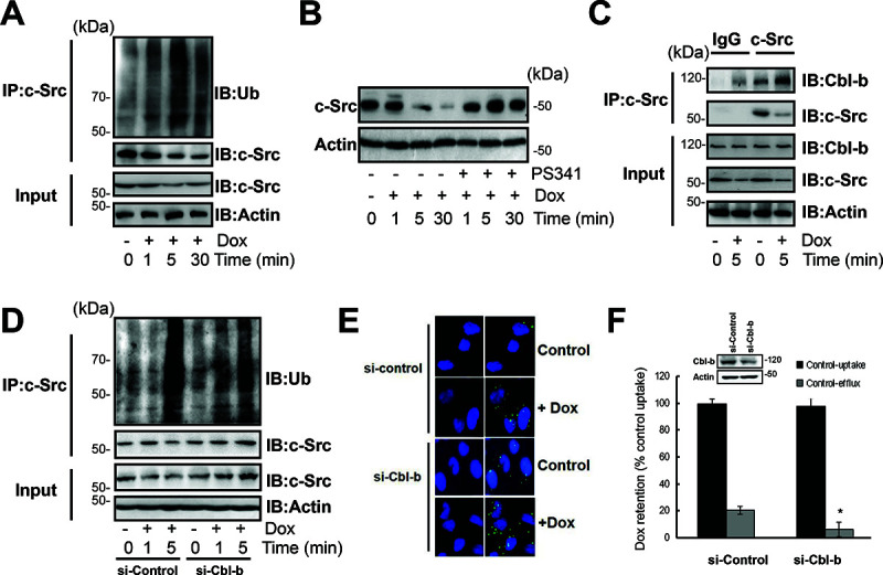
(A) SGC7901/Adr cells were exposed to 20 μg/ml Dox for 1, 5, 30 min, c-Src was immunoprecipitated and ubiquitin was analyzed by western blotting. (B) SGC7901/Adr cells were incubated with PS341 (5 nmol/l) for 12 h, then treated with 20 μg/ml Dox for 5 min, and the expression of the Src protein was analyzed by western blotting. (C) SGC7901/Adr cells were treated with or without 20 μg/ml Dox for 5, c-Src was immunoprecipitated and Cbl-b was analyzed by western blotting. (D) SGC7901/Adr cells were transiently transfected with Cbl-b siRNA (si-Cbl-b) for 48 h, followed by 20 μg/ml Dox for 1 or 5 min, c-Src was immunoprecipitated and ubiquitin was analyzed by western blotting. (E) SGC7901/Adr cells were transiently transfected with Cbl-b siRNA (si-Cbl-b) for 48 h and analyzed by PLA after incubation with 20 μg/ml Dox for 5 min. Primary mouse and rabbit antibodies against the P-gp and Cav-1 were combined with secondary PLA probes. (F) SGC7901/Adr cells were transiently transfected with Cbl-b siRNA (si-Cbl-b) for 48 h, followed by 20 μg/ml Dox and analysis of Dox uptake and efflux.
Figure 2. c-Src dependent Cav-1 phosphorylation promoted the translocation of P-gp into caveolae.
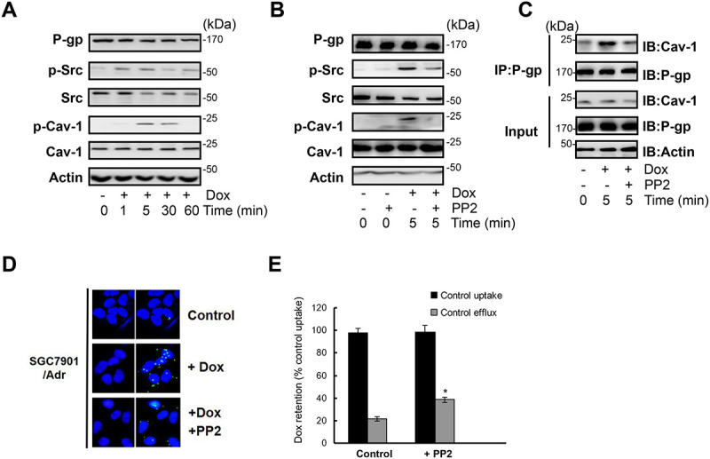
(A) SGC7901/Adr cells were treated with or without 20 μg/ml Dox for 1, 5, 30, 60 min and the expression of P-gp, p-Src, Src, p-Cav-1, Cav-1 and Actin was detected by western blotting. (B) Cells were incubated with the Src inhibitor PP2 (10 μmol/l) for 2 h, treated with 20 μg/ml Dox for 5 min, and the expression of P-gp, p-Src, Src, p-Cav-1, Cav-1 and Actin was detected by western blotting. (C) SGC7901/Adr cells were pretreated with 10 μmol/l PP2 for 2 h followed by Dox treatment, and P-gp was immunoprecipitated and Cav-1 was analyzed western blotting. (D) In situ PLA in SGC7901/Adr cells pretreated with or without 10 μmol/l PP2 for 2 h, and then incubated with 20 μg/ml Dox for 5 min. Primary mouse and rabbit antibodies against P-gp and Cav-1 were combined with secondary PLA probes. (E) SGC7901/Adr cells were pretreated with 10 μmol/l PP2 for 2 h, followed by 20 μg/ml Dox and assessment of R-123 uptake and efflux.
Table 1. Relationship between the expression of Cbl-b and clinico-pathological characteristics of P-gp-positive breast cancer patients.
| Clinico-pathological characteristics | Number | Cbl-b expression | p value | ||
| Negative | Positive | ||||
| Age (years) | |||||
| ≤ 35 | 7 | 5 | 2 | ||
| > 35 | 114 | 43 | 71 | 0.112 | |
| Tumour size (cm) | |||||
| ≤ 2 | 19 | 9 | 10 | ||
| > 2 | 102 | 39 | 63 | 0.455 | |
| pN stage | |||||
| 0 | 58 | 19 | 39 | ||
| 1–3 | 63 | 29 | 34 | 0.136 | |
| Histology grade | |||||
| 1 + 2 | 87 | 33 | 54 | ||
| 3 | 34 | 15 | 19 | 0.532 | |
| ER/PR status§ | |||||
| Negative | 59 | 19 | 40 | ||
| Positive | 62 | 29 | 33 | 0.101 | |
| HER2 status¶ | |||||
| Negative | 39 | 22 | 17 | ||
| Positive | 82 | 26 | 56 | 0.0091 | |
1Two-sided p value; values shown in bold are statistically significant.
§ER/PR status: oestrogen receptor or progesterone receptor status by immunohistochemistry (IHC); negative: ER and PR double negative; positive: ER or PR positive.
¶HER2 status: HER2-positive status is IHC 3+ or fluorescence in situ hybridisation (FISH) positive; HER2-negative status is IHC 0, 1+ or FISH negative.
HER2, Human epidermal growth factor receptor 2.
Original article: Oncotarget. 2015; 6:6737–6748. 6737-6748. https://doi.org/10.18632/oncotarget.3253