PLOS ONE logoLink to PLOS ONE
. 2021 Aug 10;16(8):e0255937. doi: 10.1371/journal.pone.0255937

Radiographic interpretation using high-resolution Cbct to diagnose degenerative temporomandibular joint disease

Jonas Bianchi 1,2,3,*, João Roberto Gonçalves 3, Antônio Carlos de Oliveira Ruellas 2,4, Júlia Vieira Pastana Bianchi 1, Lawrence M Ashman 5, Marilia Yatabe 1, Erika Benavides 6, Fabiana Naomi Soki 6, Lucia Helena Soares Cevidanes 1
Editor: Farhan Bazargani7
PMCID: PMC8354480  PMID: 34375354

Abstract

The objective of this study was to use high-resolution cone-beam computed images (hr- CBCT) to diagnose degenerative joint disease in asymptomatic and symptomatic subjects using the Diagnostic Criteria for Temporomandibular Disorders DC/TMD imaging criteria. This observational study comprised of 92 subjects age-sex matched and divided into two groups: clinical degenerative joint disease (c-DJD, n = 46) and asymptomatic control group (n = 46). Clinical assessment of the DJD and high-resolution CBCT images (isotropic voxel size of 0.08mm) of the temporomandibular joints were performed for each participant. An American Board of Oral and Maxillofacial Radiology certified radiologist and a maxillofacial radiologist used the DC/TMD imaging criteria to evaluate the radiographic findings, followed by a consensus of the radiographic evaluation. The two radiologists presented a high agreement (Cohen’s Kappa ranging from 0.80 to 0.87) for all radiographic findings (osteophyte, erosion, cysts, flattening, and sclerosis). Five patients from the c- DJD group did not present radiographic findings, being then classified as arthralgia. In the asymptomatic control group, 82.6% of the patients presented radiographic findings determinant of DJD and were then classified as osteoarthrosis or overdiagnosis. In conclusion, our results showed a high number of radiographic findings in the asymptomatic control group, and for this reason, we suggest that there is a need for additional imaging criteria to classify DJD properly in hr-CBCT images.

Introduction

Medical and dental imaging research on patient-specific diagnostics is a growing area, and the use of different imaging modalities has been reported, such as MRI [1], conventional X-rays [2], Computed Tomography, and Cone-Beam Computed Tomography (CBCT) [36]. Additionally, the development of new technologies and the improvement of existing imaging equipment facilitate the extraction of more precise and meaningful diagnostic information. Consequently, there is a need to manage and interpret these novel data to develop new therapies and diagnostic approaches [79].

In dentistry, CBCT is the exam of choice to evaluate many bone-related diseases [1012]. The temporomandibular disorders (TMD) [13] is a broad term used to describe signs and symptoms that affect muscles and joints of the temporomandibular area. The recommended “Diagnostic Criteria for TMD (DC/TMD) Axis I protocol” include diagnostic criteria for differentiating the most common pain-related TMD [14]. In the temporomandibular joint (TMJ), the degenerative joint disease (DJD) is a degenerative disorder involving the joint characterized by deterioration of articular tissue with concomitant skeletal changes in the condyle, articular fossa, and eminence. Different terminology is associated with DJD: ‘Osteoarthritis’ is used to describe for any clinical and radiographic signs and symptoms associated with pain; ‘Osteoarthrosis’ is used when no clinical signs and symptoms are present, but has radiographic findings, and the term ‘Arthralgia’ is used when clinical signs and symptoms are present, but no radiographic findings [13, 14].

In the DC/TMD guidelines, the diagnosis for TMJ disc displacement with reduction and DJD should include history in the last 30 days of any TMJ noise present with jaw movement or function, or the patient reports any noise during the exam; however, the sensitivity and specificity for this diagnosis are only 0.55 and 0.61, respectively [14]. The confirmatory and the reference standard exam for the diagnosis is the Computed Tomography (CT) criteria which should contain one of the following radiographic signs: subchondral cyst(s), erosion(s), generalized sclerosis, or osteophyte(s). Flattening and sclerosis only are considered indeterminant factors for OA. However, the study that describes the radiological/imaging criteria published by Ahmad et al. [6] used Computed Tomography (CT) of the TMJ with a slice thickness of 1mm, and two diplomates of the American Board of Oral and Maxillofacial Radiology assessed the images showing the reliability of 0.71 to evaluate the osseous changes. However, as the spatial resolution of those images was relatively large (1 mm thickness), the influence of the voxel size in the diagnostic performance to assess TMJ bony changes in CBCT images has been widely discussed in the literature. Overall conclusion is that there is a need for more studies exploring the diagnostic quality and reliability of CBCT images [4].

In this study, we used a relatively new CBCT machine [15] using a high-resolution imaging protocol to obtain high-resolution cone-beam computed images (hr-CBCT) of the TMJs (0.08 mm of isotropic voxel-size). The aim was to use high-resolution cone-beam computed images (hr-CBCT) to diagnose degenerative joint disease in asymptomatic and symptomatic subjects using the Diagnostic Criteria for Temporomandibular Disorders DC/TMD imaging criteria. We also hypothesized that there are no differences between the two radiologists for the diagnosis of the degenerative joint disease in the sample.

Material and methods

We followed the Strengthening the Reporting of Observational Studies in Epidemiology (STROBE) [16] guidelines for reporting observational studies.

Study design, setting, participants, and ethics approval

This study was reviewed and approved by the Institutional Review Board of the University of Michigan (HUM00105204 and HUM00113199). A written consent form was obtained for each participant. This observational study was composed of human subjects recruited at the University of Michigan (Ann Arbor–MI, USA) from January 2016 to July 2019. The sample comprised of prospectively recruited 46 clinically asymptomatic control patients (Control group) and 46 patients with a clinical diagnosis of degenerative joint disease (c-DJD group), resulting in 92 participants and 184 mandibular condyles.

Sample size calculation

The sample size calculation for comparing groups was done using the software G-power [17] with α of 0.05, power (1- β) of 0.80, and Cohen’s effect size given by the median and standard deviation of each group from a pilot sample. We performed the calculation for the imaging findings of osteophytes, erosion, and cysts. The size (n) necessary per group was 32, 44, and 58, respectively, and in this study, we had a final sample size of 92 per group.

Clinical diagnosis, exclusion, and inclusion criteria

All participants were between 21–70 years of age, with no history of systemic diseases, jaw joint trauma, surgery or recent jaw joint injections, pregnancy, or congenital bone or cartilage disease. They were clinically evaluated by the same temporomandibular joint specialist, using the clinical signs and symptoms of the Diagnostic Criteria for Temporomandibular Disorders (DC/TMD). To be clinically diagnosed with degenerative joint disease (c-DJD group), they must present TMJ noise during movement or function in the last 30 days, and crepitus detected during mandibular excursive movements. In addition, they must have reported TMJ pain within ten years. On the other hand, for the control group, the subjects did not present any clinical signs and symptoms of DJD.

Cone-beam computed tomography acquisition

Each participant had a high-resolution cone-beam computed tomography (CBCT) exam of each TMJ acquired using the 3D Accuitomo 170 (J. Morita MFG. CORP. Tokyo, Japan) scanner at the University of Michigan. The TMJ acquisition protocol used a field of view 40x40 mm; 90 kVp, 5 mAs, scanning time of 30.8 s, and a isotropic voxel size of 0.08 mm x 0.08 mm x 0.08 mm. The images were exported in DICOM (.dcm) format using the i- Dixel software (J. Morita MFG. CORP Tokyo, Japan) and were de-identified for further radiological evaluation. The cone-beam computed tomography (CBCT) exams for both groups were acquired respecting the ALARA and ALADA principles [17, 18]. All participants agreed and signed the informed consent term for participating in this study. In this study, the radiation dose to the patients was kept as low as possible by limiting the field of view (FOV) of the CBCT scan to 40 x 40 mm, which is enough to cover the TMJ area. The patients also wore a lead apron with a thyroid collar.

Imaging diagnostic criteria and radiographic findings agreement

A multi-planar and blinded evaluation of the CBCT scans was performed by an American Board of Oral and Maxillofacial Radiology certified radiologist and a maxillofacial radiologist from the Department of Periodontics & Oral Medicine at the University of Michigan–School of Dentistry (Ann Arbor, USA). The multi-planar cross-sectional images were assessed in sagittal and coronal planes to score the mandibular condyle using the following five categories (as defined and adapted from the DC/TMD) and four scales (0 to 3): A) Flattening: 0 = Not visualized, 1 = Mild, 2 = Moderate and 3 = Severe; B) Osteophytes: 0 = Not visualized, 1 = Mild/Small, 2 = Moderate/Medium, 3 = Severe/Large; C) Sclerosis: 0 = Not visualized, 1 = Mild/Localized, 2 = Moderate/Generalized, 3 = Severe/Generalized; D) Erosion: 0 = Not visualized, 1 = Mild/Localized, 2 = Moderate, 3 = Severe, and E) Cysts: 0 = Not visualized, 1 = one to two cysts, 2 = three to four cysts, 3 = five or more cysts. Each CBCT scan was evaluated separately by each radiologist, followed by a consensus radiographic evaluation. The consensus data was used for interpretation and radiological classification, and if the patient presents any score for erosion, osteophyte and/or cysts, he was classified as having a radiographic diagnosis of degenerative joint disease.

Statistical analysis

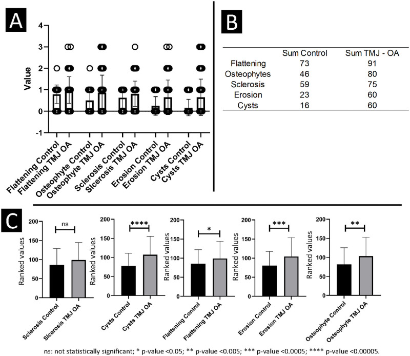
Our data showed nom-normal distribution assessed by the Shapiro-Wilk test. For this reason, the Mann–Whitney U test was used to compare the radiographic findings between the two groups (Fig 1C). To test observer concordance, an agreement matrix was calculated, and to test the inter-rater reliability, the Cohen’s kappa coefficient (κ) was conducted. A description of the main findings was evaluated and presented in plots (Fig 1A and 1B) to visualize the distribution of the radiographic findings between the groups and imaging findings (Fig 1A) and the sum of each radiographic finding (Fig 1B).

Fig 1.

Fig 1

A, B—Descriptive statistics for the radiologists’ consensus. C- Mann Whitney U test for comparison between the groups.

Results

The sample was composed of sex-age matched subjects, resulting in 2 groups: c-DJD group and Control group. The c-DJD group (46 patients and n = 92 condyles) consisted of 39 females and 7 males, average age: 38.50 (S.D: 13.26), and the Control Group (46 patients and n = 92 condyles) has 39 females and 7 males, with a mean age of 38 years (S.D: 12.34). Table 1 shows the agreement matrix between the two observers for the radiographic findings of both groups together. It can be noted that for flattening, osteophytes, sclerosis, erosion, and cysts scores. The only disagreement was one above or one below score, and that the overall agreement was excellent. Table 2 shows the Cohen’s Kappa statistic values for the measurements between the observers: the lowest κ value being 0.80 for sclerosis, and the highest κ being 0.87 for erosion.

Table 1. Agreement matrix of the radiologists (observer 1 and 2) for each radiographic finding visualized in a multi-planar HR-CBCT evaluation for both groups together.

The agreement is shown in diagonal. The numbers represent the total number of mandibular condyles in their specific category.

Flattening Observer 2 Total
Not visualized Mild Moderate Severe
Observer 1 Not visualized 30 4 0 0 34
Mild 7 124 2 0 133
Moderate 0 3 12 0 15
Severe 0 0 0 2 2
Total 37 131 14 2 184
Osteophytes Not visualized Mild / Moderate / Severe / Total
Small Medium Large
Not visualized 71 14 0 0 85
Mild / Small 5 77 0 0 82
Moderate / Medium 0 3 7 0 10
Severe / Large 0 0 2 5 7
Total 76 94 9 5 184
Sclerosis Not visualized Mild / Moderate / Severe / Total
Localized Generalized Generalized
Not visualized 49 14 0 0 63
Mild / Localized 4 110 0 0 114
Moderate / Generalized 0 1 5 0 6
Severe / Generalized 0 0 0 1 1
Total 53 125 5 1 184
Erosion Not visualized Mild Moderate Severe Total
Not visualized 114 4 0 0 118
Mild / Localized 5 45 1 0 51
Moderate 0 3 9 1 13
Severe 0 0 1 1 2
Total 119 52 11 2 184
Cysts Not visualized 1–2 3–4 5 or more Total
cysts cysts cysts
Not visualized 120 6 0 0 126
1–2 cysts 7 38 4 0 49
3–4 cysts 0 1 6 1 8
5 or more cysts 0 0 0 1 1
Total 127 45 10 2 184

Table 2. Cohen’s Kappa statistic to test inter-rater reliability between the radiologists.

Observer 1 x Observer 2 95% CI
Weighted
Kappa
SE Lower Upper Observed Agreements Agreements Expected
by Chance
Flattening 0.83 0.05 0.71 0.90 91.30% 55.81%
Osteophytes 0.82 0.04 0.69 0.86 86.96% 42.22%
Sclerosis 0.80 0.05 0.69 0.88 89.67% 52.04%
Erosion 0.87 0.04 0.76 0.91 91.85% 49.74%
Cysts 0.81 0.05 0.68 0.87 89.67% 54.02%

CI: Confidence interval; SE: Standard error of Kappa.

The agreement between the radiologists’ interpretation of the findings in each group is summarized in Fig 1A and 1B. The sum count of the classification used in this study included 0 to 3 scoring. All radiographic finding values were higher in the c-DJD group compared to the control group. As expected, flattening showed a high incidence in both groups, followed by osteophytes, sclerosis, erosion, and cysts. Thus, those findings showed that radiographic changes were observed in both groups, having a higher magnitude in the c-DJD group. Inter-group comparison shows that ‘sclerosis’ was the only radiographic finding that was statistically similar between both groups (p>0.05). The following radiographic findings showed that c-DJD group had significantly worse scores: flattening (p-value ≤ 0.05), osteophytes (p-value ≤ 0.005), erosion (p-value ≤ 0.0005), and cysts (p-value ≤ 0.00005) as illustrated in Fig 1C.

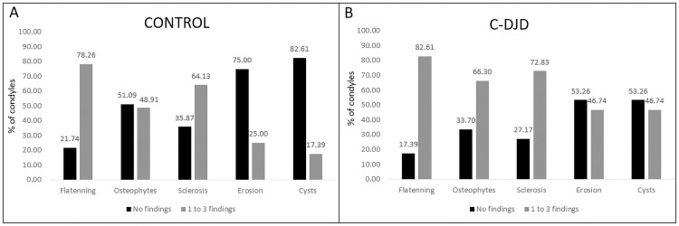
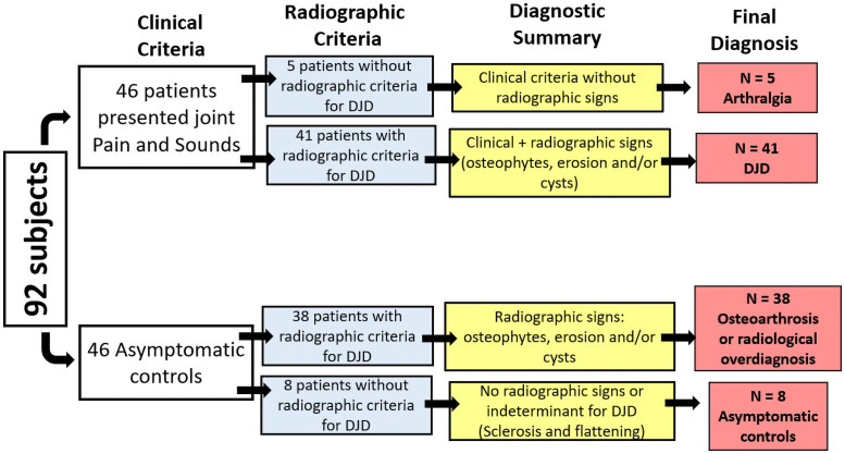
Fig 2 compares the percentage of condyles with no radiographic findings versus condyles with radiographic findings. Fig 3 summarizes this study’s sample radiographic interpretation.

Fig 2. Percentage of condyles with no radiographic findings (score 0, not visualized) compared to condyles with radiographic findings (scores 1–3 of severity grouped).

Fig 2

Fig 3. Summary of patient’s diagnosis after clinical and radiological assessment.

Fig 3

Fig 3 summarizes our main findings for patient classification. For c-DJD group, approximately 11% of the population did not show imaging signs of DJD. From 92 enrolled subjects, 46 presented clinical symptoms of DJD associated with TMJ pain, and 46 participants did not present any clinical sign and/or history of TMJ problems. In the radiographic interpretation, 41 patients showed signs of DJD whereas five did not. For these five patients, we suggested a diagnosis of Arthralgia and the other 41 patients received a diagnosis of DJD–Osteoarthritis. For the control group in the radiographic interpretation, 38 subjects showed signs of DJD, and only 8 did not, so based on the DC/TMD these patients should receive a diagnosis of Osteoarthrosis. Fig 4 shows the cross-sectional hr-CBCT and the radiographic findings: cysts (Fig 4A), localized erosions (Fig 4B), and osteophytes (Fig 4C) in both groups.

Fig 4. Radiographic findings determinants for DJD.
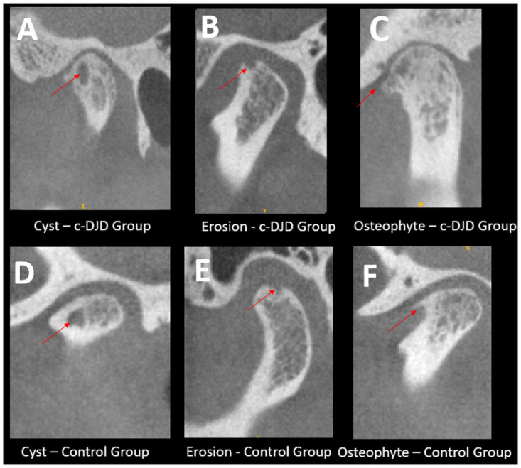
Fig 4

Each image shows the sagittal slices of the TMJs and hr-CBCT with isotropic voxel size of 0.08 mm with the arrows are pointing to the finding. A-C: Represents the DJD group and D–F the asymptomatic control group.

Discussion

In this study, we proposed to evaluate the radiological interpretation of cone-beam computed tomography images with a voxel size of 0.08mmx0.08mmx0.08mm using the DC/TMD imaging criteria for DJD diagnosis. We showed that the two radiologists presented a good agreement between them (κ = 0.80). However, over 82% of the asymptomatic control patients showed a radiographic finding (Fig 2), which is a higher incidence than the current literature [6, 1921], suggesting either a high occurrence of bone remodeling that may be over-diagnosed as osteoarthrosis or true incidence of osteoarthrosis in our study sample. It is possible that as the DC/TMD imaging criteria for DJD were validated for CT images with lower voxel size, there may be a lack of adequate imaging criteria to properly classify DJD using high-resolution CBCT images.

The validation of CT images for diagnosis of DJD was proposed in 2009 to confirm the clinical diagnosis based on the patient’s history and signs/symptoms because the clinical diagnosis only had a sensitivity of 0.55 and specificity of 0.61 [14]. The determinant imaging criteria for DJD proposed by Ahmad et al. used CT images with a slice thickness of 1mm and the following categories: subchondral cyst, and/or erosion, and/or osteophyte, and/or generalized sclerosis6. The challenge was the osseous defects had to be close to 1mm in order to be detected, making this diagnosis more reliable for late to chronic DJD stages. The greatest challenge now is to detect the disease before severe bone changes occur in the mandibular condyles, and studies indicate the key role subchondral bone plays in osteoarthritis progression [2023]. In cross-sectional evaluation of the CT images, authors found a κ = 0.71 for reliability and 86% of the agreement for TMJ OA diagnosis6. In present study, our results are based on each radiographic finding that characterizes the DJD, and has showed an inter-rater agreement of 86% for osteophytes, 89% for cysts and sclerosis, and 91% for erosion and flattening (Table 2). The Cohen’s kappa values were also high between 0.80 to 0.87 showing the radiologists were calibrated between them, and they could identify and correctly classify the imaging findings based on the DC/TMD guidelines.

Although CT imaging has contributed to the diagnosis of TMDs [14], in dentistry, most studies have used CBCT images to assess the bone changes, and the accuracy of CBCT for the detection of osseous defects remains under discussion [3, 20, 21, 2426]. A systematic review reported the reliability of studies that used CBCT images to detect the osseous changes in TMJs and concluded that voxel size is a parameter that affects the pooled sensitivity (PSS) and pooled specificity (PSC), i.e., for isotropic voxel sizes of 0.2 mm or less the PSS is 0.73 and PSC is 0.68 and the studies that used voxel size of 0.4 to 0.5 the values are 0.83 and 1.00 respectively [4]. Another study has pointed out that voxel size is not important for the improvement of diagnosis; however, an image downsizing technique was used to increase the voxel size in that study and does not represent the real patient CBCT protocol acquisition [2]. A study from Lukat et al. [27] assessed the effects of voxel size to detect osseous changes in temporomandibular joint (TMJ), and they found no statistically significant difference between the voxel sizes in detection of TMJ osteoarthritic changes. However, a limitation was that the authors acquired the images with a isotropic voxel size of 0.076 mm and computationally downsized the image to a voxel size of 0.300 mm, which does not represent a true CBCT acquisition with 0.300 mm of voxel size.

In the present study, we have shown that using CBCT images with 0.08mm isotropic voxel resolution resulted in a good agreement in the diagnostic interpretation between the two observers. Furthermore, we found a high number of osseous changes in asymptomatic control group (38 out of 46 patients). These findings correspond to approximately 82% of the control group that presented radiographic signs of DJD. However, the literature has shown a smaller percentage of asymptomatic patients. Cevidanes et al. [28] found approximately 15% of changes in control patients using a isotropic voxel size of 0.5 mm. Krisjane et al. [29] assessed osteoarthritis findings in asymptomatic patients and found the most common signs were articular surface flattening and subcortical sclerosis; however, it was found only in 43% of class ll patients without clinical signs and symptoms. Here, we also found the most common imaging features for the control group were flattening and sclerosis in initial stages, which does not correspond to OA findings; however, a significant number of patients presented cysts, erosions, or osteophytes (84%), leading to question whether the control patients truly presented with osteoarthrosis or the high-resolution of the images is what led to over-diagnosis when using the general guidelines from RDC/TMD.

This study also found the most common radiological features presented in both groups which were flattening and sclerosis. These results agree with the general literature and with the DC/TMD criteria, where those signs are not determinant for osteoarthritis/osteoarthrosis and are commonly seen due to adaptative responses and the aging process. Emshoff et al. [19] reported a rate of 21% of patients showing erosions without clinical symptoms and a study by dos Anjos et al. [30] showed that in a population treated for orthodontic purposes (n = 382) only 3% presented osteophytes, 0.5% erosions, and 0.8% cysts. In our study, we found approximately 48% with osteophytes, 25% erosions, and 17% cysts, using CBCT images with 1mm slice thickness. It is possible that this discrepancy is based on image resolution since both studies were conducted by experienced radiologists. The challenge now appears on how to propose adequate diagnostic criteria for images with higher resolutions (0.08 mm x 0.08 mm x 0.08 mm of voxel size), while trying to avoid a radiographic over-diagnosis.

Our statistical comparison suggests that erosion, osteophytes, and cysts are more robust to differentiate the groups compared to flattening, which has a significant p-value, but not as significant as the others (Fig 1C). The control group showed more than 70% of condyles having flattening, 48% showed osteophytes, 64% with sclerosis, and only 17% with cysts. For the c-DJD group, flattening was present in 82% of the condyles, osteophytes in 66%, sclerosis in72%, erosions in 46% and cysts in 46%. The DC/TMD criteria does not use flattening and localized sclerosis as determinants for DJD, and our results support this recommendation due to the higher number of these two findings in the control group (Fig 2).

Jiang et al. [5], in 2015 assessed CBCT images with a isotropic voxel size of 0.5 mm in asymptomatic patients according to the chewing-side preference. They did not evaluate the presence of erosions, osteophytes, or cysts; however, the results indicated the occurrence of morphological changes in the TMJ region, suggesting an adaptive process which occurs when patients have a chewing-side preference. In comparison to our results, approximately 78% of all condyles presented with some degree of flattening (Fig 2), and one of the reasons may also be due to those adaptative responses to abnormal functions.

As a limitation of this study, we did not perform a dosimetry study to determine and assess the X-ray absorption during our protocol for hr-CBCT acquisition. Lukat et al. [31], evaluated the effective dose of the CBCT for the TMJ region with a small FOV (5x3.7 cm); but, they used a different CBCT machine/protocol, and reported unilateral doses of 20.5 ± 1.3 μSv. Another limitation of this study is that we define as high-resolution the CBCT with a voxel size of 0.08mmx0.08mmx0.08mm; however, the spatial resolution is related to multiple factors, as described by Brulmann and Schulze [31]: “…The spatial resolution is related to the physical pixel size of the sensor, the grey-level resolution, the reconstruction technique applied…” which we have not addressed.

Conclusion

Our results suggest that additional imaging criteria for hr-CBCT may be needed to properly classify DJD since the results showed a high number of radiographic findings in the asymptomatic control group.

Supporting information

S1 File

(TXT)

Data Availability

All relevant data are within the manuscript and its Supporting information files.

Funding Statement

National Institute of Health (NIH), National Institute of Dental and Craniofacial Research (NIDCR) grant: R01DE024450.

References

  • 1.Ashinsky BGG, Coletta CEE, Bouhrara M, Lukas VAA, Boyle JMM, Reiter DAA, et al. Machine learning classification of OARSI-scored human articular cartilage using magnetic resonance imaging. Osteoarthr Cartil. 2015;23: 1704–1712. doi: 10.1016/j.joca.2015.05.028 [DOI] [PMC free article] [PubMed] [Google Scholar]
  • 2.Hirvasniemi J, Niinimäki J, Thevenot J, Saarakkala S. Bone Density and Texture from Minimally Post-Processed Knee Radiographs in Subjects with Knee Osteoarthritis. Ann Biomed Eng. 2019;47: 1181–1190. doi: 10.1007/s10439-019-02227-y [DOI] [PMC free article] [PubMed] [Google Scholar]
  • 3.Larheim TA, Abrahamsson A-K, Kristensen M, Arvidsson LZ. Temporomandibular joint diagnostics using CBCT. Dentomaxillofacial Radiol. 2015;44: 20140235. doi: 10.1259/dmfr.20140235 [DOI] [PMC free article] [PubMed] [Google Scholar]
  • 4.Ma RH, Yin S, Li G. The detection accuracy of cone beam CT for osseous defects of the temporomandibular joint: A systematic review and meta-analysis. Sci Rep. 2016;6: 1–8. [DOI] [PMC free article] [PubMed] [Google Scholar]
  • 5.Jiang H, Li C, Wang Z, Cao J, Shi X, Ma J, et al. Assessment of osseous morphology of temporomandibular joint in asymptomatic participants with chewing-side preference. J Oral Rehabil. 2015;42: 105–112. doi: 10.1111/joor.12240 [DOI] [PubMed] [Google Scholar]
  • 6.Ahmad M, Hollender L, Anderson Q, Kartha K, Ohrbach R, Truelove EL, et al. Research diagnostic criteria for temporomandibular disorders (RDC/TMD): development of image analysis criteria and examiner reliability for image analysis. Oral Surg Oral Med Oral Pathol Oral Radiol Endod. 2009;107: 844–60. doi: 10.1016/j.tripleo.2009.02.023 [DOI] [PMC free article] [PubMed] [Google Scholar]
  • 7.Alyass A, Turcotte M, Meyre D. From big data analysis to personalized medicine for all: Challenges and opportunities. BMC Med Genomics. 2015;8: 1–12. [DOI] [PMC free article] [PubMed] [Google Scholar]
  • 8.Aerts HJWL, Velazquez ER, Leijenaar RTH, Parmar C, Grossmann P, Carvalho S, et al. Decoding tumour phenotype by noninvasive imaging using a quantitative radiomics approach. Nat Commun. 2014;5: 4006. doi: 10.1038/ncomms5006 [DOI] [PMC free article] [PubMed] [Google Scholar]
  • 9.Shoukri B, Prieto JCC, Ruellas A, Yatabe M, Sugai J, Styner M, et al. Minimally Invasive Approach for Diagnosing TMJ Osteoarthritis. J Dent Res. 2019;98: 1103–1111. doi: 10.1177/0022034519865187 [DOI] [PMC free article] [PubMed] [Google Scholar]
  • 10.Kapila SD, Nervina JM. CBCT in orthodontics: assessment of treatment outcomes and indications for its use. Dentomaxillofac Radiol. 2014;44: 20140282. doi: 10.1259/dmfr.20140282 [DOI] [PMC free article] [PubMed] [Google Scholar]
  • 11.Brüllmann D, Schulze RKW, Brullmann D, Schulze RKW. Spatial resolution in CBCT machines for dental/maxillofacial applications—What do we know today? Dentomaxillofacial Radiol. 2015;44. doi: 10.1259/dmfr.20140204 [DOI] [PMC free article] [PubMed] [Google Scholar]
  • 12.De Vos W, Casselman J, Swennen GRJ. Cone-beam computerized tomography (CBCT) imaging of the oral and maxillofacial region: a systematic review of the literature. Int J Oral Maxillofac Surg. 2009;38: 609–25. [DOI] [PubMed] [Google Scholar]
  • 13.McNeill C. Management of temporomandibular disorders: Concepts and controversies. J Prosthet Dent. 1997;77: 510–522. doi: 10.1016/s0022-3913(97)70145-8 [DOI] [PubMed] [Google Scholar]
  • 14.Schiffman E, Ohrbach R, Truelove E, Look J, Anderson G, Goulet J-P, et al. Diagnostic Criteria for Temporomandibular Disorders (DC/TMD) for Clinical and Research Applications: recommendations of the International RDC/TMD Consortium Network* and Orofacial Pain Special Interest Group†. J oral facial pain headache. 2014;28: 6–27. doi: 10.11607/jop.1151 [DOI] [PMC free article] [PubMed] [Google Scholar]
  • 15.Morita CBCT Imaging. 2017. https://www.morita.com/america/en/products/diagnostic-and-imaging-equipment/cone-beam-ct-systems/3d-accuitomo-170/?tab=technical_details.
  • 16.von Elm E, Altman DG, Egger M, Pocock SJ, Gøtzsche PC, Vandenbroucke JP, et al. The Strengthening the Reporting of Observational Studies in Epidemiology (STROBE) statement: guidelines for reporting observational studies. J Clin Epidemiol. 2008;61: 344–9. doi: 10.1016/j.jclinepi.2007.11.008 [DOI] [PubMed] [Google Scholar]
  • 17.Editorial. ALARA still applies. Oral Surg Oral Med Oral Pathol Oral Radiol Endod. 2005;100: 395–397. doi: 10.1016/j.tripleo.2005.05.055 [DOI] [PubMed] [Google Scholar]
  • 18.Jaju PP, Jaju SP. Cone-beam computed tomography: Time to move from ALARA to ALADA. Imaging Sci Dent. 2015;45: 263–5. doi: 10.5624/isd.2015.45.4.263 [DOI] [PMC free article] [PubMed] [Google Scholar]
  • 19.Emshoff R, Bertram F, Schnabl D, Stigler R, Steinmaßl O, Rudisch A. Condylar Erosion in Patients With Chronic Temporomandibular Joint Arthralgia: A Cone-Beam Computed Tomography Study. J Oral Maxillofac Surg. 2016;74: 1343.e1–1343.e8. doi: 10.1016/j.joms.2016.01.029 [DOI] [PubMed] [Google Scholar]
  • 20.Shahidi S, Salehi P, Abedi P, Dehbozorgi M, Hamedani S, Berahman N. Comparison of the Bony Changes of TMJ in Patients With and Without TMD Complaints Using CBCT. J Dent (Shiraz, Iran). 2018;19: 142–149. Available: http://www.ncbi.nlm.nih.gov/pubmed/29854888%0Ahttp://www.pubmedcentral.nih.gov/articlerender.fcgi?artid=PMC5960734. [PMC free article] [PubMed] [Google Scholar]
  • 21.Bakke M, Petersson A, Wiese M, Svanholt P, Sonnesen L. Bony Deviations Revealed by Cone Beam Computed Tomography of the Temporomandibular Joint in Subjects Without Ongoing Pain. J Oral Facial Pain Headache. 2018;28: 331–337. doi: 10.11607/ofph.1255 [DOI] [PubMed] [Google Scholar]
  • 22.Burr DB, Gallant MA. Bone remodelling in osteoarthritis. Nat Publ Gr. 2012;8: 665–673. doi: 10.1038/nrrheum.2012.130 [DOI] [PubMed] [Google Scholar]
  • 23.Pritzker KPH, Gay S, Jimenez SA, Ostergaard K, Pelletier JP, Revell K, et al. Osteoarthritis cartilage histopathology: Grading and staging. Osteoarthr Cartil. 2006;14: 13–29. doi: 10.1016/j.joca.2005.07.014 [DOI] [PubMed] [Google Scholar]
  • 24.Cömert Kiliç S, Kiliç N, Sümbüllü MA. Temporomandibular joint osteoarthritis: Cone beam computed tomography findings, clinical features, and correlations. Int J Oral Maxillofac Surg. 2015;44: 1268–1274. doi: 10.1016/j.ijom.2015.06.023 [DOI] [PubMed] [Google Scholar]
  • 25.Hussain AM, Packota G, Major PW, Flores-Mir C. Role of different imaging modalities in assessment of temporomandibular joint erosions and osteophytes: A systematic review. Dentomaxillofacial Radiol. 2008;37: 63–71. doi: 10.1259/dmfr/16932758 [DOI] [PubMed] [Google Scholar]
  • 26.Alexiou KE, Stamatakis HC, Tsiklakis K. Evaluation of the severity of temporomandibular joint osteoarthritic changes related to age using cone beam computed tomography. Dentomaxillofacial Radiol. 2009;38: 141–147. doi: 10.1259/dmfr/59263880 [DOI] [PubMed] [Google Scholar]
  • 27.Fakhar HB, Mallahi M, Panjnoush M, Kashani PM. Effect of Voxel Size and Object Location in the Field of View on Detection of Bone Defects in Cone Beam Computed Tomography. J Dent (Tehran). 2016;13: 279–286. Available: http://www.ncbi.nlm.nih.gov/pubmed/28127320%0Ahttp://www.pubmedcentral.nih.gov/articlerender.fcgi?artid=PMC5253221. [PMC free article] [PubMed] [Google Scholar]
  • 28.Cevidanes LHS, Hajati A-K, Paniagua B, Lim PF, Walker DG, Palconet G, et al. Quantification of condylar resorption in temporomandibular joint osteoarthritis. Oral Surg Oral Med Oral Pathol Oral Radiol Endod. 2010;110: 110–7. doi: 10.1016/j.tripleo.2010.01.008 [DOI] [PMC free article] [PubMed] [Google Scholar]
  • 29.Krisjane Z, Urtane I, Krumina G, Neimane L, Ragovska I. The prevalence of TMJ osteoarthritis in asymptomatic patients with dentofacial deformities: A cone-beam CT study. Int J Oral Maxillofac Surg. 2012;41: 690–695. doi: 10.1016/j.ijom.2012.03.006 [DOI] [PubMed] [Google Scholar]
  • 30.dos Anjos Pontual ML, Freire JSL, Barbosa JMN, Frazão M a G, dos Anjos Pontual A. Evaluation of bone changes in the temporomandibular joint using cone beam CT. Dentomaxillofac Radiol. 2012;41: 24–9. doi: 10.1259/dmfr/17815139 [DOI] [PMC free article] [PubMed] [Google Scholar]
  • 31.Lukat TD, Wong JCM, Lam EWN. Small field of view cone beam CT temporomandibular joint imaging dosimetry. Dentomaxillofacial Radiol. 2013;42. doi: 10.1259/dmfr.20130082 [DOI] [PMC free article] [PubMed] [Google Scholar]

Decision Letter 0

Farhan Bazargani

8 Apr 2021

PONE-D-21-00635

RADIOGRAPHIC INTERPRETATION USING HIGH-RESOLUTION CBCT TO DIAGNOSE DEGENERATIVE TEMPOROMANDIBULAR JOINT DISEASE

PLOS ONE

Dear Dr. Bianchi,

Thank you for submitting your manuscript to PLOS ONE. After careful consideration, we feel that it has merit but does not fully meet PLOS ONE’s publication criteria as it currently stands. Therefore, we invite you to submit a revised version of the manuscript that addresses the points raised during the review process.

Please submit your revised manuscript by May 21st. If you will need more time than this to complete your revisions, please reply to this message or contact the journal office at plosone@plos.org. When you're ready to submit your revision, log on to https://www.editorialmanager.com/pone/ and select the 'Submissions Needing Revision' folder to locate your manuscript file.

Please include the following items when submitting your revised manuscript:

  • A rebuttal letter that responds to each point raised by the academic editor and reviewer(s). You should upload this letter as a separate file labeled 'Response to Reviewers'.

  • A marked-up copy of your manuscript that highlights changes made to the original version. You should upload this as a separate file labeled 'Revised Manuscript with Track Changes'.

  • An unmarked version of your revised paper without tracked changes. You should upload this as a separate file labeled 'Manuscript'.

If you would like to make changes to your financial disclosure, please include your updated statement in your cover letter. Guidelines for resubmitting your figure files are available below the reviewer comments at the end of this letter.

If applicable, we recommend that you deposit your laboratory protocols in protocols.io to enhance the reproducibility of your results. Protocols.io assigns your protocol its own identifier (DOI) so that it can be cited independently in the future. For instructions see: http://journals.plos.org/plosone/s/submission-guidelines#loc-laboratory-protocols. Additionally, PLOS ONE offers an option for publishing peer-reviewed Lab Protocol articles, which describe protocols hosted on protocols.io. Read more information on sharing protocols at https://plos.org/protocols?utm_medium=editorial-email&utm_source=authorletters&utm_campaign=protocols.

We look forward to receiving your revised manuscript.

Kind regards,

Farhan Bazargani, DDS, PhD

Academic Editor

PLOS ONE

Journal requirements:

When submitting your revision, we need you to address these additional requirements.

1. Please ensure that your manuscript meets PLOS ONE's style requirements, including those for file naming. The PLOS ONE style templates can be found at

https://journals.plos.org/plosone/s/file?id=wjVg/PLOSOne_formatting_sample_main_body.pdf and

https://journals.plos.org/plosone/s/file?id=ba62/PLOSOne_formatting_sample_title_authors_affiliations.pdf

2. We note that you have indicated that data from this study are available upon request. PLOS only allows data to be available upon request if there are legal or ethical restrictions on sharing data publicly. For information on unacceptable data access restrictions, please see http://journals.plos.org/plosone/s/data-availability#loc-unacceptable-data-access-restrictions.

In your revised cover letter, please address the following prompts:

a) If there are ethical or legal restrictions on sharing a de-identified data set, please explain them in detail (e.g., data contain potentially identifying or sensitive patient information) and who has imposed them (e.g., an ethics committee). Please also provide contact information for a data access committee, ethics committee, or other institutional body to which data requests may be sent.

b) If there are no restrictions, please upload the minimal anonymized data set necessary to replicate your study findings as either Supporting Information files or to a stable, public repository and provide us with the relevant URLs, DOIs, or accession numbers. Please see http://www.bmj.com/content/340/bmj.c181.long for guidelines on how to de-identify and prepare clinical data for publication. For a list of acceptable repositories, please see http://journals.plos.org/plosone/s/data-availability#loc-recommended-repositories.

We will update your Data Availability statement on your behalf to reflect the information you provide.

[Note: HTML markup is below. Please do not edit.]

Reviewers' comments:

Reviewer's Responses to Questions

Comments to the Author

1. Is the manuscript technically sound, and do the data support the conclusions?

The manuscript must describe a technically sound piece of scientific research with data that supports the conclusions. Experiments must have been conducted rigorously, with appropriate controls, replication, and sample sizes. The conclusions must be drawn appropriately based on the data presented.

Reviewer #1: Partly

Reviewer #2: Partly

Reviewer #3: Partly

**********

2. Has the statistical analysis been performed appropriately and rigorously?

Reviewer #1: No

Reviewer #2: Yes

Reviewer #3: Yes

**********

3. Have the authors made all data underlying the findings in their manuscript fully available?

The PLOS Data policy requires authors to make all data underlying the findings described in their manuscript fully available without restriction, with rare exception (please refer to the Data Availability Statement in the manuscript PDF file). The data should be provided as part of the manuscript or its supporting information, or deposited to a public repository. For example, in addition to summary statistics, the data points behind means, medians and variance measures should be available. If there are restrictions on publicly sharing data—e.g. participant privacy or use of data from a third party—those must be specified.

Reviewer #1: Yes

Reviewer #2: Yes

Reviewer #3: Yes

**********

4. Is the manuscript presented in an intelligible fashion and written in standard English?

PLOS ONE does not copyedit accepted manuscripts, so the language in submitted articles must be clear, correct, and unambiguous. Any typographical or grammatical errors should be corrected at revision, so please note any specific errors here.

Reviewer #1: Yes

Reviewer #2: Yes

Reviewer #3: No

**********

5. Review Comments to the Author

Please use the space provided to explain your answers to the questions above. You may also include additional comments for the author, including concerns about dual publication, research ethics, or publication ethics. (Please upload your review as an attachment if it exceeds 20,000 characters)

Reviewer #1: Point 01

“However, the sensitivity and specificity for this diagnosis are only 0.55 and 0.61, respectively.”

What is(are) the reference(s) for this information?

Point 02

In the “sample size calculation” it is written that “We performed a post-hoc analysis for the comparison between groups and the variables using the software G-power”

How come that did the author perform a post-hoc analysis for sample size calculation if post-hoc analyses are conducted using data that has already been collected, and the sample size is supposed to be calculated before the experimental part of the study begins?

Point 03

Still for the “sample size calculation”:

“for the sum of the flattening values, 97% - osteophytes, 70% - sclerosis; 99% erosion and 99% for the cysts”

Where did these values come from? This was not explained.

Point 04

The following paragraph is repeated in the text:

“All participants were between 21 – 70 years old, with no history of systemic diseases, jaw joint trauma, surgery or recent jaw joint injections, current pregnancy, or congenital bone or cartilage disease. They were clinically evaluated by the same temporomandibular joint specialist, using the clinical signs and symptoms of the Diagnostic Criteria for Temporomandibular Disorders (DC/TMD). To be clinically diagnosed with degenerative joint disease (c-DJD group), they must present TMJ noise during movement or function in the last 30 days, and crepitus detected during mandibular excursive movements. In addition, they must report TMJ pain for less than ten years. On the other hand, for the control group, the subjects did not present any clinical signs and symptoms of DJD.”

Point 05

“The X-rays for both groups were taken”

Since when x-rays are “taken”?

Point 06

What is the meaning of “corrected” sagittal and coronal planes?

Point 07

The authors used Mann-Whitney test to compare sums of categorical variables?

And why are the results shown in figure 1C presented in ranks?

Together with my point 02 above, I can only conclude that this study was not correctly conducted from the statistical point of view. The authors of this manuscript need to address these issues by looking for help of a professional statistician. These statistical issues may question the validity of the results.

And the sub-item “Statistical analysis” from the Materials and Methods section is extremely poorly explained.

Point 08

In the Discussion, the paragraph beginning with “Interestingly, (…)” is a mere repetition of the results.

Point 09

The limitations of the study were neither listed nor discussed.

Point 10

“This study evaluated the radiographic findings based on the DC/TMD criteria for DJD in hr-CBCT images, and we”

Remove this text from the conclusion.

Reviewer #2: 1. The authors might wish to read and cite “Small field-of-view cone beam CT temporomandibular joint imaging dosimetry” (Dentomaxillofac. Radiol. 42: 20130082; 2013) and “The effect of voxel size on cone beam CT images of the temporomandibular joints” (Oral Surg., Oral Med., Oral Pathol., Oral Radiol. 119:229-237; 2015) from the Toronto group.

2. Please clarify the voxel dimensions of the Morita CBCT device. Is the product of length, width and height of the voxel equal to 0.08 mm^3, or is each individual dimension 0.08 mm (i.e., so the voxel size is 0.08 mm x 0.08 mm x 0.08 mm, and the volume is 0.000512 mm^3]. When I look at the Morita website, it is the latter (0.08 mm per side) and not what the authors have indicated. I believe this same error propagates into the Discussion as well when they cite the work of others.

3. How were the subjects recruited? Was this sequential enrollment of all patients who were seen in a TMD clinic between Jan 2016 and July 2019? Or were these a subset of the patients? If the latter, why were they chosen to be included. Please clarify.

4. Why did clinically asymptomatic control patients receive TMJ imaging? What was the justification for this?

5. Define “radiological experts”.

6. Table 1. Can the authors please explain what a “Confusion matrix” is and how the numbers were derived? Are there units for these numbers or are the units arbitrary? Please clarify.

7. Figure 1C. Please provide units of measurement along the y-axis of the graphs (even if they are Arbitrary Units; AU).

8. Is it necessary to report frequency to one-one-hundredths of a percentage?

9. A significant weakness of this study is that the authors did not perform any dosimetry for this imaging protocol, and inappropriately cited the work of others. There is considerable variation in calculated effective radiation doses for small field size CBCT fields, and the calculation of effective dose depends on the location of the field and the tissues contained in the volume. The Ludlow meta-analysis (reference 21) cites doses of 32 uSv in the maxilla and 43 uSv in the mandible. But Pauwels (who originally reported the 43 uSv mandible value) indicates that the field was centered in the “Lower jaw molar region” and not on the TMJ. So the dose from the current study where the image volume is centered on the TMJ is more than likely not 43 uSv. If you read the Toronto paper (in DMFR 2013), they reported unilateral doses of about 10 uSv and bilateral doses of about 21 uSv; much less than what Pauwels reported for the posterior mandible. Although the Toronto group used a 5 cm x 3.7 cm field size, their volumes were centered over the joints, and so should provide much more accurate values than Pauwels. However, without having done any dosimetry work themselves for this application of the 4 cm x 4 cm field, the authors can only speculate about the dose.

10. In the last paragraph of the Discussion, the authors bring in the topic of “radiomics”, but do not expand on the topic. I’m not sure how radiomics relates to the current work.

11. I am somewhat concerned about the reliability of the radiologic interpretation of normal and disease, although the consistency appears to be very good. In Figure 4D, I would say that this arrow is simply pointing to a large marrow space. There are other such spaces on the same image throughout the imaged portion of the cancellous bone, and some are perhaps even larger; this is not a subchondral cyst. In 4E, this may be an erosion (although the shape is not quite what I would expect for an erosion. More likely, this could be a small neurovascular canal. You can only truly tell by scrolling "in and out" of this cut. In 4F, I would argue that the “Osteophyte” in the control group is simply the edge of the mandibular fovea; the raised bony border where the lateral pterygoid muscle inserts into the bone. Some individuals have more prominent fovea than others which is why you might not see this in 100% of patients.

12. I think the conclusion over-reaches the scope of the findings. The results indicate very good consistency for the detection of osseous abnormalities, but that is all. Without having data from a larger voxel size system, it is not possibly to take the next step to suggest that “hr” CBCT is any better or worse than current CBCT protocols that use larger voxel sizes (again, see the Toronto publication from 2015 in 4O).

Reviewer #3: 1. Diagnostic Criteria for Temporomandibular Disorders (DC/TMD) (Schiffman et al.) considers various factors such as pain, headache, and disc displacment. In addition, it is presented for the purpose of simpler screening of clinical factors such as pain (not diagnosis).

However, in this paper, it is based only on pain and sound, and the criteria are not clear. This paper is only thesis that confirmed the bone changes by taking CBCT in the group with pain and sound and the group without sound.

2. CT slice thickness is not a spatial resolution.

3. If you have a 4x4 cm FOV, you could not take both TMJs at a time. Is the suggested effective dose doubled? It is difficult to accurately acquire the TMJ complex. Was it filmed without a sout image?

4.0.08mm3 is high resolution? The smaller the voxel size, the greater the noise. Why did you use 0.08mm3?

5. The image interpretation does not seem to have been evaluated correctly. In particular, when c of the control grouup is about to adaptation state, when the scelrosis of the trabecular pattern begins, the existing marrow space is emphasized, but not cyst.

6.What kind of opinion did you take when consent was not reached between the reviewers of the video?

**********

6. PLOS authors have the option to publish the peer review history of their article (what does this mean?). If published, this will include your full peer review and any attached files.

If you choose “no”, your identity will remain anonymous but your review may still be made public.

Do you want your identity to be public for this peer review? For information about this choice, including consent withdrawal, please see our Privacy Policy.

Reviewer #1: No

Reviewer #2: No

Reviewer #3: No

[NOTE: If reviewer comments were submitted as an attachment file, they will be attached to this email and accessible via the submission site. Please log into your account, locate the manuscript record, and check for the action link "View Attachments". If this link does not appear, there are no attachment files.]

While revising your submission, please upload your figure files to the Preflight Analysis and Conversion Engine (PACE) digital diagnostic tool, https://pacev2.apexcovantage.com/. PACE helps ensure that figures meet PLOS requirements. To use PACE, you must first register as a user. Registration is free. Then, login and navigate to the UPLOAD tab, where you will find detailed instructions on how to use the tool. If you encounter any issues or have any questions when using PACE, please email PLOS at figures@plos.org. Please note that Supporting Information files do not need this step.

PLoS One. 2021 Aug 10;16(8):e0255937. doi: 10.1371/journal.pone.0255937.r002

Author response to Decision Letter 0


30 May 2021

Dear editor and reviewers. It is with great happiness that I’m submitting this revised version of our manuscript. The comments and observations were crucial to improve this study. We appreciate the time spent by the reviewers. We hope to have provided answers to your questions, and we did the modifications as requested in our revised manuscript.

Reviewer #1: Point 01

“However, the sensitivity and specificity for this diagnosis are only 0.55 and 0.61, respectively.” What is(are) the reference(s) for this information?

Thank you for this observation. We forgot to add this reference, and we appreciate your attention. We have updated the manuscript with the reference as follows:

14: Schiffman E, Ohrbach R, Truelove E, Look J, Anderson G, Goulet J-P, et al. Diagnostic Criteria for Temporomandibular Disorders (DC/TMD) for Clinical and Research Applications: recommendations of the International RDC/TMD Consortium Network* and Orofacial Pain Special Interest Group†. J oral facial pain headache. 2014;28: 6–27. doi:10.11607/jop.1151

In the above manuscript, the authors state on page 12: “… Validity of the Newly Recommended DC/TMD Axis I Diagnostic Algorithms Sufficient: The sensitivity of the recommended clinical criteria for DJD was 0.55 (0.47, 0.62) and specificity was 0.61 (0.56, 0.65)….”

Point 02

In the “sample size calculation” it is written that “We performed a post-hoc analysis for the comparison between groups and the variables using the software G-power”

How come that did the author perform a post-hoc analysis for sample size calculation if post-hoc analyses are conducted using data that has already been collected, and the sample size is supposed to be calculated before the experimental part of the study begins?

Thank you again for this valuable observation. We added the proper sample size calculation and excluded the post-hoc analysis after consulting a statistician.

Point 03

Still for the “sample size calculation”:

“for the sum of the flattening values, 97% - osteophytes, 70% - sclerosis; 99% erosion and 99% for the cysts” Where did these values come from? This was not explained.

We have fixed this statement after adding our sample size calculation.

Point 04

The following paragraph is repeated in the text:

“All participants were between 21 – 70 years old, with no history of systemic diseases, jaw joint trauma, surgery or recent jaw joint injections, current pregnancy, or congenital bone or cartilage disease. They were clinically evaluated by the same temporomandibular joint specialist, using the clinical signs and symptoms of the Diagnostic Criteria for Temporomandibular Disorders (DC/TMD). To be clinically diagnosed with degenerative joint disease (c-DJD group), they must present TMJ noise during movement or function in the last 30 days, and crepitus detected during mandibular excursive movements. In addition, they must report TMJ pain for less than ten years. On the other hand, for the control group, the subjects did not present any clinical signs and symptoms of DJD.”

Sorry for this typo. We have removed it from our corrected paper.

Point 05

“The X-rays for both groups were taken”

Since when x-rays are “taken”?

We have not noticed this informal language during our initial submission. The corrected manuscript is corrected as follows: “… The cone-beam computed tomography exams for both groups were acquired…”

Point 06

What is the meaning of “corrected” sagittal and coronal planes?

The radiologist's experts performed a spatial orientation in the images when needed (sectional views) to best fit the TMJ region. We have removed the term “corrected” from our manuscript since it does not add valuable information. Thank you for noticing this.

Point 07

The authors used Mann-Whitney test to compare sums of categorical variables?

And why are the results shown in figure 1C presented in ranks?

Together with my point 02 above, I can only conclude that this study was not correctly conducted from the statistical point of view. The authors of this manuscript need to address these issues by looking for help of a professional statistician. These statistical issues may question the validity of the results.

And the sub-item “Statistical analysis” from the Materials and Methods section is extremely poorly explained.

Thank you for this observation; we have consulted a statistician as well as requested. We are sorry that we did not express how we treated the data for the Mann-Whitney test. In this revised version, we hope that we could clarify your concerns and why the values are presented in Ranks. Also, we realized that our title for figure 1 was wrong. We have stated that we used the sum of the variables, which did not occur for the Mann-Whitney test (we used the actual values – quantitative ordinal variables). We have corrected the title of figure 1 as well.

As an additional explanation, in our study, for the Mann-Whitney analysis specifically, we have used the variables as quantitative ordinal variables (0 to 3 on an ordinal scale) for each group. The Mann-Whitney results display the ‘Ranks' (that’s why the bar shows values from 0 to 200. Those values are “ranked values” given by the Mann-Whitney test and not the variables' values nor the sum of them. In summary, the logic behind the Mann-Whitney test is to rank the data for each condition and then see how different the two rank totals are. If there is a systematic difference between the two conditions, then most of the high ranks will belong to one condition, and most of the low ranks will belong to the other one. As a result, the rank totals will be quite different. On the other hand, if the two conditions are similar, then high and low ranks will be distributed fairly evenly between the two conditions, and the rank totals will be fairly similar.

Point 08

In the Discussion, the paragraph beginning with “Interestingly, (…)” is a mere repetition of the results.

You are right. We moved this paragraph to our results section because it summarizes important findings that are in figure 3 as well. Thank you for the observation and careful reading of our paper.

Point 09

The limitations of the study were neither listed nor discussed.

We have added limitations in our discussion as suggested by you and others reviewers. Thank you.

Point 10

“This study evaluated the radiographic findings based on the DC/TMD criteria for DJD in hr-CBCT images, and we”

Remove this text from the conclusion.

It was removed, thank you.

Reviewer #2: 1. The authors might wish to read and cite “Small field-of-view cone beam CT temporomandibular joint imaging dosimetry” (Dentomaxillofac. Radiol. 42: 20130082; 2013) and “The effect of voxel size on cone beam CT images of the temporomandibular joints” (Oral Surg., Oral Med., Oral Pathol., Oral Radiol. 119:229-237; 2015) from the Toronto group.

Thank you. We read the papers and added them to our manuscript (discussion section).

2. Please clarify the voxel dimensions of the Morita CBCT device. Is the product of length, width and height of the voxel equal to 0.08 mm^3, or is each individual dimension 0.08 mm (i.e., so the voxel size is 0.08 mm x 0.08 mm x 0.08 mm, and the volume is 0.000512 mm^3]. When I look at the Morita website, it is the latter (0.08 mm per side) and not what the authors have indicated. I believe this same error propagates into the Discussion as well when they cite the work of others.

You are right. The voxel size is 0.08 mm x 0.08 mm x 0.08 mm (each individual dimension). We have corrected through our text this important information. Thank you for your observation.

3. How were the subjects recruited? Was this sequential enrollment of all patients who were seen in a TMD clinic between Jan 2016 and July 2019? Or were these a subset of the patients? If the latter, why were they chosen to be included. Please clarify.

This was a sequential enrollment of the patients with the TMD specialist. We have added this information to the text.

4. Why did clinically asymptomatic control patients receive TMJ imaging? What was the justification for this?

They were recruited as part of a large prospective study; the hr-CBCT was just one procedure; they also collected saliva, blood sample, and clinical parameters. This manuscript aims to assess only the hr-CBCT images (and clinical parameters for inclusion criteria). All participants have signed an informed consent term to participate in the research, and ethical approval was obtained from the University of Michigan.

5. Define “radiological experts.”

They are two dental Professors with a degree (residency/masters) in radiology (Radiologists). We added into the manuscript the information below, summarized in our methods section. Thank you for noticing this. We agree that this information is essential as well to the readers.

Expert 1: Clinical Associate Professor in the Department of Periodontics and Oral Medicine at the University of Michigan School of Dentistry and a Diplomate and Vice-President of the American Board of Oral and Maxillofacial Radiology (ABOMR)

Expert 2: Clinical Assistant Professor of Oral and Maxillofacial Radiology in the Department of Periodontics and Oral Medicine and Division of Oral Pathology/Medicine/Radiology. Ph.D. in Oral Health Sciences and residency/master’s degree in Oral and Maxillofacial Radiology - University of Connecticut

6. Table 1. Can the authors please explain what a “Confusion matrix” is and how the numbers were derived? Are there units for these numbers or are the units arbitrary? Please clarify.

The correct term is Agreement Matrix. Sorry for this mistake. We have corrected it in our new version. The number showed in the matrix is the number of condyles with the imaging finding and its category. We added this information in the Table Title.

7. Figure 1C. Please provide units of measurement along the y-axis of the graphs (even if they are Arbitrary Units; AU).

Thank you again for this observation. We have added the information as Ranked Values. For the Mann-Whitney U test, the values (y-axis) are transformed into ranked values for comparison purposes (this is the statistical approach that this specific test used to treat ordinal data); for this reason, the y-axis represents Ranked values; without a unit of measurement.

8. Is it necessary to report frequency to one-one-hundredths of a percentage?

We tried to be consistent with our decimal values, but we agree that there is no clinical relevance for one-one-hundredths of a percentage. We kept this in the text since it does not modify the results, but we appreciate your concerns.

9. A significant weakness of this study is that the authors did not perform any dosimetry for this imaging protocol, and inappropriately cited the work of others. There is considerable variation in calculated effective radiation doses for small field size CBCT fields, and the calculation of effective dose depends on the location of the field and the tissues contained in the volume. The Ludlow meta-analysis (reference 21) cites doses of 32 uSv in the maxilla and 43 uSv in the mandible. But Pauwels (who originally reported the 43 uSv mandible value) indicates that the field was centered in the “Lower jaw molar region” and not on the TMJ. So the dose from the current study where the image volume is centered on the TMJ is more than likely not 43 uSv. If you read the Toronto paper (in DMFR 2013), they reported unilateral doses of about 10 uSv and bilateral doses of about 21 uSv; much less than what Pauwels reported for the posterior mandible. Although the Toronto group used a 5 cm x 3.7 cm field size, their volumes were centered over the joints, and so should provide much more accurate values than Pauwels. However, without having done any dosimetry work themselves for this application of the 4 cm x 4 cm field, the authors can only speculate about the dose.

Thank you so very much for noticing this weakness and limitation of this study. Our goal was not to assess the radiation dose; however, we agree that our paper should define how we obtained this dosage and the limitations. We added this information/references in our discussion as limitation. We also add the information in our methodology so that the reader can see the information in the methods section.

10. In the last paragraph of the Discussion, the authors bring in the topic of “radiomics”, but do not expand on the topic. I’m not sure how radiomics relates to the current work.

We removed this sentence from the manuscript since it was not our main goal here to discuss radiomics. Thank you for observing this.

11. I am somewhat concerned about the reliability of the radiologic interpretation of normal and disease, although the consistency appears to be very good. In Figure 4D, I would say that this arrow is simply pointing to a large marrow space. There are other such spaces on the same image throughout the imaged portion of the cancellous bone, and some are perhaps even larger; this is not a subchondral cyst. In 4E, this may be an erosion (although the shape is not quite what I would expect for an erosion. More likely, this could be a small neurovascular canal. You can only truly tell by scrolling "in and out" of this cut. In 4F, I would argue that the “Osteophyte” in the control group is simply the edge of the mandibular fovea; the raised bony border where the lateral pterygoid muscle inserts into the bone. Some individuals have more prominent fovea than others which is why you might not see this in 100% of patients.

Thank you for this comment. We are sorry that we have chosen those “borderline” images. Your comments go towards the same comments that the radiologists' experts had when they were doing the radiological analysis. Unfortunately, Fig.4 is a statistic screenshot that has limitations for diagnosis purposes if only assessed by that single cross-sectional view; we agree that some findings may be related to anatomical variations, but at the same time, the radiologists have spent several hours scrolling the image up and down to make the final diagnosis, leading to the good agreement. We agree with your suggestions, and we changed figure 4 to show the imaging findings more clear.

12. I think the conclusion over-reaches the scope of the findings. The results indicate very good consistency for the detection of osseous abnormalities, but that is all. Without having data from a larger voxel size system, it is not possibly to take the next step to suggest that “hr” CBCT is any better or worse than current CBCT protocols that use larger voxel sizes (again, see the Toronto publication from 2015 in 4O).

We agree. Our results do not support the previous conclusion. A comparative analysis with a large voxel size would be a future study for our group that may be helpful to make additional conclusions. We have adaptated our conclusion to be more concise with our results. Thank you.

Reviewer #3:

1. Diagnostic Criteria for Temporomandibular Disorders (DC/TMD) (Schiffman et al.) considers various factors such as pain, headache, and disc displacment. In addition, it is presented for the purpose of simpler screening of clinical factors such as pain (not diagnosis).

However, in this paper, it is based only on pain and sound, and the criteria are not clear. This paper is only thesis that confirmed the bone changes by taking CBCT in the group with pain and sound and the group without sound.

Thank you so very much for your comment. Here, the patients were prior screening with pain, headache, TMJ sounds (such as disc displacement) to be part of the degenerative joint disease group, as we stated in the Clinical diagnosis, exclusion, and inclusion criteria section. In this paper, we aimed to show how a high-resolution CBCT can influence the imaging diagnosis, and that can be observed mainly based on the large number of imaging findings in the control group.

2. CT slice thickness is not a spatial resolution.

Sorry for this mistake. We have corrected this in the updated version of the paper. We have simplified the use of spatial resolution as slice thickness, but according to Brullmann and Schulze: “… The spatial resolution is related to the physical pixel size of the sensor, the grey-level resolution, the reconstruction technique applied, and some other factors…” We also added this information to our discussion.

3. If you have a 4x4 cm FOV, you could not take both TMJs at a time. Is the suggested effective dose doubled? It is difficult to accurately acquire the TMJ complex. Was it filmed without a sout image?

Yes, we have acquired one TMJ per time; we have added this missing information. We believe the CBCT technician has used one scout for positioning of the head. We added more information about the radiation dose in the discussion, limitations of the study.

4.0.08mm3 is high resolution? The smaller the voxel size, the greater the noise. Why did you use 0.08mm3?

Thank you for this question. The noise here was reduced using a limited FOV and adequate CBCT sensor/parameters. Also, we used the term high-resolution because it is higher resolution than conventional CBCT (0.5 mm voxel size), but we have added in our discussion this topic. We understand that many factors affect spatial resolution, and we hope to provide more information to the reader in this revised manuscript version.

5. The image interpretation does not seem to have been evaluated correctly. In particular, when c of the control grouup is about to adaptation state, when the scelrosis of the trabecular pattern begins, the existing marrow space is emphasized, but not cyst.

Thank you for this comment. We are sorry that we have chosen those “borderline” images. Your comments go towards the same comments that the radiologists' experts had when they were doing the radiological analysis. Unfortunately, Fig.4 is a statistic screenshot that has limitations for diagnosis purposes if only assessed by that single cross-sectional view; we agree that some findings may be related to anatomical variations, but at the same time, the radiologists have spent several hours scrolling the image up and down to make the final diagnosis, leading to the good agreement. We agree with your suggestions, and we changed figure 4 to show the imaging findings more clear.

6.What kind of opinion did you take when consent was not reached between the reviewers of the video?

Thank you for these comments. We did not take an opinion from them; after the blinded and separated radiological classification, the radiologists re-evaluated all the images that they disagreed with and got a final consensus based on their expertise and agreement.

Attachment

Submitted filename: Response to reviewers.docx

Decision Letter 1

Farhan Bazargani

2 Jul 2021

PONE-D-21-00635R1

RADIOGRAPHIC INTERPRETATION USING HIGH-RESOLUTION CBCT TO DIAGNOSE DEGENERATIVE TEMPOROMANDIBULAR JOINT DISEASE

PLOS ONE

Dear Dr. BIANCHI,

Thank you for submitting your manuscript to PLOS ONE. After careful consideration, we feel that it has merit but does not fully meet PLOS ONE’s publication criteria as it currently stands. Therefore, we invite you to submit a revised version of the manuscript that addresses the points raised during the review process.

Please submit your revised manuscript by Aug 16 2021 11:59PM. If you will need more time than this to complete your revisions, please reply to this message or contact the journal office at plosone@plos.org. When you're ready to submit your revision, log on to https://www.editorialmanager.com/pone/ and select the 'Submissions Needing Revision' folder to locate your manuscript file.

Please include the following items when submitting your revised manuscript:

  • A rebuttal letter that responds to each point raised by the academic editor and reviewer(s). You should upload this letter as a separate file labeled 'Response to Reviewers'.

  • A marked-up copy of your manuscript that highlights changes made to the original version. You should upload this as a separate file labeled 'Revised Manuscript with Track Changes'.

  • An unmarked version of your revised paper without tracked changes. You should upload this as a separate file labeled 'Manuscript'.

If you would like to make changes to your financial disclosure, please include your updated statement in your cover letter. Guidelines for resubmitting your figure files are available below the reviewer comments at the end of this letter.

If applicable, we recommend that you deposit your laboratory protocols in protocols.io to enhance the reproducibility of your results. Protocols.io assigns your protocol its own identifier (DOI) so that it can be cited independently in the future. For instructions see: http://journals.plos.org/plosone/s/submission-guidelines#loc-laboratory-protocols. Additionally, PLOS ONE offers an option for publishing peer-reviewed Lab Protocol articles, which describe protocols hosted on protocols.io. Read more information on sharing protocols at https://plos.org/protocols?utm_medium=editorial-email&utm_source=authorletters&utm_campaign=protocols.

We look forward to receiving your revised manuscript.

Kind regards,

Lucinda Shen

Staff Editor 

on behalf of 

Farhan Bazargani, DDS, PhD

Academic Editor

PLOS ONE

Journal Requirements:

Please review your reference list to ensure that it is complete and correct. If you have cited papers that have been retracted, please include the rationale for doing so in the manuscript text, or remove these references and replace them with relevant current references. Any changes to the reference list should be mentioned in the rebuttal letter that accompanies your revised manuscript. If you need to cite a retracted article, indicate the article’s retracted status in the References list and also include a citation and full reference for the retraction notice.

Additional Editor Comments:

Dear Dr. Bianchi and co-workers,

It's my pleasure to inform you that your manuscript has been accepted for publication in the PLOS ONE.

[Note: HTML markup is below. Please do not edit.]

Reviewers' comments:

Reviewer's Responses to Questions

Comments to the Author

1. If the authors have adequately addressed your comments raised in a previous round of review and you feel that this manuscript is now acceptable for publication, you may indicate that here to bypass the “Comments to the Author” section, enter your conflict of interest statement in the “Confidential to Editor” section, and submit your "Accept" recommendation.

Reviewer #1: All comments have been addressed

Reviewer #2: (No Response)

**********

2. Is the manuscript technically sound, and do the data support the conclusions?

The manuscript must describe a technically sound piece of scientific research with data that supports the conclusions. Experiments must have been conducted rigorously, with appropriate controls, replication, and sample sizes. The conclusions must be drawn appropriately based on the data presented.

Reviewer #1: Yes

Reviewer #2: Partly

**********

3. Has the statistical analysis been performed appropriately and rigorously?

Reviewer #1: Yes

Reviewer #2: Yes

**********

4. Have the authors made all data underlying the findings in their manuscript fully available?

The PLOS Data policy requires authors to make all data underlying the findings described in their manuscript fully available without restriction, with rare exception (please refer to the Data Availability Statement in the manuscript PDF file). The data should be provided as part of the manuscript or its supporting information, or deposited to a public repository. For example, in addition to summary statistics, the data points behind means, medians and variance measures should be available. If there are restrictions on publicly sharing data—e.g. participant privacy or use of data from a third party—those must be specified.

Reviewer #1: Yes

Reviewer #2: Yes

**********

5. Is the manuscript presented in an intelligible fashion and written in standard English?

PLOS ONE does not copyedit accepted manuscripts, so the language in submitted articles must be clear, correct, and unambiguous. Any typographical or grammatical errors should be corrected at revision, so please note any specific errors here.

Reviewer #1: Yes

Reviewer #2: No

**********

6. Review Comments to the Author

Please use the space provided to explain your answers to the questions above. You may also include additional comments for the author, including concerns about dual publication, research ethics, or publication ethics. (Please upload your review as an attachment if it exceeds 20,000 characters)

Reviewer #1: The manuscript now seems to be suitable for publication. The authors were able to address all the issues raised.

Reviewer #2: Comments from the original review:

1. Comment 5. The word “expert” is a difficult one to quantify and so should be avoided. When referring to the individuals who viewed the CBCT images, I would suggest you the term “board-certified oral and maxillofacial radiologist”.

2. Comment 9. I would remove any and all references to dosimetry since i) it was never done; and ii) you cannot apply the dosimetry of one system to another, particularly if the center of the imaging volume is not the same. I believe I made mention of this in my original comments.

New comments:

1. The use of English in this revision reads more poorly than the initial submission. It requires some work.

2. Abstract. ‘k’ is Cohen’s kappa? If ‘yes’, then “kappa” should be spelled out (with the name Cohen) or the appropriate Greek letter should be used.

3. If you tell us the voxel is 0.08 mm, you should add a word or phrase to indicate that the voxel is isotopic since you only give 1 dimension of the 3.

4. There should be alignment between the Objective/Aim and what is being concluded. This does not occur in the Abstract.

5. Introduction. CBCT imaging is not a “dental research” tool, so the first sentence in the second paragraph is a strange assertion.

6. In the last paragraph of the Introduction, an Aim or Objective, or better yet, a hypothesis should be included. Providing insight” is not scientific.

7. Materials and Methods. What is “STROBE”?

8. Again, reference is incorrectly made to 43 uSv as the dose for each TMJ in the Materials and Methods section in the paragraph “High-resolution CBCT radiation information”. For the reasons I gave in the original review about dosimetric calculations, 43 uSv is misleading and erroneous. I don't know why such references continue in this revised version.

9. To be clear, this is the Department of [MEDICAL] Radiology at the University of Michigan?

10. The references numbers do not align with the correct references in the text.

11. I am still suspicious of the validity of the radiologic observations. In Figure 4A, the “Cyst” is not round or hydraulic or smooth (as cysts should be). This is not a cyst but likely a marrow space like the other similarly appearing marrow spaces in the condylar neck.

12. In Figure 4B, how can an erosion be located along the anterior surface of the condylar head since there is no biomechanical loading in this area.

13. Figure 4E is a nice example of a subchondral cyst; it is not an erosion because I can resolve a cortex that is intact.

**********

7. PLOS authors have the option to publish the peer review history of their article (what does this mean?). If published, this will include your full peer review and any attached files.

If you choose “no”, your identity will remain anonymous but your review may still be made public.

Do you want your identity to be public for this peer review? For information about this choice, including consent withdrawal, please see our Privacy Policy.

Reviewer #1: No

Reviewer #2: No

[NOTE: If reviewer comments were submitted as an attachment file, they will be attached to this email and accessible via the submission site. Please log into your account, locate the manuscript record, and check for the action link "View Attachments". If this link does not appear, there are no attachment files.]

While revising your submission, please upload your figure files to the Preflight Analysis and Conversion Engine (PACE) digital diagnostic tool, https://pacev2.apexcovantage.com/. PACE helps ensure that figures meet PLOS requirements. To use PACE, you must first register as a user. Registration is free. Then, login and navigate to the UPLOAD tab, where you will find detailed instructions on how to use the tool. If you encounter any issues or have any questions when using PACE, please email PLOS at figures@plos.org. Please note that Supporting Information files do not need this step.

PLoS One. 2021 Aug 10;16(8):e0255937. doi: 10.1371/journal.pone.0255937.r004

Author response to Decision Letter 1


23 Jul 2021

We thank you to all the three reviewers and editors that spent time and effort helping to improve this manuscript. We certainly have increased the quality of our data after your suggestions and comments.

Sincerely,

Authors.

Reviewer #2:

We thank you so much for your comments and observations. They improved the quality of our work. We greatly appreciate your time and careful analysis of our study, and we hope to provide the answers to your questions/suggestions.

1. Comment 5. The word "expert" is a difficult one to quantify and so should be avoided. When referring to the individuals who viewed the CBCT images, I would suggest you the term "board-certified oral and maxillofacial radiologist".

We have addressed the changes as requested. We are sorry that we have not corrected this properly in the previous review.

2. Comment 9. I would remove any and all references to dosimetry since i) it was never done; and ii) you cannot apply the dosimetry of one system to another, particularly if the center of the imaging volume is not the same. I believe I made mention of this in my original comments.

We agree with the reviewer, we have added as a limitation previously, but we agree that using this in our methodology can lead the readers to the wrong perception. Therefore, as we have not performed a dosimetry study, we have removed the information from our manuscript, as suggested.

1. The use of English in this revision reads more poorly than the initial submission. It requires some work.

We did a general English correction in the entire paper with our department's native English speaker staff; the changes are tracked.

2. Abstract. 'k' is Cohen's kappa? If 'yes', then "kappa" should be spelled out (with the name Cohen) or the appropriate Greek letter should be used.

Thank you for this critical observation. We have corrected this not only in the abstract but in the entire manuscript as well.

3. If you tell us the voxel is 0.08 mm, you should add a word or phrase to indicate that the voxel is isotopic since you only give 1 dimension of the 3.

Thank you. In this revised version, we added this information.

4. There should be alignment between the Objective/Aim and what is being concluded. This does not occur in the Abstract.

We wrote the conclusion again, and we hope to be now better aligned with our aim.

5. Introduction. CBCT imaging is not a "dental research" tool, so the first sentence in the second paragraph is a strange assertion.

We changed this paragraph as follows: "In dentistry, CBCT is the exam of choice to evaluate many bone-related diseases."

6. In the last paragraph of the Introduction, an Aim or Objective, or better yet, a hypothesis should be included. Providing insight" is not scientific.

We agree; we have now stated our hypothesis.

7. Materials and Methods. What is "STROBE"?

We have added this information in the material and methods section. (Strengthening the Reporting of Observational Studies in Epidemiology (STROBE))

8. Again, reference is incorrectly made to 43 uSv as the dose for each TMJ in the Materials and Methods section in the paragraph "High-resolution CBCT radiation information". For the reasons I gave in the original review about dosimetric calculations, 43 uSv is misleading and erroneous. I don't know why such references continue in this revised version.

Thank you again for this observation; we now have removed this information since this was not our aim, and we did not perform a dosimetry study. However, we also have included the manuscript provided by you from the Toronto group in our discussion as additional information.

9. To be clear, this is the Department of [MEDICAL] Radiology at the University of Michigan?

This was the Department of Periodontics & Oral Medicine at the University of Michigan – School of Dentistry. We have corrected this in our manuscript. Thank you for this observation; we have not noticed this typo before because both professors (radiologists) teach radiology at the school.

10. The references numbers do not align with the correct references in the text.

We did an overall review of the references/numbers. The problems occurred during the track version changes / clean version and reference manager.

11. I am still suspicious of the validity of the radiologic observations. In Figure 4A, the "Cyst" is not round or hydraulic or smooth (as cysts should be). This is not a cyst but likely a marrow space like the other similarly appearing marrow spaces in the condylar neck.

Thank you for your observation; you are right. I have consulted the two radiologists who agreed with your comments. For this reason, I have chosen another case to illustrate the radiologist's findings.

12. In Figure 4B, how can an erosion be located along the anterior surface of the condylar head since there is no biomechanical loading in this area.

I'm sorry, I have added the arrow in the wrong direction. As I do not have the original PowerPoint to modify the arrow position, I choose another image to replace 4B. Thank you.

13. Figure 4E is a nice example of a subchondral cyst; it is not an erosion because I can resolve a cortex that is intact.

After consulting the radiologists, they also agree with you. For this reason, I have chosen another case to illustrate the erosion in the control group.

Attachment

Submitted filename: Reply to the reviewers v3.0.docx

Decision Letter 2

Farhan Bazargani

28 Jul 2021

RADIOGRAPHIC INTERPRETATION USING HIGH-RESOLUTION CBCT TO DIAGNOSE DEGENERATIVE TEMPOROMANDIBULAR JOINT DISEASE

PONE-D-21-00635R2

Dear Dr. Bianchi,

We’re pleased to inform you that your manuscript has been judged scientifically suitable for publication and will be formally accepted for publication once it meets all outstanding technical requirements.

Within one week, you’ll receive an e-mail detailing the required amendments. When these have been addressed, you’ll receive a formal acceptance letter and your manuscript will be scheduled for publication.

An invoice for payment will follow shortly after the formal acceptance. To ensure an efficient process, please log into Editorial Manager at http://www.editorialmanager.com/pone/, click the 'Update My Information' link at the top of the page, and double check that your user information is up-to-date. If you have any billing related questions, please contact our Author Billing department directly at authorbilling@plos.org.

If your institution or institutions have a press office, please notify them about your upcoming paper to help maximize its impact. If they’ll be preparing press materials, please inform our press team as soon as possible -- no later than 48 hours after receiving the formal acceptance. Your manuscript will remain under strict press embargo until 2 pm Eastern Time on the date of publication. For more information, please contact onepress@plos.org.

Kind regards,

Farhan Bazargani, DDS, PhD

Academic Editor

PLOS ONE

Acceptance letter

Farhan Bazargani

2 Aug 2021

PONE-D-21-00635R2

Radiographic Interpretation Using High-Resolution Cbct to Diagnose Degenerative Temporomandibular Joint Disease.

Dear Dr. Bianchi:

I'm pleased to inform you that your manuscript has been deemed suitable for publication in PLOS ONE. Congratulations! Your manuscript is now with our production department.

If your institution or institutions have a press office, please let them know about your upcoming paper now to help maximize its impact. If they'll be preparing press materials, please inform our press team within the next 48 hours. Your manuscript will remain under strict press embargo until 2 pm Eastern Time on the date of publication. For more information please contact onepress@plos.org.

If we can help with anything else, please email us at plosone@plos.org.

Thank you for submitting your work to PLOS ONE and supporting open access.

Kind regards,

PLOS ONE Editorial Office Staff

on behalf of

Dr. Farhan Bazargani

Academic Editor

PLOS ONE

Associated Data

    This section collects any data citations, data availability statements, or supplementary materials included in this article.

    Supplementary Materials

    S1 File

    (TXT)

    Attachment

    Submitted filename: Response to reviewers.docx

    Attachment

    Submitted filename: Reply to the reviewers v3.0.docx

    Data Availability Statement

    All relevant data are within the manuscript and its Supporting information files.


    Articles from PLoS ONE are provided here courtesy of PLOS

    RESOURCES