Abstract
目的
比较全关节镜下与改良开放式 Broström 锚钉修复距腓前韧带(anterior talofibular ligament,ATFL)治疗踝关节外侧不稳(lateral instability of the ankle,LIA)的临床疗效。
方法
回顾分析 2014 年 1 月—2017 年 1 月行 ATFL 修复的 65 例 LIA 患者临床资料,其中全关节镜下锚钉修复 35 例(关节镜组),改良开放式 Broström 锚钉修复 30 例(开放组)。两组患者年龄、性别、损伤侧别、受伤至手术时间以及术前卡尔森踝关节功能(KAF)评分、美国矫形足踝协会(AOFAS)评分、日本足踝外科学会(JSSF)量表评分、距骨前移、距骨倾斜角等一般资料比较,差异均无统计学意义(P>0.05),具有可比性。记录 2 组术中出血量、手术时间、术后恢复正常生活时间,随访期间并发症发生情况。术前以及术后 2 周、3 个月、末次随访时,X 线片测量距骨前移、距骨倾斜角,KAF 评分、AOFAS 评分及 JSSF 量表评分评价踝关节功能。
结果
两组患者均获随访,随访时间 24~30 个月,平均 26 个月。关节镜组手术时间、术中出血量及术后恢复正常生活时间均明显优于开放组(P<0.05)。关节镜组术后出现暂时性脚踝及足背麻木 2 例、线头反应 1 例,开放组上述并发症各发生 2 例。术后 2 周,关节镜组 AOFAS 评分、KAF 评分及 JSSF 量表评分均优于开放组(P<0.05);术后 3 个月及末次随访两组上述评分差异均无统计学意义(P>0.05)。X 线片测量术后 2 周、3 个月及末次随访时两组距骨前移和距骨倾斜角差异均无统计学意义(P>0.05)。
结论
与改良开放式 Broström 锚钉修复相比,全关节镜下锚钉修复作为一种微创技术不仅能取得相似疗效,而且具有手术时间短、术中出血量少及早期疼痛轻等优势。
Keywords: 踝关节不稳, 距腓前韧带, 关节镜, 改良 Broström 技术, 锚钉, 韧带修复
Abstract
Objective
To compare the effectiveness of all-arthroscopic technique and modified open Broström technique in repair of anterior talofibular ligament (ATFL) for lateral instability of the ankle (LIA).
Methods
A retrospective analysis was made on 65 patients who underwent ATFL repair with anchors for LIA between January 2014 and January 2017. The ATFL was repaired by all-arthroscopic technique in 35 patients (arthroscopic group) and modified open Broström technique in 30 patients (open group). There was no significant difference in age, gender, the side of injured ankle, the time from injury to operation, and preoperative anterior displacement of talus, tilt angle of talus, the Karlsson Ankle Functional (KAF) score, American Orthopaedic Foot and Ankle Society (AOFAS) score, and Japanese Society for Surgery of the foot ankle-hindfoot (JSSF) scale score between the two groups (P>0.05). The operation time, the intraoperative bleeding volume, and the length of time for surgery recovery were recorded. The anterior displacement of talus, the tilt angle of talus, KAF score, AOFAS score, and JSSF scale score were evaluated at 2 weeks, 3 months, and the last follow-up.
Results
All patients were followed up 24-30 months, with an average of 26 months. The operation time, intraoperative bleeding volume, and the length of time for surgery recovery of arthroscopic group were superior to open group (P<0.05). There were 2 cases of temporary ankle and dorsum numbness and 1 case of thread reaction in arthroscopic group; and there were 2 cases of temporary ankle and dorsum numbness and 2 cases of thread reaction in open group. The AOFAS score, KAF score, and JSSF scale score in arthroscopic group were significantly higher than those in open group (P<0.05) at 2 weeks after operation; there was no significant difference between the two groups at 3 months and the last follow-up (P>0.05). There was no significant difference in the anterior displacement of talus and the tilt angle of talus between the two groups at 2 weeks, 3 months, and last follow-up (P>0.05).
Conclusion
Compared with the modified open Broström technique, the all-arthroscopic technique, as a minimally invasive technique, can achieve the same effectiveness, and has the advantages of shorter operation time, less intraoperative bleeding, and less pain in the early stage.
Keywords: Ankle instability, anterior talofibular ligament, arthroscopy, modified Broström technique, anchor, ligament repair
踝关节扭伤属于常见运动损伤,常累及距腓前韧带(anterior talofibular ligament,ATFL),导致 ATFL 损伤甚至完全断裂。ATFL 损伤后,大多数患者经康复、理疗等保守治疗有效,但有少数患者保守治疗失败,进而发展为慢性踝关节外侧不稳(lateral instability of the ankle,LIA),严重影响关节功能。因此,对于保守治疗无效患者需手术治疗,旨在重建踝关节稳定性和功能。目前临床主要采用残端韧带修复和移植物(自体或同种异体肌腱)重建两种术式[1-2],若患者 ATFL 残端充足,首选残端韧带修复。
目前残端韧带修复术式较多,其中改良开放式 Broström 锚钉修复术是公认的治疗 LIA 金标准[3-5]。术后患者可早期负重锻炼、恢复时间短,甚至能满足运动员恢复至术前高运动水平的需求[5]。随着关节镜和微创技术的发展,近年有报道关节镜技术用于修复 ATFL 治疗 LIA 操作简便、效果好[1, 6-9],但目前鲜有全关节镜下与改良开放式 Broström 锚钉修复技术的对比研究,为此我们对收治的 LIA 患者临床资料进行回顾分析,探讨两种术式疗效及优势,以期为临床治疗提供参考。报告如下。
1. 临床资料
1.1. 一般资料
纳入标准:① 年龄 18~60 岁;② 反复发作并经保守治疗无效的踝关节扭伤;③ 术前经专科体征检查、踝关节应力位 X 线片及 MRI 检查提示 ATFL 损伤,符合 LIA 诊断;④ 踝关节镜检查明确腓骨端 ATFL 损伤;⑤ 获定期随访且随访时间超过 24 个月。排除标准:① 因 ATFL 残端不足需行肌腱重建术;② 伴其他严重踝关节疾病或踝关节畸形;③ 伴踝关节周围骨折;④ 翻修手术;⑤ 因重大疾病或体质等因素不宜手术者。2014 年 1 月—2017 年 1 月,共 65 例患者符合选择标准纳入研究。35 例行全关节镜下锚钉修复(关节镜组),30 例行改良开放式 Broström 锚钉修复(开放组)。
关节镜组:男 24 例,女 11 例;年龄 18~56 岁,平均 39.3 岁。左踝 16 例,右踝 19 例。受伤至手术时间 1~5 个月,平均 3.1 个月。开放组:男 22 例,女 8 例;年龄 22~54 岁,平均 37.3 岁。左踝 12 例,右踝 18 例。受伤至手术时间 1~4 个月,平均 2.8 个月。
两组患者年龄、性别、损伤侧别、受伤至手术时间以及术前卡尔森踝关节功能(KAF)评分、美国矫形足踝协会(AOFAS)评分、日本足踝外科学会(JSSF)量表评分、距骨前移、距骨倾斜角等一般资料比较,差异均无统计学意义(P>0.05),具有可比性。
1.2. 手术方法
两组手术均由同一组医生完成。采用全麻或蛛网膜下腔阻滞麻醉联合持续硬膜外麻醉,患者取仰卧位,患侧大腿绑气压止血带。
1.2.1. 关节镜组
踝关节固定于跖屈中立位,以内侧中线入路为视觉入路、辅助前外侧入路为操作入路。其中,内侧中线入路处于胫骨前肌肌腱外侧边缘,辅助前外侧入路在腓骨尖前方约 1.5 cm 处。关节镜下探查并评估关节内软骨及 ATFL 等组织损伤情况,予以骨赘磨除、滑膜清理、距骨软骨面修复(3 例Ⅰ、Ⅱ 型软骨损伤行软骨修整术,7 例 Ⅲ、Ⅳ 型行微骨折处理)等处理。镜下检查进一步明确患者均为腓骨端 ATFL 损伤且残留足量组织用于修复。
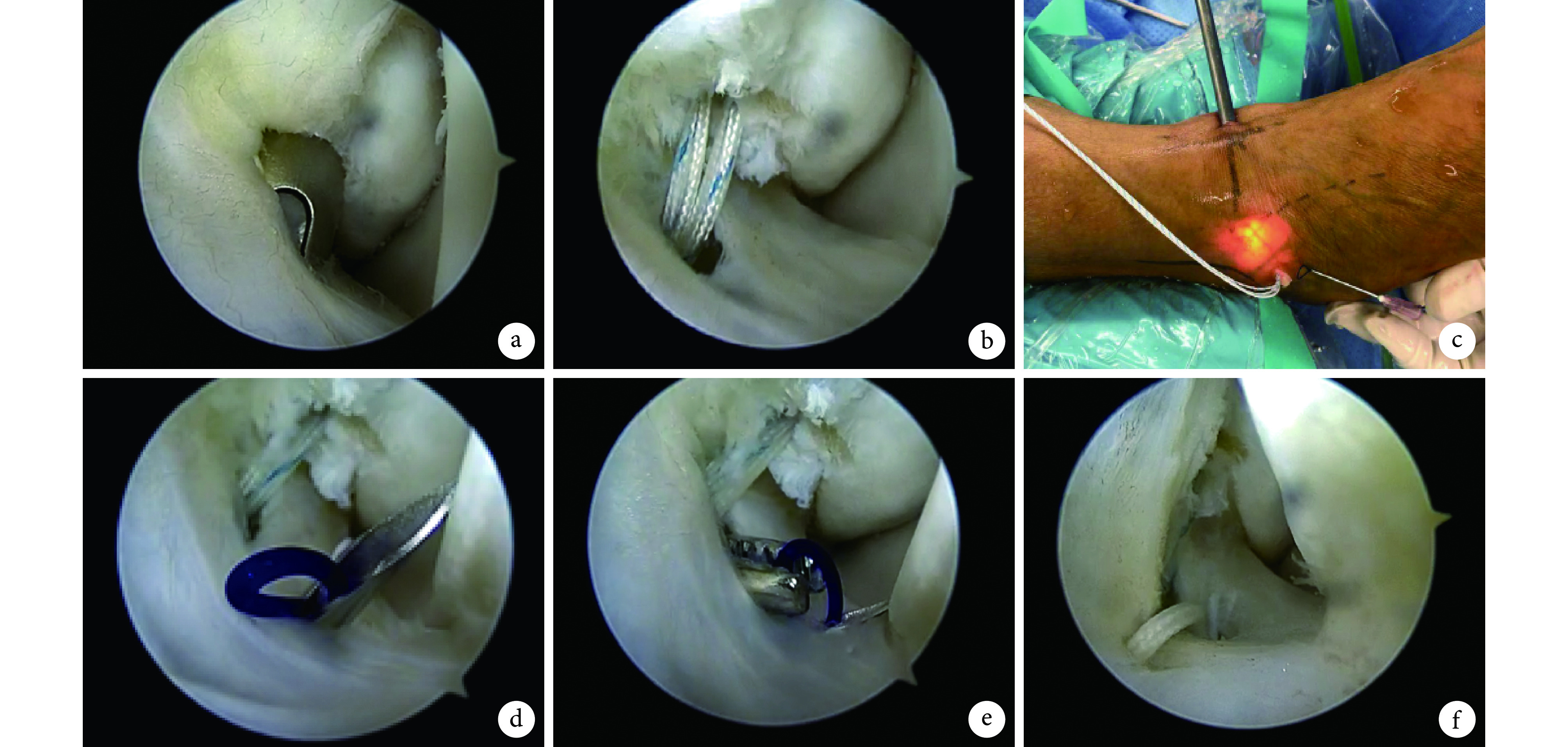
ATFL 缝合修复分为两步:第 1 步植入带缝线锚钉,在内侧中线入路辅助下,从辅助前外侧入路于 ATFL 腓骨附着点植入 1 枚双缝线可吸收骨锚钉(Smith & Nephew 公司,美国),锚钉直径 2.9 mm,并携带 2 根 2 号 ULTRABRAID 超强缝线;第 2 步将锚钉 2 根缝线穿过 ATFL 并打结固定于腓骨。具体操作步骤:将一针头从辅助前外侧入路前下方约 1 cm 处穿入,该针头携带 1 根 2-0 尼龙环,可在后续操作中引导锚钉缝线顺利穿过 ATFL。针头依次穿过皮肤、皮下组织进入踝关节腔,从下往上穿过 ATFL,然后用一抓握钳通过辅助前外侧入路进入夹住尼龙环后拔出,尼龙环引导锚钉 1 根缝线穿过 ATFL 后从辅助前外侧入路拔出;重复上述步骤,使锚钉另 1 根缝线穿过 ATFL 并加固伸肌下支持带(inferior extensor retinaculum,IER),最后缝线打结固定于 ATFL 腓骨附着点,注意两缝线间保持适当距离。探查踝关节稳定后缝合切口。见 图 1、2。
图 1.
The approaches of all-arthroscopic technique
全关节镜下 ATFL 锚钉修复手术入路示意图
a~c. 手术入路 MM:内侧中线入路 ACAL:辅助前外侧入路 TA:胫骨前肌腱 LM:外踝;d. 带缝线锚钉植入
a-c. Approaches MM: Medial midline approach ACAL: Accessory anterolateral approach TA: Tibialis anterior LM: Lateral malleolus; d. Implantation of anchor

图 2.
The procedure of all-arthroscopic repair of ATFL with anchors
全关节镜下 ATFL 锚钉修复操作示意图
a、b. 通过辅助前外侧入路于 ATFL 腓骨附着点植入带线锚钉;c. 于辅助前外侧入路前下方穿入针头;d. 针头进入关节腔后穿透 ATFL;e. 通过辅助前外侧入路拔出尼龙环;f. 将另一缝线穿透 ATFL 后于腓骨上打结固定
a, b. The anchor was placed at the fibular attachment point of ATFL through the accessory anterolateral approach; c. The needle was placed via the accessory anterolateral approach; d. The needle was penetrated the ATFL after entering the articular cavity; e. The nylon ring was pulled out via the accessory anterolateral approach; f. The other suture was penetrated the ATFL and fixed with top knot

1.2.2. 开放组
关节镜下探查并行骨赘磨除、滑膜清理及距骨软骨面修复(2 例 Ⅰ、Ⅱ型损伤行软骨修整术,5 例 Ⅲ、Ⅳ 型损伤行微骨折处理)等相应处理,患者体位及操作方法与关节镜组一致。镜下检查进一步明确患者均为腓骨端 ATFL 损伤且残留足量组织用于修复。然后采用改良开放式 Broström 入路,于腓骨远端前作一长 3~4 cm 切口,逐层切开,注意分离 ATFL 和 IER。在与关节镜组相同的腓骨远端位置(ATFL 腓骨附着点)植入 1 枚双缝线可吸收骨锚钉,直视下缝线固定 ATFL 于腓骨,加强固定 IER。
1.3. 术后处理及疗效评价指标
术后采用短腿石膏固定术肢,维持踝关节中立位。术后第 2 天开始等长收缩训练肌肉群;2 周后拆除石膏,更换为踝关节功能锻炼支具,开始部分负重,在康复师指导下行肌力训练及主、被动活动锻炼;6 周后开始平衡训练和耐力训练,并过渡至完全负重。
记录 2 组术中出血量、手术时间、术后恢复正常生活时间,以及随访期间并发症发生情况。术前以及术后 2 周、3 个月、末次随访时,摄踝关节正侧位 X 线片,测量前抽屉试验时距骨前移、内翻应力测试时距骨倾斜角;采用 KAF 评分、AOFAS 评分、JSSF 量表评分评价踝关节功能,末次随访时按各评级系统评分标准进行分级。
1.4. 统计学方法
采用 SPSS20.0 统计软件进行分析。计量资料以均数±标准差表示,组间比较采用独立样本 t 检验;计数资料组间比较采用 χ2 检验,等级资料采用秩和检验;检验水准 α=0.05。
2. 结果
两组患者均获随访,随访时间 24~30 个月,平均 26 个月。关节镜组手术时间、术中出血量及术后恢复正常生活时间均明显优于开放组,差异有统计学意义(P<0.05)。见表 1。关节镜组术后出现暂时性脚踝及足背麻木 2 例、线头反应 1 例,开放组上述并发症各发生 2 例;患者脚踝及足背麻木症状均自行缓解,线头反应经切口清创、取出线头后消失。其余患者切口均Ⅰ期愈合,无早期手术相关并发症发生。
表 1.
Comparison of clinical indicators between the two groups (
 )
)
两组患者临床指标比较(
 )
)
| 组别
Group |
例数
n |
手术时间(min)
Operation time (minutes) |
术中出血量(mL)
Intraoperative bleeding volume (mL) |
术后恢复正常生活时间(周)
Length of time for surgery recovery (weeks) |
| 关节镜组
Arthroscopic group |
35 | 24.6±5.8 | 8.7±2.6 | 5.4±1.7 |
| 开放组
Open group |
30 | 41.5±7.2 | 32.8±9.5 | 7.2±2.3 |
| 统计值
Statistic |
t=10.479
P=0.000 |
t=14.409
P=0.000 |
t=3.620
P=0.001 |
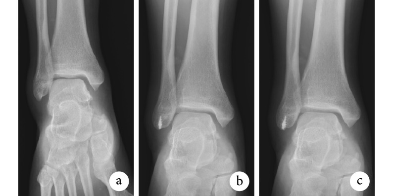
术后 2 周,关节镜组 AOFAS 评分、KAF 评分及 JSSF 量表评分均优于开放组,差异有统计学意义(P<0.05);术后 3 个月及末次随访时两组上述评分差异均无统计学意义(P>0.05)。见表 2。末次随访时,两组 KAF 评分、AOFAS 评分及 JSSF 量表评分等级差异均无统计学意义(P>0.05)。见表 3。X 线片测量,术后 2 周、3 个月及末次随访时两组距骨前移和距骨倾斜角差异均无统计学意义(P>0.05)。见表 4。随访期间两组均未出现锚钉拔出、踝关节不稳及其他并发症。见图 3。
表 2.
Comparison of functional scores at pre- and post-operation between the two groups (
 )
)
手术前后两组踝关节功能评分比较(
 )
)
| 组别
Group |
例数
n |
AOFAS 评分
AOFAS score |
KAF 评分
KAF score |
JSSF 量表评分
JSSF scale score |
|||||||||||
| 术前
Preoperative |
术后 2 周
Two weeks after operation |
术后 3
个月 Three months after operation |
末次随访
Last follow-up |
术前
Preoperative |
术后 2 周
Two weeks after operation |
术后 3
个月 Three months after operation |
末次随访
Last follow-up |
术前
Preoperative |
术后 2 周
Two weeks after operation |
术后 3
个月 Three months after operation |
末次随访
Last follow-up |
||||
| 关节镜组
Arthroscopic group |
35 | 63.8±11.3 | 79.5±6.4 | 87.7±4.2 | 93.4±5.7 | 67.9±12.2 | 77.2±8.8 | 83.5±6.9 | 89.3±9.8 | 70.6±14.7 | 87.8±8.4 | 90.8±7.6 | 97.2±6.6 | ||
| 开放组
Open group |
30 | 65.2±13.8 | 76.3±4.9 | 86.5±6.4 | 91.8±7.3 | 68.3±11.8 | 73.4±5.3 | 82.1±7.3 | 88.9±7.1 | 69.8±15.4 | 83.5±7.6 | 90.3±9.0 | 95.6±5.2 | ||
| 统计值
Statistic |
t=0.450
P=0.655 |
t=2.234
P=0.029 |
t=0.905
P=0.369 |
t=0.992
P=0.305 |
t=0.134
P=0.894 |
t=2.065
P=0.043 |
t=0.794
P=0.430 |
t=0.186
P=0.853 |
t=0.214
P=0.831 |
t=2.149
P=0.035 |
t=0.243
P=0.809 |
t=1.072
P=0.288 |
|||
表 3.
Comparison of functional grading at last follow-up between the two groups
两组末次随访时踝关节功能评分等级结果比较
| 组别
Group |
例数
n |
AOFAS 评分
AOFAS score |
KAF 评分
KAF score |
JSSF 量表评分
JSSF scale score |
|||||||||||
| 优
Excellent |
良
Good |
可
Poor |
差
Bad |
优
Excellent |
良
Good |
中
Poor |
差
Bad |
优
Excellent |
良
Good |
中
Poor |
差
Bad |
||||
| 关节镜组
Arthroscopic group |
35 | 24 | 9 | 2 | 0 | 15 | 18 | 2 | 0 | 25 | 9 | 1 | 0 | ||
| 开放组
Open group |
30 | 21 | 8 | 1 | 0 | 14 | 12 | 4 | 0 | 20 | 7 | 3 | 0 | ||
| 统计值
Statistic |
Z=−0.179
P=0.858 |
Z=−0.058
P=0.953 |
Z=−0.570
P=0.569 |
||||||||||||
表 4.
Comparison of the anterior displacement and the tilt angle of talus at pre- and post-operation between the two groups (
 )
)
手术前后两组距骨前移及距骨倾斜角比较(
 )
)
| 组别
Group |
例数
n |
距骨前移(mm)
Anterior displacement of talus (mm) |
距骨倾斜角(°)
Tilt angle of talus (°) |
|||||||
| 术前
Preoperative |
术后 2 周
Two weeks after operation |
术后 3 个月
Three months after operation |
末次随访
Last follow-up |
术前
Preoperative |
术后 2 周
Two weeks after operation |
术后 3 个月
Three months after operation |
末次随访
Last follow-up |
|||
| 关节镜组
Arthroscopic group |
35 | 8.6±2.2 | 2.9±0.4 | 3.1±0.5 | 3.2±0.4 | 8.8±2.9 | 3.2±0.6 | 3.3±0.9 | 3.3±0.8 | |
| 开放组
Open group |
30 | 8.9±2.7 | 2.8±0.4 | 3.2±0.3 | 3.3±0.5 | 9.5±3.3 | 3.0±0.5 | 3.1±0.3 | 3.1±0.4 | |
| 统计值
Statistic |
t=0.494
P=0.623 |
t=1.005
P=0.319 |
t=0.957
P=0.342 |
t=0.895
P=0.374 |
t=0.910
P=0.366 |
t=1.445
P=0.153 |
t=1.162
P=0.250 |
t=1.242
P=0.219 |
||
图 3.
Anteroposterior X-ray films of a 32-year-old female patient with right LIA in arthroscopic group
关节镜组患者,女,32 岁,右侧 LIA 踝关节正位 X 线片
a. 术前;b. 术后 2 周;c. 术后 1 年
a. Before operation; b At 2 weeks after operation; c. At 1 year after operation
3. 讨论
临床 LIA 手术治疗目的是最大程度恢复踝关节稳定性及功能,对于 ATFL 残留充分的患者可选择韧带修复。ATFL 修复不仅能尽可能维持原踝关节的解剖结构及生物力学[10-12],而且无需取用自体肌腱重建,减少创伤和术后并发症。近年国内外大量研究表明,开放及关节镜下 ATFL 修复术均安全、可行且效果良好[13-17],但是鲜有两者疗效对比报道。为此,我们进行了两种术式的临床比较研究。
全关节镜下手术为微创术式,无需在腓骨远端前作一长 3~4 cm 切口,仅作两个关节镜切口,有效减小了血管损伤,减少术中出血,同时也缩短了手术时间。本研究结果显示关节镜组手术时间及术中出血量均显著低于开放组。既往关节镜技术至少需要 3 个切口[7, 9, 18],在 ATFL 周围最小面积清创且保证全关节镜下手术能顺利完成前提下,结合以往经皮技术及皮肤切口延伸技术报道,本研究关节镜组选择了辅助前外侧和内侧中线两个手术入路,手术均顺利完成。切口小,ATFL 周围组织损伤也相应减小,术后粘连发生率降低,有利于患者关节功能恢复及尽早恢复正常生活。本研究关节镜组患者术后恢复正常生活时间较开放组明显缩短,术后 2 周时 AOFAS 评分、KAF 评分及 JSSF 量表评分均显著优于开放组。ATFL 修复术后存在失败风险,Petrera 等[19]对 49 例改良开放式 Broström 锚钉修复术患者进行随访,发现手术失败率约为 6%。Maffulli 等[20]对 38 例接受改良开放式 Broström 锚钉修复术的 LIA 患者进行平均 8.7 年的随访,结果 5 例(13%)患者复发。本研究两组患者均在关节镜下进一步明确为韧带残留量充足、状况良好,最终确定行残端韧带修复术,随访期间无手术失败及 LIA 复发。
经临床应用,我们认为两手术入路全关节镜下锚钉修复术也存在一定不足。首先,关节镜手术有一定学习曲线,术者达到熟练完成手术需要一定时间及经验积累。关节镜组患者中早期手术的 10 例手术时间略长(26~45 min),之后收治的 25 例患者手术时间显著缩短为 15~28 min。此外,全关节镜下 ATFL 锚钉修复有其适应证,患者合并严重疾病时常无法实施。全关节镜技术也存在损伤腓浅神经风险,术后关节镜组 2 例腓浅神经损伤,表现为暂时性脚踝及足背麻木,分析原因为辅助前外侧入路与腓浅神经走行较近。但有研究报道开放性手术切口也与腓浅神经靠近,易引起腓浅神经损伤[21-22],本研究开放组术后也有 2 例患者出现腓浅神经损伤症状,分析可能为 IER 在手术过程中对该神经造成了压迫或刺激。但是两组手术操作均在以往研究报道的安全区域[23]内进行,因此如何避免腓浅神经损伤有待进一步明确。既往研究认为加强缝合 IER 能增强韧带修复的稳定性,本研究两组均行 IER 加强固定。但 Maffulli 等[20]通过 9 年临床随访发现在未行 IER 加强情况下,无神经损伤发生。因此,全关节镜或开放手术中是否加强固定 IER 以及相应疗效也是下一步研究重点。
综上述,在 ATFL 残端充足前提下,全关节镜下与改良开放式 Broström 锚钉修复治疗 LIA 均能取得良好疗效。与改良开放式 Broström 锚钉修复相比,全关节镜下锚钉修复手术时间短、创伤小、恢复快,但当无法顺利行关节镜修复时,要及时调整手术方案,避免手术失败。另外,本研究随访时间较短,需要进一步随访评估远期疗效。
作者贡献:易刚负责实验设计及实施、数据收集整理及统计分析、起草文章,扶世杰负责实验设计及实施、对文章知识性内容作批评性审阅,杨静负责数据收集整理、图片处理及统计分析,汪国友起草文章、对文章的知识性内容作批评性审阅,刘洋、郭晓光、石杰负责数据和图片的收集整理,张磊负责实验设计、对文章的知识性内容作批评性审阅、相关经费支持。
利益冲突:所有作者声明,在课题研究和文章撰写过程中不存在利益冲突。经费支持没有影响文章观点和对研究数据客观结果的统计分析及其报道。
机构伦理问题:研究方案经西南医科大学附属中医医院医学伦理审查委员会批准(KY201806121)。
Funding Statement
四川省中医药管理局中医药科研专项(2018YY003);泸州市人民政府-西南医科大学科技战略合作项目(2018LZXNYD-ZK43);西南医科大学应用基础研究项目(2018-ZRQN-104);西南医科大学科技成果转化类项目(0903-00050003);泸州市院士工作站在建项目(20180101)
Chinese Medicine Research of Sichuan Provincial Administration of Traditional Chinese Medicine (2018YY003); Luzhou Municipal People’s Government-Southwest Medical University Science and Technology Strategic Cooperation Project (2018LZXNYD-ZK43); Applied Basic Research Project of Southwest Medical University (2018-ZRQN-104); Scientific and Technological Achievements Conversion Project of Southwest Medical University (0903-00050003); Academician Workstation Construction Project of Luzhou (20180101)
References
- 1.Pellegrini MJ, Sevillano J, Ortiz C, et al Knotless modified arthroscopic-Broström technique for ankle instability. Foot Ankle Int. 2019;40(4):475–483. doi: 10.1177/1071100718820341. [DOI] [PubMed] [Google Scholar]
- 2.Higashiyama R, Aikawa J, Iwase D, et al Anatomical arthroscopic anterior talofibular ligament and calcaneofibular ligament reconstruction using an autogenic hamstring tendon: safe creation of anatomical fibular tunnel. Arthrosc Tech. 2019;8(3):e215–e222. doi: 10.1016/j.eats.2018.10.016. [DOI] [PMC free article] [PubMed] [Google Scholar]
- 3.张言, 梁晓军 慢性踝关节外侧不稳的解剖修复治疗进展. 中国修复重建外科杂志. 2016;30(12):1551–1555. doi: 10.7507/1002-1892.20160319. [DOI] [PubMed] [Google Scholar]
- 4.Buerer Y, Winkler M, Burn A, et al Evaluation of a modified Broström-Gould procedure for treatment of chronic lateral ankle instability: A retrospective study with critical analysis of outcome scoring. Foot Ankle Surg. 2013;19(1):36–41. doi: 10.1016/j.fas.2012.10.005. [DOI] [PubMed] [Google Scholar]
- 5.Hassan S, Thurston D, Sian T, et al Clinical outcomes of the modified Broström technique in the management of chronic ankle instability after early, intermediate, and delayed presentation. J Foot Ankle Surg. 2018;57(4):685–688. doi: 10.1053/j.jfas.2017.12.003. [DOI] [PubMed] [Google Scholar]
- 6.江东, 胡跃林, 焦晨, 等 改良辅助入路全关节镜下修复踝关节外侧副韧带. 中国微创外科杂志. 2019;19(1):11–14. doi: 10.3969/j.issn.1009-6604.2019.01.004. [DOI] [Google Scholar]
- 7.Arroyo-Hernández M, Mellado-Romero M, Páramo-Díaz P, et al Chronic ankle instability: Arthroscopic anatomical repair. Rev Esp Cir Orthop Traumatol. 2017;61(2):104–110. doi: 10.1016/j.recot.2016.10.003. [DOI] [PubMed] [Google Scholar]
- 8.Yoo JS, Yang EA Clinical results of an arthroscopic modified Brostrom operation with and without an internal brace. J Orthop Traumatol. 2016;17(4):353–360. doi: 10.1007/s10195-016-0406-y. [DOI] [PMC free article] [PubMed] [Google Scholar]
- 9.Vega J, Guelfi M, Malagelada F, et al Arthroscopic all-inside anterior talofibular ligament repair through a three-portal and no-ankle-distraction technique. JBJS Essent Surg Tech. 2018;8(3):e25. doi: 10.2106/JBJS.ST.18.00026. [DOI] [PMC free article] [PubMed] [Google Scholar]
- 10.Dong P, Gu S, Jiang Y, et al All arthroscopic remnant-preserving reconstruction of the lateral ligaments of the ankle: A biomechanical study and clinical application. Biochem Biophys Res Commun. 2018;505(4):985–990. doi: 10.1016/j.bbrc.2018.10.041. [DOI] [PubMed] [Google Scholar]
- 11.Cottom JM, Baker JS, Richardson PE, et al A biomechanical comparison of 3 different arthroscopic lateral ankle stabilization techniques in 36 cadaveric ankles. J Foot Ankle Surg. 2016;55(6):1229–1233. doi: 10.1053/j.jfas.2016.07.025. [DOI] [PubMed] [Google Scholar]
- 12.Willegger M, Benca E, Hirtler L, et al Biomechanical stability of tape augmentation for anterior talofibular ligament (ATFL) repair compared to the native ATFL. Knee Surg Sports Traumatol Arthrosc. 2016;24(4):1015–1021. doi: 10.1007/s00167-016-4048-7. [DOI] [PMC free article] [PubMed] [Google Scholar]
- 13.Nakasa T, Nakashiro J, Adachi N Arthroscopic repair of anterior talofibular ligament using the pull-out technique for chronic ankle instability: Case report. J Foot Ankle Surg. 2019;58(1):156–160. doi: 10.1053/j.jfas.2018.05.016. [DOI] [PubMed] [Google Scholar]
- 14.Lopes R, Andrieu M, Cordier G, et al Arthroscopic treatment of chronic ankle instability: Prospective study of outcomes in 286 patients. Orthop Traumatol Surg Res. 2018;104(8S):S199–S205. doi: 10.1016/j.otsr.2018.09.005. [DOI] [PubMed] [Google Scholar]
- 15.Weng PW, Chen CY, Tsuang YH, et al A modified Broström repair with transosseous fixation for chronic ankle instability: a midterm followup study in soldiers. Indian J Orthop. 2018;52(3):315–321. doi: 10.4103/ortho.IJOrtho_265_16. [DOI] [PMC free article] [PubMed] [Google Scholar]
- 16.陈守勃, 华英汇 前距腓韧带修复术治疗踝关节外侧不稳定疗效分析. 中国修复重建外科杂志. 2017;31(4):427–431. doi: 10.7507/1002-1892.201611022. [DOI] [PMC free article] [PubMed] [Google Scholar]
- 17.张言, 梁晓军, 李毅, 等 改良 Brostöm-Gould 术单纯解剖修复距腓前韧带治疗慢性踝关节外侧不稳的中短期随访研究. 中华骨与关节外科杂志. 2016;9(5):427–431. [Google Scholar]
- 18.Batista JP, Del Vecchio JJ, Patthauer L Arthroscopic lateral ligament repair through two portals in chronic ankle instability. Open Orthop J. 2017;11:617–632. doi: 10.2174/1874325001711010617. [DOI] [PMC free article] [PubMed] [Google Scholar]
- 19.Petrera M, Dwyer T, Theodoropoulos JS, et al Short- to medium-term outcomes after a modified Broström repair for lateral ankle instability with immediate postoperative weightbearing. Am J Sports Med. 2014;42(7):1542–1548. doi: 10.1177/0363546514530668. [DOI] [PubMed] [Google Scholar]
- 20.Maffulli N, Del Buono A, Maffulli GD, et al Isolated anterior talofibular ligament Broström repair for chronic lateral ankle instability: 9-year follow-up. Am J Sports Med. 2013;41(4):858–864. doi: 10.1177/0363546512474967. [DOI] [PubMed] [Google Scholar]
- 21.Li H, Hua Y, Li H, et al Activity level and function 2 years after anterior talofibular ligament repair: A comparison between arthroscopic repair and open repair procedures. Am J Sports Med. 2017;45(9):2044–2051. doi: 10.1177/0363546517698675. [DOI] [PubMed] [Google Scholar]
- 22.de Leeuw PA, Golanó P, Sierevelt IN, et al The course of the superficial peroneal nerve in relation to the ankle position: anatomical study with ankle arthroscopic implications. Knee Surg Sports Traumatol Arthrosc. 2010;18(5):612–617. doi: 10.1007/s00167-010-1099-z. [DOI] [PMC free article] [PubMed] [Google Scholar]
- 23.Santos FF, Santos NR. Arthroscopic treatment of lateral ankle instability. Is there a safe zone? An anatomic study. Foot and Ankle Surgery, 2018. https://doi.org/10.1016/j.fas.2018.11.011.


