Keywords: action potential generation, A-fiber, C-fiber, potassium channel, vagal afferent nerve
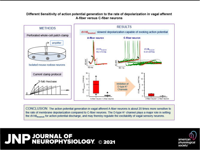
This study demonstrates that the action potential discharge in vagal afferent A-fiber neurons is about 20 times more sensitive to the rate of membrane depolarization compared to C-fiber neurons. The sensitivity of action potential generation to the depolarization rate in vagal sensory neurons is independent of the intensity of current stimuli but nearly abrogated by inhibiting the D-type potassium channel. These findings help better understand the mechanisms that control the activation of vagal afferent nerves.
Abstract
The vagal afferent nerves innervate the visceral organs and convey sensory information from the internal environment to the central nervous system. A better understanding of the mechanisms controlling the activation of vagal afferent neurons bears physiological and pathological significance. Although it is generally believed that the magnitude and the rising rate of membrane depolarization are both critical for the action potential generation, no direct or quantitative evidence has been documented so far for the sensitivity of vagal afferent neuron activation to the rate of depolarization and for its underlying ionic mechanisms. Here, by measuring the response of mouse nodose neurons to the suprathreshold current stimuli of varying rising rates, the slowest depolarization capable of evoking action potentials, the rate-of-depolarization threshold (dV/dtthreshold), was determined and found to be ∼20 fold higher in the A-fiber neurons compared to the C-fiber neurons classified based on the capsaicin responsiveness and characteristics of action potential waveforms. Moreover, although the dV/dtthreshold varied substantially among individual neurons, it was not different in any one neuron in response to different intensities of current stimuli. Finally, inhibition of low-threshold activated D-type potassium current (IK.D) by α-dendrotoxin or low concentration of 4-aminopyrydine nearly abrogated the sensitivity of action potential generation to the depolarization rate. Thus, the depolarization rate is an important independent factor contributing to the control of action potential discharge, which is particularly effective in the vagal afferent A-fiber neurons. The IK.D channel may regulate the excitability of vagal sensory neurons by setting the dV/dtthreshold for action potential discharge.
NEW & NOTEWORTHY This study demonstrates that the action potential discharge in vagal afferent A-fiber neurons is about 20 times more sensitive to the rate of membrane depolarization compared to C-fiber neurons. The sensitivity of action potential generation to the depolarization rate in vagal sensory neurons is independent of the intensity of current stimuli but nearly abrogated by inhibiting the D-type potassium channel. These findings help better understand the mechanisms that control the activation of vagal afferent nerves.
INTRODUCTION
The vagal afferent nerves innervate the visceral organs and convey sensory information from the internal environment to the central nervous system to initiate homeostatic response and reflexes (1). Accordingly, the vagal sensory system plays an important role in the dynamic regulation of visceral functions and in the host defense in response to inhaled or ingested harmful chemicals and pathogens. In visceral inflammatory diseases, however, sustained and exaggerated activation of vagal afferent nerves by various nociceptive stimuli can lead to unpleasant symptoms including diffusive pain, dyspnea and cough, and aberrant reflex activities (2). A better understanding of the mechanisms that control the activation of vagal afferent nerves may help develop novel therapeutics for more effective relief of patients’ suffering.
The vagal afferent nerves are broadly divided into fast-conducting myelinated A-fibers and slowly conducting unmyelinated C-fibers. It is often the case that the terminals of A-fibers are low-threshold mechanosensors, whereas the C-fibers are chemosensors (e.g., sensitive to inflammatory mediators) and high-threshold mechanosensors, exhibiting properties consistent with nociceptors (3, 4). The cell somas of vagal afferent neurons are situated in the vagal sensory ganglia (nodose and jugular ganglia). For a sensory neuron to be activated (i.e., firing action potentials or spikes), the membrane depolarization caused by various stimuli (i.e., the generator potential) must reach the threshold voltage at which a fast and regenerative Na+ influx via the voltage-gated Na+ channels occurs and initiates the upstroke of the action potential (4). Although it is generally believed that the magnitude and the rising rate of the membrane depolarization are both critical for evoking action potential discharge, no direct or quantitative experimental evidence has been documented so far pertaining to whether and how much the activation of vagal afferent neurons is sensitive to the rising rate of depolarization. In fact, only a few studies carried out in other types of neurons, the large amphibian nerve fibers (5, 6) and brainstem auditory neurons (7, 8), have investigated quantitatively and directly the dependence of action potential generation on the rate of depolarization. One of these studies has revealed that not all types of neurons were sensitive to the rate of depolarization (7).
The D-type potassium current (IK.D) is mediated by members of Kv1 family of voltage-gated potassium channels and highly sensitive to α-dendrotoxin (α-DTX) and 4-aminopyridine (4-AP) (9). Because of its unique biophysical properties (low-threshold activation, fast activation, and very slow inactivation), IK.D plays a key role in regulating the neuronal excitability, spike firing pattern, and synaptic transmission (10–12). Studies in mouse brainstem auditory neurons have also shown that the IK.D channel is the main ion channel responsible for the strong dependence of action potential generations on the rate of depolarization observed in octopus and bushy cells (7, 8).
The present study aimed to investigate quantitatively the sensitivity of action potential generation to the rising rate of depolarization in mouse vagal afferent A-fiber and C-fiber neurons classified based on the capsaicin responsiveness and characteristics of action potential waveforms and to examine whether IK.D channels play a role in determining the dependence of vagal sensory neuron activation on the rate of depolarization.
MATERIALS AND METHODS
Ethical Approval
The experiments in mice were carried out in accordance with the protocol approved by The Johns Hopkins Animal Care and Use Committee.
Isolation of Mouse Nodose Neurons
C57BL/6 mice (8–14 wk, male) from The Jackson Laboratory were used in this study. Mice were euthanized by CO2 inhalation and subsequent exsanguinations. Both sides of jugular/nodose ganglia were dissected and cleared of adhering connective tissues in ice-cold calcium- and magnesium-free phosphate-buffered saline (pH 7.4). Since the jugular and nodose neurons are distinct in embryonic origin and phenotypes (13), this study will focus on nodose neurons. Accordingly, the lower 2/3 of nodose ganglion (free of jugular component, Ref. 13) were cut out and put into 1 mL of HBSS containing 1.5–2 mg type 1 collagenase and 2 mg dispase II. The enzymatic digestion proceeded at 37°C for 60 min. At the end of 30, 45, and 60 min, the ganglial tissue was gently triturated for a few times using fire-polished Pasteur pipettes. To end the digestion, the 1-mL enzymatic solution containing the dissociated neurons was transferred to 10 mL of prewarmed Leibovitz’s L-15 medium supplemented with 10% of FBS and centrifuged at 600 g for 2 min. After one to two more washes with the same medium, the pellet was resuspended in 200 µL of the medium, then pipetted onto eight cover glasses (25 µL each) pretreated with poly-d-lysine (0.1 mg·mL−1) and laminin (0.005 mg·mL−1) and incubated at 37°C for 2 h. After the neurons attached to the cover glass, 2 mL of fresh L-15 medium supplemented with 10% of FBS was added to the cover glass, each placed in one 35-mm Petri dish. The isolated neurons were maintained at 37°C overnight and used for recordings within 24 h after isolation.
Electrophysiological Recordings
Amphotericin B-perforated whole cell patch clamp technique was employed to record the membrane potential and currents using an Axopatch 200B amplifier interfaced with Axon Digidata 1550 A and driven by pCLAMP 10 software (Molecular Devices, Sunnyvale, CA). The recording pipettes were pulled (P-97, Sutter) and fire-polished (MF-830, Narishige) to have a resistance between 1.8 to 2.4 MΩ. Bath solution contained (mM): NaCl 136, KCl 5.4, MgCl2 1, CaCl2 1.5, HEPES 10, and glucose 10 with pH adjusted to 7.35 with NaOH. Pipette solution contained (mM): KCl 30, K-gluconate 115, and HEPES 10 with pH adjusted to 7.2 with KOH. The amphotericin B stock was freshly prepared in DMSO (3%) and sonicated for 10 min before experiments. The well-dissolved amphotericin B was then added to the pipette solution to have the final concentration of 300 μg·mL−1 and used within 2 h. The junction potential (−13.3 mV estimated using Clampex calculator) was corrected offline. The membrane potential in response to the current clamp protocols (detailed in result section where appropriate) was recorded on I-clamp fast mode at least 5 min after an adequate whole cell (access resistance < 20 MΩ) was established. To measure the whole cell capacitance (WCC) and input resistance (Rinput), the neuron was held at −65 mV on voltage-clamp mode and the current responses to 5 mV depolarization or hyperpolarization were recorded. At the end of experiments, the responsiveness of neurons to 1 µM capsaicin was examined by recording the current response from a holding potential of −70 mV. Capsaicin usually generates a large inward current in capsaicin-sensitive neurons. The signals were sampled at 10 kHz and filtered at 2 kHz. Experiments were performed at room temperature.
Chemicals and Reagents
HBSS, Leibovitz’s L-15 medium, and heat-inactivated FBS were purchased from Gibco/life Technologies. Dispase II was from Roche Diagnostics. Type 1 collagenase, amphotericin-B, 4-AP, and capsaicin were from Sigma. 4-AP was directly dissolved in the bath solution before adjusting the pH to 7.35. Capsaicin stock (10−2 M) was prepared in ethanol. α-DTX was purchased from Alomone Labs and the stock solution (10−4 M) was prepared in Millipore water. The α-DTX and capsaicin stocks were stored at −20°C and added to the bath solution before experiments.
Data Analyses
Data were analyzed using Clampfit 10 of the pCLAMP 10 software. The rising phase of depolarization preceding the onset of the action potential was fitted to a linear function, and the slope gave the rate of depolarization (dV/dt). The WCC was obtained by dividing the integral of the capacitance transient by the voltage step (from −65 to −60 or to −70 mV). The Rinput was calculated by dividing the voltage step (from −65 to −60 mV) by the amplitude of steady-state current measured at the end of 10-ms voltage pulse. The action potential firing threshold (APthreshold) was measured by differentiating the action potential with respect to time and defined as the voltage at which the deflection of the differentiation signal is greater than zero. The action potential amplitude (APA) was measured as the voltage difference between the resting membrane potential (RMP) and the peak of the upstroke. The amplitude of AHP was measured as the voltage difference between the RMP and the peak of afterhyperpolarization (AHP). The action potential duration (APD100) was measured as the time period from the onset of action potential to the point where the repolarization was complete (i.e., returning to RMP). APD50 was measured as the action potential duration at the point of 50% height of APA.
Statistical Analyses
All statistical analyses were performed using SigmaPlot software. Pooled data are expressed as mean ± SD. The statistical significance of differences between two means was determined by using either paired or unpaired Student's t test, as appropriate. In the cases that the normality test failed, Wilcoxon signed-rank test or Mann–Whitney rank sum test was used as appropriate. The significance of differences between multiple means was evaluated by one-way ANOVA or one-way repeated-measures ANOVA. Holm–Sidak or Dunn’s test as a post hoc analysis was performed for multiple pairwise comparisons.
RESULTS
Generation of Action Potentials in Mouse Nodose Neurons Depends on the Rate of Depolarization
The dependence of action potential generation on the rising rate of depolarization of the generator potential was investigated in patch-clamped nodose neurons on current clamp mode. The different rates of depolarization were produced by applying a family of depolarizing current ramps from 0 to a constant suprathreshold level with varying slopes (by varying the duration of ramps systemically as depicted in Fig. 1A). Since the minimal depolarizing current needed to evoke an action potential (defined as rheobase) is different in different neurons, we first measured the rheobase in each neuron by applying 25-ms depolarizing current steps at increasing intensity. We then set the amplitude of the slope-varying current ramps to 2-, 3-, 5- or 10-fold rheobase and examined the membrane potential response to the injected depolarizing currents with different slopes and amplitudes. One example of such experiments is given in Fig. 1. The four families of voltage response to the slope-varying current ramp sets of four different amplitudes shown in Fig. 1A were recorded from the same neuron. Ramps produced linearly rising depolarizations that led to action potential firings or tapered off. Regardless of the amplitude of the current ramp commands, faster depolarizations resulted in action potential discharge while slower depolarizations did not. The linear fits for the slowest depolarization that was capable of eliciting an action potential (the thickened black traces) in each family of voltage response are illustrated in the corresponding insets. The slopes of the curve (also given in the insets) represent the minimal rate of depolarization needed to evoke action potential firing in this neuron, which is defined as the rate-of-depolarization threshold (dV/dtthreshold). It is evident that the dV/dtthreshold is quite similar when the intensity of the current stimulus varied from 2- to 10-fold rheobase. The same type of experiments was performed in a total of six nodose neurons (Fig. 1B). Although the dV/dtthreshold varied substantially among individual neurons, it was not significantly different in any one neuron responding to different magnitudes of depolarizing currents. The dV/dtthreshold independent of the stimulus intensity was further confirmed in a larger population of neurons (n = 18 neurons) tested with current ramps set to 2-, 3-, and 5-fold rheobase (Fig. 1C). These results suggest that the rate-of-depolarization threshold is an intrinsic property of mouse nodose neurons.
Figure 1.
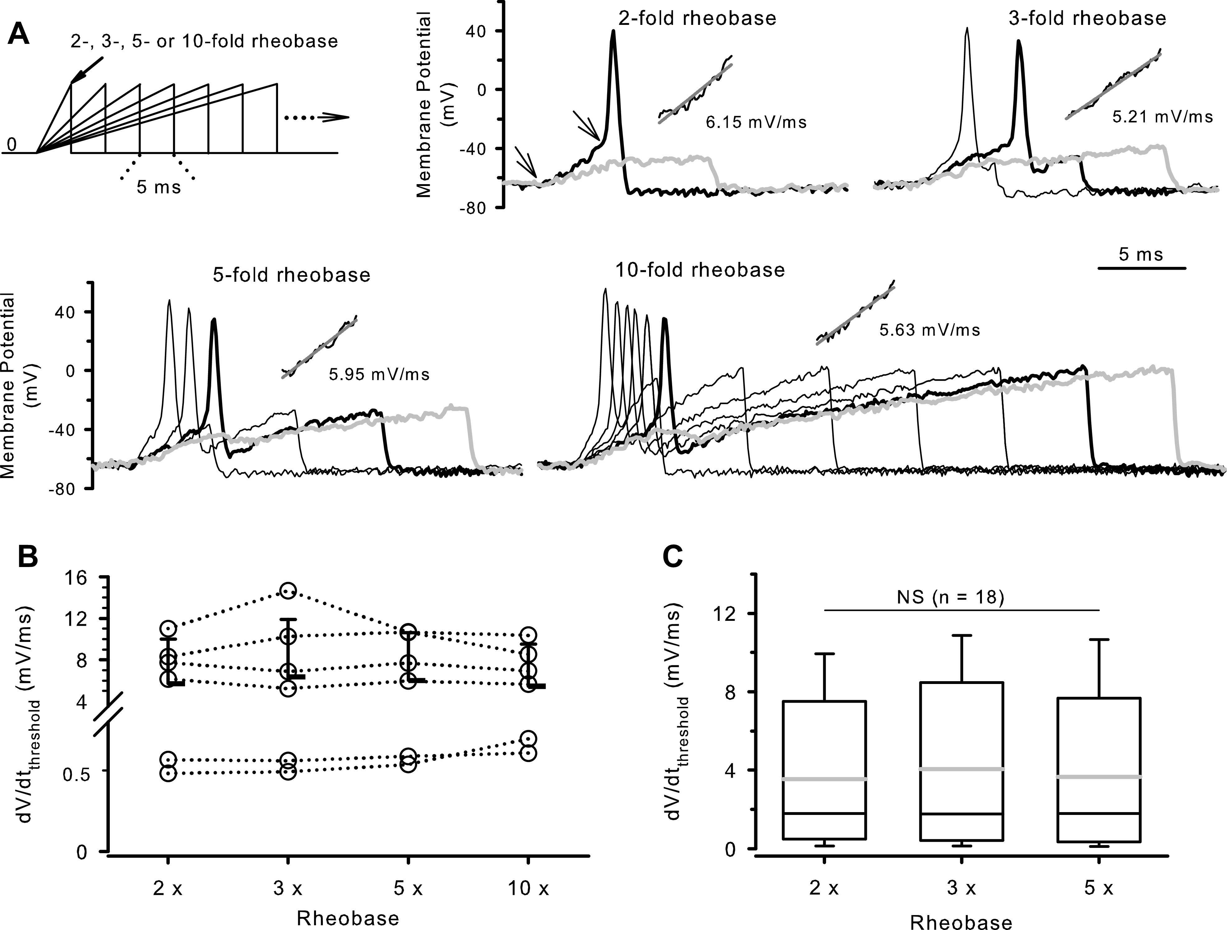
The rate-of-depolarization threshold in mouse nodose neurons is independent of the depolarizing current amplitude. A: schematic current clamp protocol and representative recordings of membrane potential obtained from one nodose neuron in response to slope-varying current ramps from zero to 2-, 3-, 5-, or 10-fold rheobase as indicated. The rheobase in this neuron is 150 pA. The current ramp duration started at 5 ms and increased with an increment of 5 ms so that the slope of the ramps was decreasing systemically. The ramp commands were applied at 0.33 Hz until at least three consecutive ramps were unable to elicit an action potential. For each family of voltage responses, the thickened black trace represents the slowest depolarization capable of eliciting an action potential, and the gray trace the first of the three consecutive depolarizations that failed to evoke an action potential. The inset in each panel depicts a fit of the depolarizing phase preceding the onset of action potentials (the region between two arrows illustrated on the top middle panel) to the linear function for the corresponding black thickened traces. The slopes of the curves (gray solid lines), also given in the insets, represent the minimal rates of depolarization needed to evoke an action potential (dV/dtthreshold) in response to different amplitudes of depolarizing currents in this neuron. B: dV/dtthreshold plotted against the amplitude of current ramps between 2- and 10-fold rheobase. The interconnected open circles show the results obtained from individual neurons. The mean ± SD pooled from the six neurons are shown with horizontal bars (P = 0.615 by Friedman repeated-measures analysis of variance on ranks). C: box and whisker plot of dV/dtthreshold obtained from a larger population of neurons (n = 18 neurons) examined with the current ramp amplitude varying between 2- and 5-fold rheobase. The gray lines inside the boxes indicate the mean. NS: P = 0.411 by Friedman repeated-measures analysis of variance on ranks. dV/dtthreshold, rate-of-depolarization threshold.
Different Rate-of-Depolarization Threshold in Nodose A-Fiber and C-Fiber Neurons
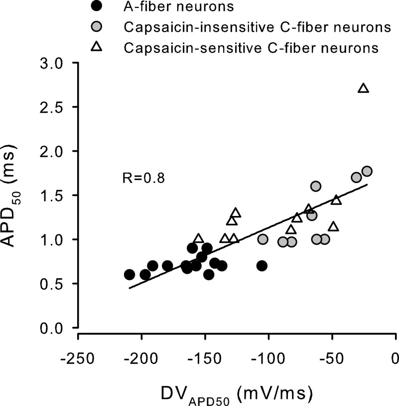
Since capsaicin is a well-known direct chemical stimulator of C-fibers (but not A-fibers in the mouse vagal afferent nerves), we classified the nodose neurons on which the capsaicin responsiveness was tested at the end of experiments into the capsaicin-insensitive and capsaicin-sensitive groups to examine whether the large variability in the dV/dtthreshold observed among nodose neurons is at least partially due to the phenotypic difference of different neuron types. The dV/dtthreshold was measured using a family of current ramps from zero to 3-fold rheobase with varying slopes. We found that the dV/dtthreshold was significantly higher in the capsaicin-insensitive neurons compared to the capsaicin-sensitive neurons (4.84 ± 4.69 mV/ms, n = 23 neurons, vs. 0.30 ± 0.29 mV/ms, n = 11 neurons. P = 0.003 by t test). In other words, the capsaicin-sensitive nodose neurons (presumably C-fiber neurons) can be activated by depolarizations with much slower rates compared to the capsaicin-insensitive neurons. Moreover, we noticed that the value of dV/dtthreshold exhibited a wider range (from 0.09 to 13.8 mV/ms) among the capsaicin-insensitive neurons compared to the capsaicin-sensitive C-fiber neurons (from 0.02 to 0.51 mV/ms). It has been reported that, in mouse, a large portion of nodose nerve fibers innervating the lungs are not sensitive to capsaicin. These include, as one might expect, vagal A-fibers, but C-fibers were also found in this capsaicin-insensitive group (14). It is therefore likely that some neurons in our capsaicin-insensitive group may in fact be C-fiber neurons. To explore this possibility, we performed a detailed analysis of the action potential waveform which has been shown to correlate well with the vagal afferent conduction velocities and fiber types in rats (15). The bone fide C-fiber neurons have a broader action potential than A-fiber neurons, mainly due to a slower repolarization as indicated by a lower downstroke velocity (DV) of action potentials (15, 16). Consistent with this, we found that the capsaicin-sensitive (C-fiber) neurons isolated from mouse nodose ganglia had broad action potentials, with the APD50 being consistently ≥1 ms. In contrast, 14 of the 23 capsaicin-insensitive neurons had narrow action potentials with an APD50 ≤ 0.9 ms. These neurons also had a significantly greater repolarizing velocity compared to capsaicin-sensitive neurons: −176.4 ± 23.5 versus −122.7 ± 31.2 mV/ms for DVmax, and −161.2 ± 26.9 versus −92.9 ± 43.3 mV/ms for DVAPD50 (Table 1). The other nine of the 23 capsaicin-insensitive neurons exhibited broader action potentials with APD50 ranging from 0.97 to 1.77 ms, and a slower repolarization with DVAPD50 being in the range of that for capsaicin-sensitive neurons as illustrated in Fig. 2. We therefore classified the capsaicin-insensitive neurons that had both broader action potentials and a lower downstroke velocity as capsaicin-insensitive C-fiber neurons (Fig. 2) and named the capsaicin-insensitive neurons exhibiting briefer action potentials and a fast repolarization the A-fiber neurons by convention. This classification was further supported by the observation that other properties like RMP, APthreshold, and rheobase in the capsaicin-insensitive and capsaicin-sensitive C-fiber neurons were closely similar but significantly different from those found in the A-fiber neurons (Table 1).
Table 1.
Passive electrical properties and characteristics of action potential waveform in mouse nodose A-fiber and C-fiber neurons
| Parameters | CAP-Insensitive A-Fiber Neurons | CAP-Insensitive C-Fiber Neurons | CAP-Sensitive C-Fiber Neurons | One-Way ANOVA |
|---|---|---|---|---|
| Number of neurons | 14 | 9 | 11 | |
| WCC, pF | 28.5 ± 7.6 | 27.5 ± 7.1 | 28.6 ± 8.8 | P = 0.945 |
| Rinput, MΩ | 313.2 ± 297.0 | 642.2 ± 418.4 | 1003.1 ± 842.8* | P = 0.017 |
| Rheobase, pA | 366 ± 243 | 125 ± 108* | 89 ± 144* | P = 0.001 |
| RMP, mV | −66.7 ± 3.3 | −63.4 ± 1.7* | −63.2 ± 2.0* | P = 0.002 |
| APA, mV | 106.2 ± 12.0 | 114.6 ± 15.0 | 121.5 ± 10.8* | P = 0.017 |
| AHP amplitude, mV | 12.7 ± 5.3 | 13.1 ± 3.7 | 15.9 ± 2.4 | P = 0.166 |
| APthreshold, mV | −38.9 ± 3.5 | −34.4 ± 4.3* | −33.2 ± 4.0* | P = 0.002 |
| APD100, ms | 1.57 ± 0.23 | 3.20 ± 1.09* | 3.19 ± 0.95* | P < 0.001 |
| APD50, ms | 0.71 ± 0.09 | 1.25 ± 0.34* | 1.31 ± 0.48* | P < 0.001 |
| UVmax, mV/ms | 252.7 ± 37.3 | 220.8 ± 39.9 | 203.6 ± 50.6* | P = 0.023 |
| DVmax, mV/ms | −176.4 ± 23.5 | −104.2 ± 12.8* | −122.7 ± 31.2* | P < 0.001 |
| UVAPD50, mV/ms | 143.8 ± 41.7 | 134.4 ± 26.2 | 135.9 ± 36.3 | P = 0.796 |
| DVAPD50, mV/ms | −161.2 ± 26.9 | −64.0 ± 26.1* | −92.9 ± 43.3* | P < 0.001 |
The data are expressed as means ± SD. AHP, afterhyperpolarization; APA, action potential amplitude; APD50, action potential duration at the point of 50% height of action potential amplitude; APD100, time period from the onset of action potential to the point where the repolarization was complete; APthreshold, action potential firing threshold; CAP, capsaicin; DVAPD50, downstroke velocity at the point of APD50; DVmax, maximal downstroke velocity; Rinput, input resistance; RMP, resting membrane potential; UVAPD50, upstroke velocity at the point of APD50; UVmax, maximal upstroke velocity; WCC, whole cell capacitance. All action potential parameters are averaged from three consecutive action potentials.
*P < 0.05 vs. capsaicin-insensitive A-fiber neuron group (post hoc Holm–Sidak test for pairwise comparison).
Figure 2.
Scatter plot of APD50 against downstroke velocity at APD50 (DVAPD50). Each symbol represents one mouse nodose neuron (n = 34 neurons). Different symbols represent different classes of nodose neurons as indicated. The solid line presents the result of fitting all data points to a linear function (R = 0.8). APD50, action potential duration at the point of 50% height of action potential amplitude.
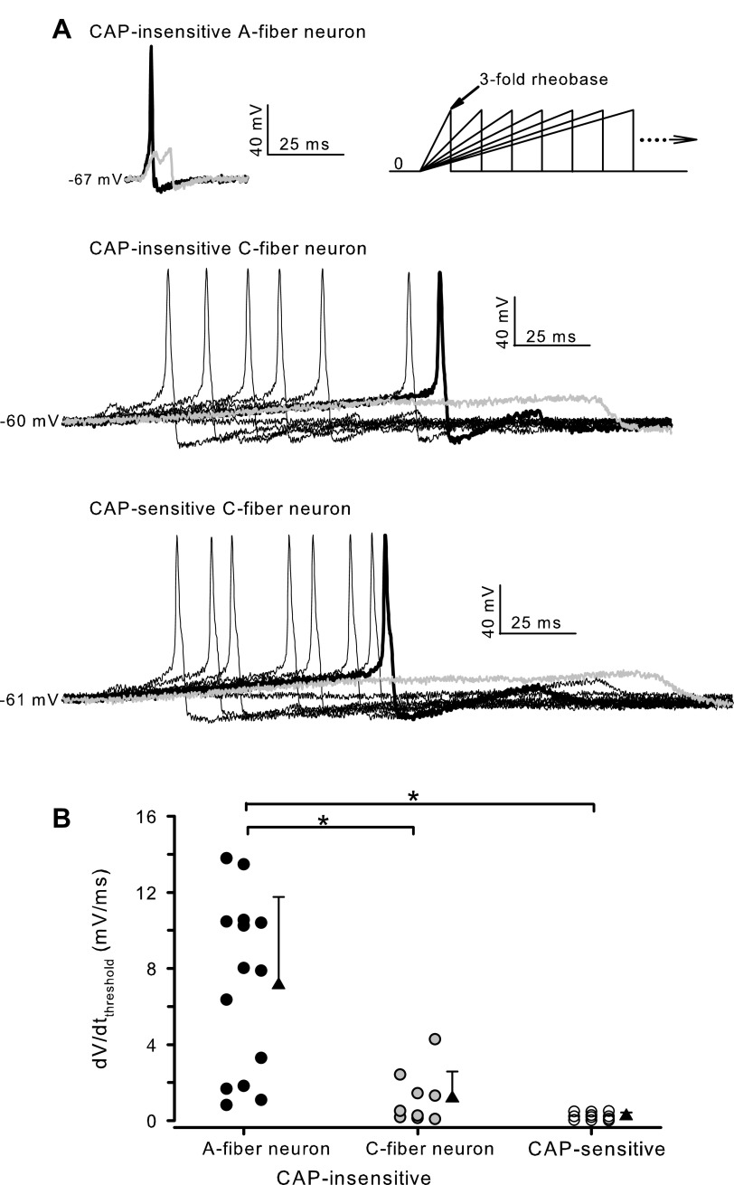
Figure 3A shows the representative recordings of membrane potentials obtained from one capsaicin-insensitive A-fiber, one capsaicin-insensitive C-fiber, and one capsaicin-sensitive C-fiber neuron in response to the current ramp protocol. The dV/dtthreshold in these three neurons was found to be 5.35 mV/ms, 0.196 mV/ms, and 0.229 mV/ms, respectively, with the A-fiber neuron having a much higher threshold. It is evident from Fig. 3B that most capsaicin-insensitive neurons that had lower dV/dtthreshold belonged to the capsaicin-insensitive C-fiber neuron group. The averaged dV/dtthreshold was significantly lower in C-fiber neurons, irrespective of the responsiveness to capsaicin, compared to that found in A-fiber neurons. The pooled dV/dtthreshold was 7.14 ± 4.63 mV/ms (n = 14 neurons), 1.18 ± 1.4 mV (n = 9 neurons), and 0.23 ± 0.19 mV/ms (n = 11 neurons) for A-fiber, capsaicin-insensitive C-fiber, and capsaicin-sensitive C-fiber neurons, respectively.
Figure 3.
Rate-of-depolarization threshold in different neuron types. A: representative recordings of membrane potential obtained from two capsaicin (CAP)-insensitive and one CAP-sensitive nodose neurons in response to the current clamp protocol schematically illustrated on the top right. For the CAP-insensitive A-fiber neuron, the duration of current ramps started at 5 ms and increased systemically with an increment of 5 ms. For the CAP-insensitive C-fiber neuron and the CAP-sensitive neuron, the duration of currents ramps started from 10 ms and increased with an increment of 20 ms. For all recordings, the thickened black trace represents the slowest depolarization capable of eliciting an action potential, and the gray trace the first of the three consecutive depolarizations that failed to evoke an action potential. B: dV/dtthreshold obtained in three different groups of neurons as indicated. For each group, the circles on the left give the values obtained from individual neurons, and the filled triangles and error bars give the mean ± SD (n = 14, 9, and 11 neurons for A-fiber, Cap-insensitive C-fiber, and Cap-sensitive C-fiber group, respectively). *P < 0.001 (one-way analysis of variance followed by Holm–Sidak test for pairwise comparisons). dV/dtthreshold, rate-of-depolarization threshold.
Longer suprathreshold current ramps sometimes induced multiple action potentials and this was more frequently observed in the capsaicin-sensitive neurons (see Fig. 4A). Five of 11 capsaicin-sensitive neurons responded with three to four action potentials to longer current stimuli and these neurons had a lower dV/dtthreshold compared to the other six neurons showing one to two action potentials (0.08 ± 0.08 vs. 0.36 ± 0.14 mV/ms, P = 0.004 by t test). Suprathreshold current ramps induced double action potentials in six of 14 A-fiber neurons and six of nine capsaicin-insensitive C-fiber neurons, but only two of 14 A-fiber neurons and one of nine capsaicin-insensitive C-fiber neurons generated three to four action potentials in response to longer stimuli.
Figure 4.
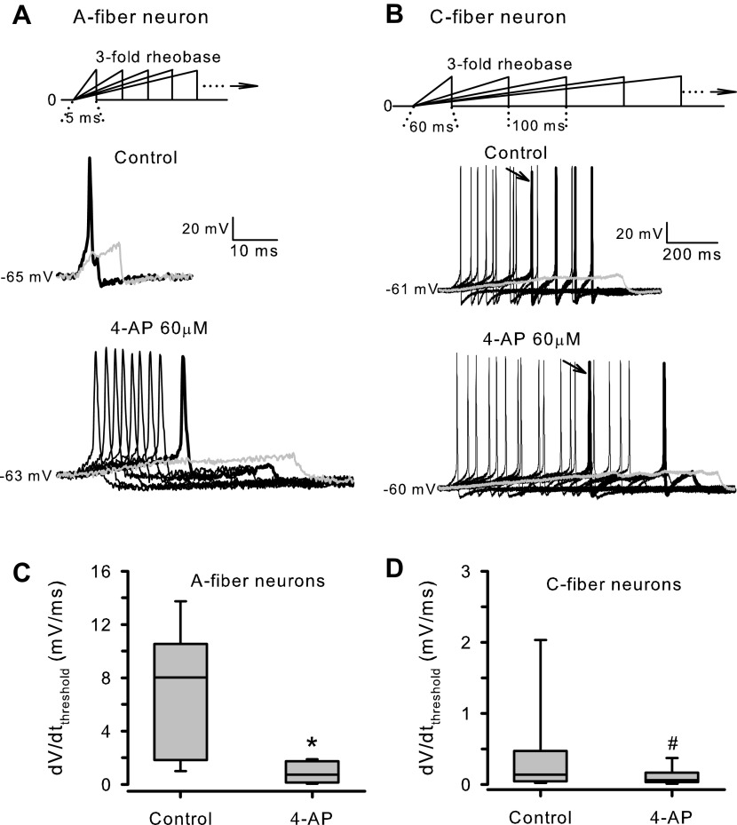
Effects of 60 µM 4-AP on the rate-of-depolarization threshold. A and B: representative membrane potential response recorded from one CAP-insensitive A-fiber neuron and one CAP-sensitive C-fiber neuron, respectively, at baseline (Control) and in the presence of 60 µM 4-AP. The current clamp protocols shown above the recordings consist of a series of current ramps to threefold rheobase with varying durations. For the A-fiber neuron, the ramp duration started at 5 ms and increased systematically with an increment of 5 ms. For the C-fiber neuron, the recordings were made with the current ramp duration stating at 10 ms then increasing with an increment of 20 ms, but only the responses to several current ramps (with starting duration and increments being indicated in the protocol) are shown for the sake of clarity. In this C-fiber neuron longer ramps evoked two to four action potentials. Please note the different time scales used to present the recordings shown in A and B. For each recording, the gray trace depicts the first of the three consecutive depolarizations that failed to elicit an action potential, and the thickened black trace the slowest depolarization capable of evoking action potential firing. The arrows in B indicate the first action potential in a train induced by the slowest depolarization. C: box and whisker plot of dV/dtthreshold measured from A-fiber neurons (n = 11 neurons) in the absence (Control) and presence of 60 µM 4-AP. *P < 0.001 (paired t test). D: box and whisker plot of dV/dtthreshold measured from C-fiber neurons (n = 11 neurons) in the absence (Control) and presence of 60 µM 4-AP. #P < 0.001 (Wilcoxon signed rank test). CAP, capsaicin; dV/dtthreshold, rate-of-depolarization threshold; 4-AP, 4-aminopyridine.
Effects of IK.D Inhibition by 4-AP on the Rate-of-Depolarization Threshold in Mouse Nodose Neurons
The low-threshold, fast-activating IK.D is characterized by its sensitivity to α-DTX and micro molar concentrations of 4-AP. 4-AP at 30–100 µM has been used to successfully and specifically inhibit this current in sensory neurons including nodose neurons (11, 17). Here, we examined the effects of 60 µM 4-AP on the dV/dtthreshold to determine whether and how much IK.D is responsible for the sensitivity of action potential generation to the rate of depolarization observed in mouse nodose neurons (Fig. 4). In this analysis, we pooled the data obtained from the capsaicin-insensitive C-fiber neurons (n = 3) and the capsaicin-sensitive C-fiber neurons (n = 8) into one C-fiber neuron group. In the presence of 4-AP, more slowly rising depolarizations became effective in evoking action potentials in both A-fiber and C-fiber neurons (Fig. 4A). In the A-fiber neurons, 4-AP led to nearly a 10-fold decrease in the sensitivity to the rate of depolarization; the dV/dtthreshold decreased from 7.46 ± 4.79 to 0.82 ± 0.73 mV/ms. In the C-fiber neurons, the dV/dtthreshold was reduced from 0.38 ± 0.69 to 0.12 ± 0.12 mV/ms (Fig. 4B).
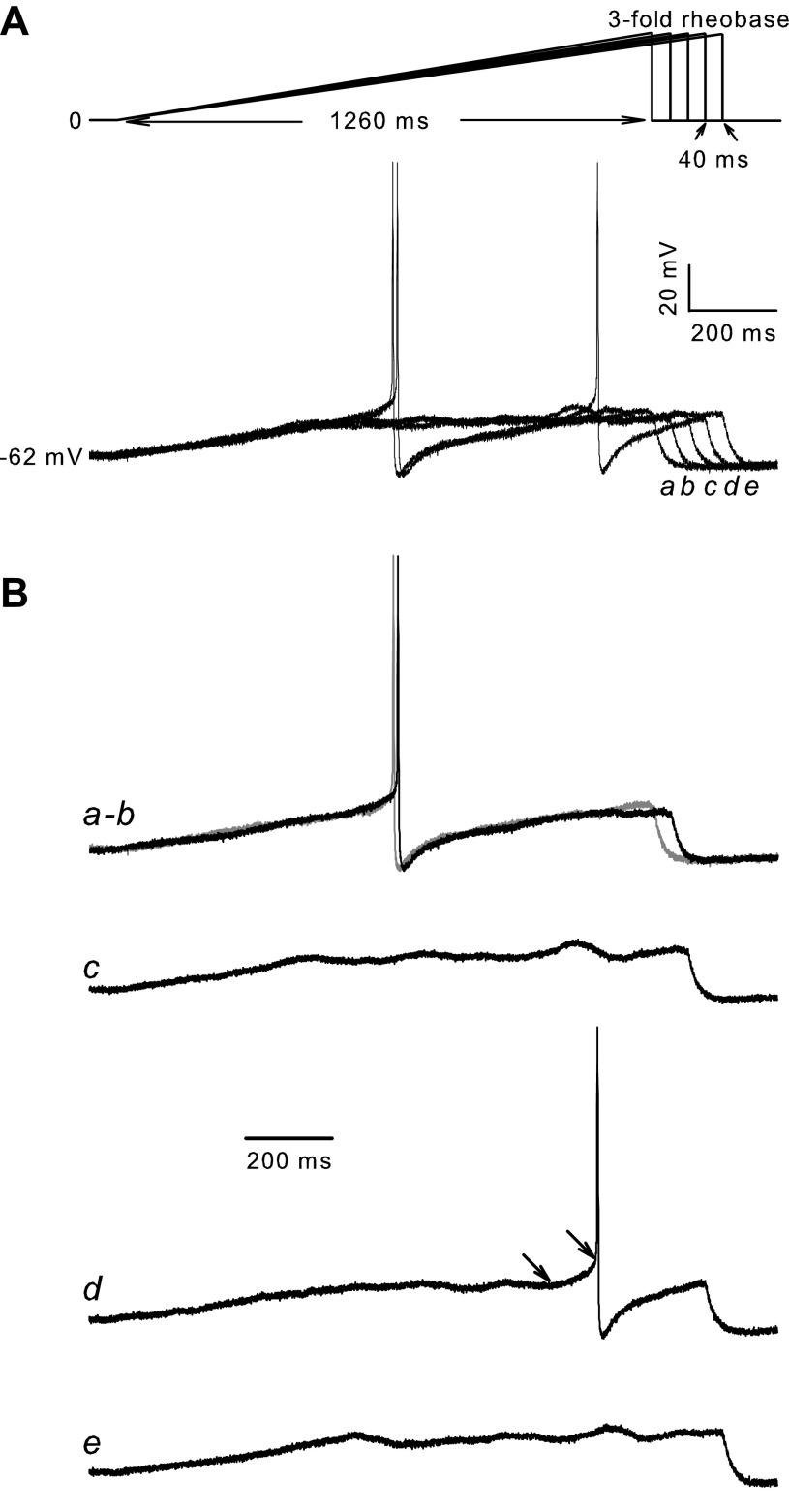
We also noticed that 4-AP rendered the subthreshold membrane potential highly unstable in most C-fiber neurons in response to slow and long current ramps. This sustained and oscillating membrane depolarization may sometimes lead to late firing of action potentials. To illustrate, Fig. 5 shows several membrane potential responses to the slow current ramps obtained from one capsaicin-sensitive neuron in the presence of 60 µM 4-AP. Tracing a and b show the two slowest linear depolarization capable of evoking action potential discharge in this neuron. A linear fit to the depolarizing phase preceding the action potential in trace b gave a dV/dtthresohld of 0.032 mV/ms. The subsequent three slower current ramps elicited an initial slow depolarization that failed to initiate an action potential, followed by a sustained and oscillating depolarization (traces c through e). The sustained depolarization in tracing d eventually led to a second rapid depolarizing phase (indicated by the arrows) that evoked an action potential. The rate of this second depolarization was found to be 0.094 mV/ms, faster than the dV/dtthreshold in this neuron. This randomly occurred action potentials during the prolonged and sustained depolarization evoked by slow depolarizing current injections was occasionally observed at baseline in three of 14 C-fiber neurons and more frequently seen following the application of 4-AP in seven of 11 C-fiber neurons. In A-fiber neurons, it was only occasionally observed in the presence of 4-AP in a smaller portion of neurons (4/11 neurons). These observations indicate that 4-AP not only reduced the threshold of depolarization rate for action potential generation but also removed an important inhibitory current that counteracts against the sustained depolarizing force in nodose neurons, particularly in C-fiber neurons.
Figure 5.
Membrane potential response to long and slow current ramp stimulations in a C-fiber neuron in the presence of 4-AP. A: current clamp protocol and the corresponding membrane potential response recorded from a capsaicin-sensitive C-fiber neuron. B: membrane potential response shown in A is replotted separately to better illustrate the linear depolarization leading to action potential discharge in traces a and b, and the oscillation of membrane potential in traces c–e. The traces a through e correspond to those indicated in A. 4-AP, 4-aminopyridine.
Effects of IK.D Inhibition by α-DTX on the Rate-of-Depolarization Threshold in Mouse Nodose Neurons
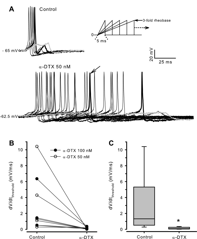
Although 4-AP at low concentrations can be considered to be relatively selective for IK.D channels at the subthreshold voltages, a role for IK.D channels in regulating the dV/dtthreshold in mouse nodose neurons was further confirmed using α-DTX, a selective IK.D inhibitor (12). As shown in Fig. 6, 50 nM of α-DTX reduced the dV/dtthreshold in every of six neurons tested. Similar effects were observed with 100 nM of α-DTX (n = 3 neurons). Pooled data obtained from nine neurons (3 A-fiber, 4 capsaicin-insensitive C-fiber, and 2 capsaicin-sensitive C-fiber neurons) showed that the effect of α-DTX on dV/dtthreshold was highly significant (Fig. 6C), closely similar to what was found with 4-AP. Moreover, slower and longer current ramps often elicited more action potentials in the presence of α-DTX (Fig. 6A). The action potential number was increased from 1–2 to 2–11 in six out of nine neurons by α-DTX treatment.
Figure 6.
Effects of α-DTX on the rate-of-depolarization threshold. A: representative membrane potential response to the current ramp protocol (inset) obtained from one capsaicin-insensitive C-fiber neuron at baseline (Control) and in the presence of 50 nM α-DTX. The thickened black trace represents the slowest depolarization capable of eliciting an action potential, and the gray trace the first of the three consecutive depolarizations that failed to evoke an action potential. Longer ramps trigger double action potentials in the presence of α-DTX. The arrow indicates the first action potential induced by the slowest depolarization. B: each pair of interconnected symbols presents the dV/dtthreshold measured from each of nine nodose neurons before (Control) and after bath application of 50 nM or 100 nM α-DTX as indicated. C: box and whisker plot of dV/dtthreshold data shown in B. *P = 0.004 (Wilcoxon signed rank test). dV/dtthreshold, rate-of-depolarization threshold; α-DTX, α-dendrotoxin.
Effects of Membrane Depolarization on the Rate-of-Depolarization Threshold in Mouse Nodose Neurons
Bath perfusion of 4-AP or α-DTX caused a modest, but significant, membrane depolarization [from −64.8 ± 3.1 to −62.1 ± 3.6 mV by 4-AP, n = 24 neurons, P < 0.001 (paired t test); or from −64.7 ± 2.8 to −61.8 ± 3.0 mV by α-DTX, n = 9 neurons, P < 0.001 (paired t test)] and an increase in input resistance measured around RMP [from 691.6 ± 433.7 to 1,037.3 ± 698.1 MΩ by 4-AP, n = 14 neurons, P = 0.002 (paired t test); or from 367.2 ± 214.6 to 809.1 ± 367.8 MΩ by α-DTX, n = 8 neurons, P = 0.002 (paired t test)] in mouse nodose neurons, indicating that IK.D channels contribute to setting the RMP and to the control of excitability. Since 4-AP and α-DTX produced similar effects, the data obtained with these inhibitors were pooled to evaluate the effects of IK.D inhibition on Rinput and RMP in different classes of nodose neurons (Table 2). It is clear that inhibition of IK.D depolarized A-fiber and C-fiber neurons (by 2.6–3.0 mV) and increased their Rinput (by ∼1.6 fold) to the same extent. The difference between A-fiber and C-fiber neurons with respect to RMP and Rinput maintained, however, after IK.D channels were blocked.
Table 2.
Effects of IK.D inhibition on RMP and Rinput in different classes of mouse nodose neurons
| RMP, mV |
Rinput, MΩ |
|||||||
|---|---|---|---|---|---|---|---|---|
| Neuron Type | n | Control | 4-AP/α-DTX | P | n | Control | 4-AP/α-DTX | P |
| A-fiber | 14 | −66.7 ± 3.4 | −64.0 ± 3.4 | <0.001 | 8 | 312 ± 310 | 504 ± 333 | <0.001 |
| Cap− C-fiber | 9 | −63.8 ± 1.0 | −60.8 ± 2.7 | 0.003 | 5 | 796 ± 432 | 1505 ± 700 | 0.013 |
| Cap+ C-fiber | 10 | −62.9 ± 1.8 | −60.3 ± 2.7 | <0.001 | 9 | 683 ± 348 | 1049 ± 447 | 0.002 |
| C-fiber (Cap− and Cap+) | 19 | −63.3 ± 1.5 | −60.5 ± 2.6* | <0.001 | 14 | 723 ± 367 | 1211 ± 570† | <0.001 |
The data are expressed as means ± SD. Cap− C-fiber, capsaicin-insensitive C-fiber neurons; Cap+ C-fiber, capsaicin-sensitive C-fiber neurons; IK.D, D-type potassium current; Rinput, input resistance; RMP, resting membrane potential; α-DTX, α-dendrotoxin; 4-AP, 4-aminopyridine. All P values were determined by paired t test for control vs. 4-AP/α-DTX.
*P = 0.002 and †P = 0.005 vs. A-fiber neurons treated with 4-AP/α-DTX (by unpaired t test).
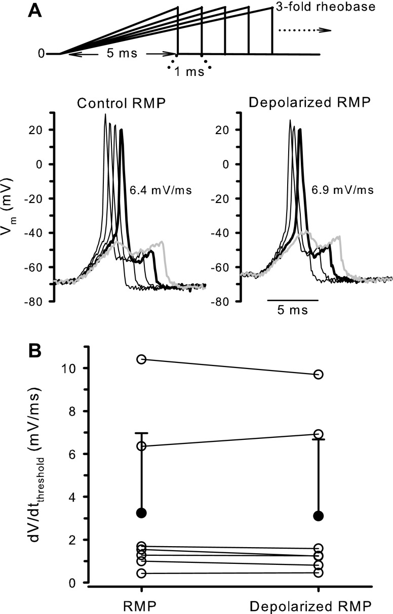
Sustained depolarization caused by IK.D inhibition, albeit modest, may alter the gating and availability of other ion channels, leading to changes in the rate-of-depolarization threshold. To test this possibility, the dV/dtthreshold was measured in a total of seven neurons before and after the neuron was depolarized by 3–4 mV (averaged 3.3 mV) via injection of a depolarizing holding current. As shown in Fig. 7, such magnitude of membrane depolarization did not have significant impact on the rate-of-depolarization threshold.
Figure 7.
Effects of modest depolarization on dV/dtthreshold. A: representative membrane potential response to the current ramp protocol obtained from one A-fiber neuron at control RMP and when a 4-mV depolarization was induced by injecting a depolarizing holding current. The thickened black trace represents the slowest depolarization capable of eliciting an action potential, and the gray trace the first depolarization that failed to evoke an action potential. B: each pair of interconnected open circles represents the dV/dtthreshold measured from each of seven nodose neurons (3 A-fiber and 4 C-fiber neurons) at control RMP and at depolarized RMP (by 3-4 mV, averaged 3.3 mV). The filled circles and the error bars represent the means ± SD obtained from the seven neurons (P = 0.445 by paired t test). dV/dtthreshold, rate-of-depolarization threshold; RMP, resting membrane potential; Vm, membrane potential.
DISCUSSION
To the best of our knowledge, this is the first study investigating quantitatively the sensitivity of action potential firing to the rate of depolarization in vagal sensory neurons. The main findings include: 1) the dependence of action potential generation on the rate of depolarization is an intrinsic property of mouse nodose neurons, regardless the intensity of depolarizing currents; 2) the dV/dtthreshold is significantly lower in C-fiber neurons compared to A-fiber neurons: to fire an action potential, A-fiber neurons must be depolarized faster than averaged 7 mV/ms, whereas C-fiber neurons can respond even when the depolarization rate is <0.4 mV/ms; and 3) inhibition of IK.D by low concentration of 4-AP or α-DTX strongly reduced the dV/dtthreshold in both A-fiber and C-fiber neurons, indicating that this current plays a key role in setting the rate-of-depolarization threshold for the action potential generation in vagal sensory neurons. Processes that reduced the dV/dtthreshold may contribute to increased excitability of vagal afferent nerves by rendering effective stimuli that might otherwise cause depolarizations too slow to evoke action potential discharge.
The dV/dtthreshold varied considerably among different nodose neurons, but it was not significantly different in any one neuron in response to different amplitudes of depolarizing currents although stronger stimuli usually led to faster depolarization. This observation suggests that the rate of depolarization is an important independent factor contributing to the control of action potential generation and represents an intrinsic property of mouse nodose neurons. In octopus cells of the mouse ventral cochlear nucleus that exhibit high sensitivity of action potential firing to the rate of depolarization, the dV/dtthreshold was also found to be constant regardless the stimulation current intensity or whether the depolarization was evoked synaptically or by the intracellular current injection (8).
A consistent finding in this study is that the nodose C-fiber neurons responded to much slower depolarizations with action potential discharge compared to the A-fiber neurons. Patch clamp recordings on the isolated neurons provide the well-controlled studies of biophysical and pharmacological properties of ion channels and membrane potential but lose the ability to identify neuron subtypes according to the conduction velocity which is the gold standard for afferent fiber classification. In order to classify the isolated nodose neurons, several alternative approaches have been explored and described, such as by examining the responsiveness to capsaicin (or the expression of capsaicin receptor TRPV1) (15, 18), the characteristics of action potential waveforms (15), the differential expression of tetrodotoxin (TTX)-sensitive (TTX-S) and TTX-resistant (TTX-R) Na+ currents (19, 20), or the neuron shape and microscopic structural features (21). A comparative analysis of action potential waveforms recorded from nodose neurons with intact afferent fibers (therefore with known conduction velocity) and those recorded from isolated nodose neurons has demonstrated that a combined use of metrics like APD50, APthreshold, upstroke and downstroke velocities at the point of APD50 is a reliable indicator of afferent fiber types in rat nodose ganglia (15). Through these analyses, the authors identified a group of fast-conducting A-fiber neurons that had the measurements of above-mentioned action potential features closer to those shown by the slowly conducting C-fiber neurons and named them Ah neurons. Ah-type neurons with similar action potential characteristics have also been described in rabbit nodose ganglia (16). The capsaicin sensitivity is a well-accepted marker of vagal afferent C-fibers, but it has also been shown that ∼40% of mouse bronchopulmonary vagal afferent fibers that conduct in the C-fiber range were insensitive to capsaicin (14). Thus, we used a combination of capsaicin test and action potential waveform analyses to classify the mouse nodose neurons in the present study. We found that about one third of the capsaicin-insensitive neurons exhibited the action potential waveform characteristic of the capsaicin-sensitive neurons, i.e., longer APD50 and APD100, slower repolarization indicated by lower UVmax and UVAPD50, and more depolarized APthreshold. This group of neurons may correspond to the previously found capsaicin-insensitive C-fibers (14) and was therefore named capsaicin-insensitive C-fiber neurons in this study. Our results further showed that the capsaicin-insensitive C-fiber neurons had a dV/dtthreshold closely similar to that found in the capsaicin-sensitive neurons but significantly lower than that recorded in the A-fiber neurons (capsaicin-insensitive neurons with a brief action potential).
The peripheral terminals of vagal A-fibers are often low-threshold mechanosensors but rarely respond directly to inflammatory mediators. Vagal C-fiber terminals, on the other hand, are stimulated by a variety of inflammatory mediators (3, 4). The lack of an A-fiber response to an inflammatory mediator may be explained by a lack of expression of the receptor for the mediator in A-fiber neurons. However, based on the present findings, even if the mediator receptor was expressed by the A-fiber neurons, the high dV/dtthreshold may render the relatively slow generator potentials evoked by the inflammatory mediator ineffective at evoking action potential discharge in these neurons.
We noticed that the A-fiber neurons have a significantly lower input resistance (therefore a higher rheobase) compared to C-fiber neurons of mouse nodose ganglia. A lower input resistance can cause a larger shunting of depolarizing currents and reduce the magnitude and kinetics of membrane depolarization in response to a given intensity of stimulation. To minimize the interference of variations in the input resistance with the determination of dV/dtthreshold, the current ramps with intensities relative to the rheobase measured in each neuron were used in this study so that similar amplitudes and kinetics of membrane depolarizations can be obtained in different neurons with different input resistance. Therefore, the differential dV/dtthreshold between A- and C-fiber neurons observed under our experimental conditions is unlikely to be due to the large difference in the input resistance between these two neuron groups. The finding that inhibition of IK.D channels by 4-AP or α-DTX minimized the difference in dV/dtthreshold but did not change the difference in the input resistance between A- and C-fiber neurons supports this assertion. Moreover, our results indicate that the slightly more depolarized RMP found in C-fiber neurons or in the presence of IK.D inhibitors in both A- and C-fiber neurons has little impact on the dV/dtthreshold.
The ionic mechanisms underlying the sensitivity of action potential generation to the rate of depolarization in mouse nodose neurons remain to be fully elucidated. Potassium channels that activate at the resting and/or subthreshold membrane potentials may counterbalance the depolarizing force and prevent the depolarization from reaching the action potential firing threshold. In nodose neurons, these K+ channels include the slowly/noninactivating IK.D channels (22), KCNQ/M-channels (23), and two-pore domain potassium (K2P) channels (24), as well as the rapidly inactivating IA channels made of Kv1.4- or Kv4 subunits (25). In this study, we found that bath application of 60 µM 4-AP lowered the minimal rate of depolarization needed to evoke an action potential by ∼10 folds in A-fiber neurons and by ∼3 fold in C-fiber neurons. It is known that IK.D channels are highly sensitive to 4-AP (11, 26) whereas M-channels and K2P channels are 4-AP-resistant (27, 28), and Kv1.4/or Kv4/IA channels are only modestly sensitive to this inhibitor (IC50 ∼1 mM) (29, 30). Therefore, 60 µM of 4-AP used in this study is expected to be relatively selective for IK.D channels at the subthreshold and threshold voltages, and its effect on the dV/dtthreshold can be attributed to the inhibition of IK.D channels. This conclusion is further strengthened by the same effects of α-DTX, a selective IK.D inhibitor, on the dV/dtthreshold in nodose neurons observed in this study. The finding that the IK.D channel is a key determinant of the dV/dtthreshold in mouse nodose neurons is consistent with the unique biophysical properties of IK.D channels, i.e., low-threshold activation, fast activation, and very slow inactivation (9–12). The activation time constant of IK.D has been reported to be voltage-dependent, being 5.4–9.2 ms at −10 mV (31) and 17 ± 3.2 ms at −20 mv (22) in rat nodose neurons. Therefore, activation of a substantial amount of IK.D during current ramps ≥ 10–20 ms is expected, which can counterbalance the rising depolarizing current and preclude action potential firing. This is particularly easier to appreciate in A-fiber neurons (Fig. 3 and 4). Downregulation of Kv1 channels and/or reduced IK.D have been reported in sensory neurons associated with chronic inflammation induced by nerve injury (17, 32–34) or irritable bowel syndrome (35) and shown to be associated with peripheral hyperexcitability and pain. The results obtained in this study suggest that a reduction in IK.D may contribute to enhanced activation of vagal afferent nerves observed in various inflammatory diseases in visceral organs (36, 37) by lowering the dV/dtthreshold for action potential generation.
Among the other low-threshold-activated K+ channels mentioned above, M-channels activate slowly with an activation time constant being ∼200 ms at −30mV and −20 mV in nodose neurons (23, 27). Thus, M-channels may become important in regulating the response to longer current ramps, such as those that failed to elicit an action potential in C-type neurons or in A-fiber neurons following inhibition of IK.D. K2P channels are mainly activated by various mechanical, physical, and chemical stimuli but may also generate an background conductance in neurons (38). The latter is expected to increase linearly as the membrane depolarizes (unless at very positive potentials) due to increasing driving force for the K+ efflux. As a result, this instantaneous component of K2P current would increase most rapidly in response to short current ramps and exert its inhibitory function. This is, however, inconsistent with our observation that short current ramps were more effective in triggering action potential firing in nodose neurons. Low threshold-activated IA channels are also likely not to have a role in setting the kinetic threshold for action potential generation observed in this study, since Kv4/IA channels are largely inactivated at the resting potential (39), and Kv1.4/IA channels inactivate very rapidly with an inactivation time constant < 10 ms in sensory neurons (40).
The mechanisms underlying the impressive difference in dV/dtthreshold identified in nodose A-fiber versus C-fiber neurons in this study remain elusive. Given that the inhibition of IK.D by α-DTX or low concentration of 4-AP nearly abrogated the dependence of action potential discharge on the rate of depolarization, it is reasonable to hypothesize that the IK.D current may be different in A-fiber versus C-fiber neurons. The IK.D current has been recorded in both A-fiber and C-fiber neurons isolated from rat nodose ganglia (11, 22, 41). In one study where the rat nodose neurons were classified based on the expression pattern of TTX-R Na+ currents, the authors have reported that IK.D measured at +20 mV represents ∼20% of the total outward K+ current in both A-fiber (lacking TTX-R) and C-fiber (expressing TTX-R) neurons, but whether the total K+ current had the same amplitude in both cell types was not clear nor the properties of IK.D at the subthreshold voltages in different types of neurons were described (22). It is known that in native neurons the IK.D channels are primarily heterotetramers composed of two or three different types of Kv1α-subunits (α-DTX-sensitive Kv1.1, Kv1.2, Kv1.6, and α-DTX-insensitive Kv1.4) (42–45). The heterotetrameric IK.D channels with different α-subunit compositions and/or different stoichiometry of contributing subunits exhibit different biophysical properties (46–49). For example, KV1.2/1.6 channels activate at more negative voltages than KV1.1/1.2 channels (49). A complete and detailed comparative study on the identification of major α-subunit composition of IK.D channels and the biophysical properties of the current at the subthreshold voltages such as the voltage threshold of activation, current density, and kinetics in nodose A-fiber and C-fiber neurons will be needed to address this hypothesis.
Although we focused here on IK.D it is likely that the inactivation of voltage-gated Na+ channels during slower depolarizations also contributes to setting the dV/dtthreshold as suggested by a study in the large myelinated nerve fibers of frogs (6). The mammalian vagal afferent nerves mainly expressed Nav1.7, Nav1.8, and Nav1.9 (50, 51). The Nav1.7 generates TTX-S, whereas the Nav1.8 and Nav1.9 currents are TTX-resistant. Both TTX-S (mainly Nav1.7) and Nav1.8/TTX-R can generate inward currents sufficiently large to initiate the upstroke of action potentials in vagal afferent neurons (52). The function of Nav1.9 channels in nodose neurons remains unknown. Given that Nav1.9 generates a noninactivating inward current at membrane potentials between −60 and −30 mV in sensory neurons (53), it may exert influence on the subthreshold depolarization and on the action potential firing threshold in nodose neurons. The spike-generating TTX-S and Nav1.8/TTX-R have different voltage-dependent inactivation properties. The TTX-S inactivates from −90 mV with the 50% of inactivation around −66 mV and complete inactivation by −30 mV. The Nav1.8/TTX-R inactivates at more positive voltages, with 50% of inactivation occurring around −31 mV (20, 54). Thus, due to an accumulating inactivation of TTX-S following a slow depolarization, the major spike-generating Na+ channel available at the threshold voltages (−38 to −32 mV) would be the Nav1.8/TTX-R channel. It has been reported that the TTX-S Na+ current was present in all mouse nodose neurons, whereas the TTX-R Na+ current (recorded at 0 mV, presumably via Nav1.8) was recorded in all capsaicin-sensitive C-fiber neurons but not found in nearly half (21 out of 44) of capsaicin-insensitive nodose neurons (19). It is tempting to speculate that the differential expression pattern of Nav1.8/TTX-R Na+ channels in A-fiber and C-fiber neurons might contribute to the distinct dV/dtthreshold observed in these neurons. A detailed comparative study on the expression and voltage-dependent properties of main Nav subtypes and their response to the depolarization of different kinetics in A- and C-fiber neurons is required to test this hypothesis.
In summary, this study has provided the direct evidence that the rate of an evoked depolarization (or generator potential) is an independent factor in controlling the initiation of action potentials in vagal sensory neurons, and it exerts a particularly strong influence on the A-fiber neuron activation. The data also support that the IK.D current is the main player in determining the rate-of-depolarization threshold in vagal afferent neurons. The symptoms of many visceral inflammatory diseases are consistent with excessive activation of visceral C-fibers. One approach to treating these symptoms is to decrease C-fiber activity by antagonizing the receptor for the activating chemical mediator. Hoverer, at the site of inflammation there may be several mediators capable of redundantly activating the C-fibers. In these cases a more rational approach would be to focus on “choke” points down stream of the receptors that could limit activation. One such choke point might be the ion channels involved in regulating the low dV/dtthreshold of C-fibers, i.e., therapeutically manipulating ion channels that would render the dV/dtthreshold of C-fibers more similar to A-fibers.
GRANTS
This work is supported by Johns Hopkins Blaustein Pain Research Funds Grant 80048814 and NIH Grant R01 HL137807/R35 HL155671.
DISCLOSURES
No conflicts of interest, financial or otherwise, are declared by the author.
AUTHOR CONTRIBUTIONS
H.S. conceived and designed research; H.S. performed experiments; H.S. analyzed data; H.S. interpreted results of experiments; H.S. prepared figures; H.S. drafted manuscript; H.S. edited and revised manuscript; H.S. approved final version of manuscript.
ACKNOWLEDGMENTS
The author thanks Dr. Bradley J. Undem for support, inspiring discussions, and providing valuable comments to the manuscript.
REFERENCES
- 1.Berthoud HR, Neuhuber WL. Functional and chemical anatomy of the afferent vagal system. Auton Neurosci 85: 1–17, 2000. doi: 10.1016/S1566-0702(00)00215-0. [DOI] [PubMed] [Google Scholar]
- 2.Undem BJ, Taylor-Clark T. Mechanisms underlying the neuronal-based symptoms of allergy. J Allergy Clin Immunol 133: 1521–1534, 2014. doi: 10.1016/j.jaci.2013.11.027. [DOI] [PMC free article] [PubMed] [Google Scholar]
- 3.Lee LY, Yu J. Sensory nerves in lung and airways. Compr Physiol 4: 287–324, 2014[Erratum inCompr Physiol5: 1971, 2015]. doi: 10.1002/cphy.c130020. [DOI] [PubMed] [Google Scholar]
- 4.Undem BJ, Sun H. Molecular/ionic basis of vagal bronchopulmonary C-fiber activation by inflammatory mediators. Physiology 35: 57–68, 2020. doi: 10.1152/physiol.00014.2019. [DOI] [PMC free article] [PubMed] [Google Scholar]
- 5.Tasaki I. Electrical excitation of the nerve fiber. Part 1. Excitation by linearly increasing currents. Jpn J Physiol 1: 1–6, 1950. doi: 10.2170/jjphysiol.1.1. [DOI] [Google Scholar]
- 6.Vallbo AB. Accommodation related to inactivation of the sodium permeability in single myelinated nerve fibres from Xenopus laevis. Acta Physiol Scand 61: 429–444, 1964. [PubMed] [Google Scholar]
- 7.McGinley MJ, Oertel D. Rate thresholds determine the precision of temporal integration in principal cells of the ventral cochlear nucleus. Hear Res 216-217: 52–63, 2006. doi: 10.1016/j.heares.2006.02.006. [DOI] [PubMed] [Google Scholar]
- 8.Ferragamo MJ, Oertel D. Octopus cells of the mammalian ventral cochlear nucleus sense the rate of depolarization. J Neurophysiol 87: 2262–2270, 2002. doi: 10.1152/jn.00587.2001. [DOI] [PubMed] [Google Scholar]
- 9.Ovsepian SV, LeBerre M, Steuber V, O'Leary VB, Leibold C, Oliver Dolly J. Distinctive role of KV1.1 subunit in the biology and functions of low threshold K+ channels with implications for neurological disease. Pharmacol Ther 159: 93–101, 2016. doi: 10.1016/j.pharmthera.2016.01.005. [DOI] [PubMed] [Google Scholar]
- 10.Higgs MH, Spain WJ. Kv1 channels control spike threshold dynamics and spike timing in cortical pyramidal neurones. J Physiol 589: 5125–5142, 2011. doi: 10.1113/jphysiol.2011.216721. [DOI] [PMC free article] [PubMed] [Google Scholar]
- 11.Stansfeld CE, Marsh SJ, Halliwell JV, Brown DA. 4-Aminopyridine and dendrotoxin induce repetitive firing in rat visceral sensory neurones by blocking a slowly inactivating outward current. Neurosci Lett 64: 299–304, 1986. doi: 10.1016/0304-3940(86)90345-9. [DOI] [PubMed] [Google Scholar]
- 12.Harvey AL, Robertson B. Dendrotoxins: structure-activity relationships and effects on potassium ion channels. Curr Med Chem 11: 3065–3072, 2004. doi: 10.2174/0929867043363820. [DOI] [PubMed] [Google Scholar]
- 13.Nassenstein C, Taylor-Clark TE, Myers AC, Ru F, Nandigama R, Bettner W, Undem BJ. Phenotypic distinctions between neural crest and placodal derived vagal C-fibres in mouse lungs. J Physiol 588: 4769–4783, 2010. doi: 10.1113/jphysiol.2010.195339. [DOI] [PMC free article] [PubMed] [Google Scholar]
- 14.Kollarik M, Dinh QT, Fischer A, Undem BJ. Capsaicin-sensitive and -insensitive vagal bronchopulmonary C-fibres in the mouse. J Physiol 551: 869–879, 2003. doi: 10.1113/jphysiol.2003.042028. [DOI] [PMC free article] [PubMed] [Google Scholar]
- 15.Li BY, Schild JH. Electrophysiological and pharmacological validation of vagal afferent fiber type of neurons enzymatically isolated from rat nodose ganglia. J Neurosci Methods 164: 75–85, 2007. doi: 10.1016/j.jneumeth.2007.04.003. [DOI] [PMC free article] [PubMed] [Google Scholar]
- 16.Stansfeld CE, Wallis DI. Properties of visceral primary afferent neurons in the nodose ganglion of the rabbit. J Neurophysiol 54: 245–260, 1985. doi: 10.1152/jn.1985.54.2.245. [DOI] [PubMed] [Google Scholar]
- 17.Gonzalez A, Ugarte G, Restrepo C, Herrera G, Pina R, Gomez-Sanchez JA, Pertusa M, Orio P, Madrid R. Role of the excitability brake potassium current IKD in cold allodynia induced by chronic peripheral nerve injury. J Neurosci 37: 3109–3126, 2017. doi: 10.1523/JNEUROSCI.3553-16.2017. [DOI] [PMC free article] [PubMed] [Google Scholar]
- 18.Kwong K, Carr MJ, Gibbard A, Savage TJ, Singh K, Jing J, Meeker S, Undem BJ. Voltage-gated sodium channels in nociceptive versus non-nociceptive nodose vagal sensory neurons innervating guinea pig lungs. J Physiol 586: 1321–1336, 2008. doi: 10.1113/jphysiol.2007.146365. [DOI] [PMC free article] [PubMed] [Google Scholar]
- 19.Fernandez-Fernandez D, Cadaveira-Mosquera A, Rueda-Ruzafa L, Herrera-Perez S, Veale EL, Reboreda A, Mathie A, Lamas JA. Activation of TREK currents by riluzole in three subgroups of cultured mouse nodose ganglion neurons. PloS One 13: e0199282, 2018. doi: 10.1371/journal.pone.0199282. [DOI] [PMC free article] [PubMed] [Google Scholar]
- 20.Schild JH, Kunze DL. Experimental and modeling study of Na+ current heterogeneity in rat nodose neurons and its impact on neuronal discharge. J Neurophysiol 78: 3198–3209, 1997. doi: 10.1152/jn.1997.78.6.3198. [DOI] [PubMed] [Google Scholar]
- 21.Lu XL, Xu WX, Yan ZY, Qian Z, Xu B, Liu Y, Han LM, Gao RC, Li JN, Yuan M, Zhao CB, Qiao GF, Li BY. Subtype identification in acutely dissociated rat nodose ganglion neurons based on morphologic parameters. Int J Biol Sci 9: 716–727, 2013. doi: 10.7150/ijbs.7006. [DOI] [PMC free article] [PubMed] [Google Scholar]
- 22.Glazebrook PA, Ramirez AN, Schild JH, Shieh CC, Doan T, Wible BA, Kunze DL. Potassium channels Kv1.1, Kv1.2 and Kv1.6 influence excitability of rat visceral sensory neurons. J Physiol 541: 467–482, 2002. doi: 10.1113/jphysiol.2001.018333. [DOI] [PMC free article] [PubMed] [Google Scholar]
- 23.Sun H, Lin AH, Ru F, Patil MJ, Meeker S, Lee LY, Undem BJ. KCNQ/M-channels regulate mouse vagal bronchopulmonary C-fiber excitability and cough sensitivity. JCI Insight 4: e124467, 2019. doi: 10.1172/jci.insight.124467. [DOI] [PMC free article] [PubMed] [Google Scholar]
- 24.Cadaveira-Mosquera A, Perez M, Reboreda A, Rivas-Ramirez P, Fernandez-Fernandez D, Lamas JA. Expression of K2P channels in sensory and motor neurons of the autonomic nervous system. J Mol Neurosci 48: 86–96, 2012. doi: 10.1007/s12031-012-9780-y. [DOI] [PubMed] [Google Scholar]
- 25.Matsumoto S, Yoshida S, Ikeda M, Kadoi J, Takahashi M, Tanimoto T, Kitagawa J, Saiki C, Takeda M, Shima Y. Effects of acetazolamide on transient K+ currents and action potentials in nodose ganglion neurons of adult rats. CNS Neurosci Ther 17: 66–79, 2011. doi: 10.1111/j.1755-5949.2010.00133.x. [DOI] [PMC free article] [PubMed] [Google Scholar]
- 26.Storm JF. Temporal integration by a slowly inactivating K+ current in hippocampal neurons. Nature 336: 379–381, 1988. doi: 10.1038/336379a0. [DOI] [PubMed] [Google Scholar]
- 27.Wladyka CL, Kunze DL. KCNQ/M-currents contribute to the resting membrane potential in rat visceral sensory neurons. J Physiol 575: 175–189, 2006. doi: 10.1113/jphysiol.2006.113308. [DOI] [PMC free article] [PubMed] [Google Scholar]
- 28.Lotshaw DP. Biophysical, pharmacological, and functional characteristics of cloned and native mammalian two-pore domain K+ channels. Cell Biochem Biophys 47: 209–256, 2007. doi: 10.1007/s12013-007-0007-8. [DOI] [PubMed] [Google Scholar]
- 29.Song WJ, Tkatch T, Baranauskas G, Ichinohe N, Kitai ST, Surmeier DJ. Somatodendritic depolarization-activated potassium currents in rat neostriatal cholinergic interneurons are predominantly of the A type and attributable to coexpression of Kv4.2 and Kv4.1 subunits. J Neurosci 18: 3124–3137, 1998. doi: 10.1523/JNEUROSCI.18-09-03124.1998. [DOI] [PMC free article] [PubMed] [Google Scholar]
- 30.Yao JA, Tseng GN. Modulation of 4-AP block of a mammalian A-type K channel clone by channel gating and membrane voltage. Biophys J 67: 130–142, 1994. doi: 10.1016/S0006-3495(94)80462-X. [DOI] [PMC free article] [PubMed] [Google Scholar]
- 31.McFarlane S, Cooper E. Kinetics and voltage dependence of A-type currents on neonatal rat sensory neurons. J Neurophysiol 66: 1380–1391, 1991. doi: 10.1152/jn.1991.66.4.1380. [DOI] [PubMed] [Google Scholar]
- 32.Zhao X, Tang Z, Zhang H, Atianjoh FE, Zhao JY, Liang L, Wang W, Guan X, Kao SC, Tiwari V, Gao YJ, Hoffman PN, Cui H, Li M, Dong X, Tao YX. A long noncoding RNA contributes to neuropathic pain by silencing Kcna2 in primary afferent neurons. Nat Neurosci 16: 1024–1031, 2013. doi: 10.1038/nn.3438. [DOI] [PMC free article] [PubMed] [Google Scholar]
- 33.Kim DS, Choi JO, Rim HD, Cho HJ. Downregulation of voltage-gated potassium channel alpha gene expression in dorsal root ganglia following chronic constriction injury of the rat sciatic nerve. Brain Res Mol Brain Res 105: 146–152, 2002. doi: 10.1016/S0169-328X(02)00388-1. [DOI] [PubMed] [Google Scholar]
- 34.Yang EK, Takimoto K, Hayashi Y, de Groat WC, Yoshimura N. Altered expression of potassium channel subunit mRNA and alpha-dendrotoxin sensitivity of potassium currents in rat dorsal root ganglion neurons after axotomy. Neuroscience 123: 867–874, 2004. doi: 10.1016/j.neuroscience.2003.11.014. [DOI] [PubMed] [Google Scholar]
- 35.Luo JL, Qin HY, Wong CK, Tsang SY, Huang Y, Bian ZX. Enhanced excitability and down-regulated voltage-gated potassium channels in colonic drg neurons from neonatal maternal separation rats. J Pain 12: 600–609, 2011. doi: 10.1016/j.jpain.2010.11.005. [DOI] [PubMed] [Google Scholar]
- 36.Trankner D, Hahne N, Sugino K, Hoon MA, Zuker C. Population of sensory neurons essential for asthmatic hyperreactivity of inflamed airways. Proc Natl Acad Sci USA 111: 11515–11520, 2014. doi: 10.1073/pnas.1411032111. [DOI] [PMC free article] [PubMed] [Google Scholar]
- 37.Zaccone EJ, Lieu T, Muroi Y, Potenzieri C, Undem BE, Gao P, Han L, Canning BJ, Undem BJ. Parainfluenza 3-induced cough hypersensitivity in the guinea pig airways. PLoS One 11: e0155526, 2016. doi: 10.1371/journal.pone.0155526. [DOI] [PMC free article] [PubMed] [Google Scholar]
- 38.Enyedi P, Czirják G. background of leak K+ currents: two-pore domain potassium channels. Physiol Rev 90: 559–605, 2010. doi: 10.1152/physrev.00029.2009. [DOI] [PubMed] [Google Scholar]
- 39.Jerng HH, Pfaffinger PJ, Covarrubias M. Molecular physiology and modulation of somatodendritic A-type potassium channels. Mol Cell Neurosci 27: 343–369, 2004. doi: 10.1016/j.mcn.2004.06.011. [DOI] [PubMed] [Google Scholar]
- 40.Zemel BM, Ritter DM, Covarrubias M, Muqeem T. A-type KV channels in dorsal root ganglion neurons: diversity, function, and dysfunction. Front Mol Neurosci 11: 253, 2018. doi: 10.3389/fnmol.2018.00253. [DOI] [PMC free article] [PubMed] [Google Scholar]
- 41.Stansfeld CE, Marsh SJ, Parcej DN, Dolly JO, Brown DA. Mast cell degranulating peptide and dendrotoxin selectively inhibit a fast-activating potassium current and bind to common neuronal proteins. Neuroscience 23: 893–902, 1987. doi: 10.1016/0306-4522(87)90166-7. [DOI] [PubMed] [Google Scholar]
- 42.Scott VE, Muniz ZM, Sewing S, Lichtinghagen R, Parcej DN, Pongs O, Dolly JO. Antibodies specific for distinct Kv subunits unveil a heterooligomeric basis for subtypes of alpha-dendrotoxin-sensitive K+ channels in bovine brain. Biochemistry 33: 1617–1623, 1994. doi: 10.1021/bi00173a001. [DOI] [PubMed] [Google Scholar]
- 43.Shamotienko OG, Parcej DN, Dolly JO. Subunit combinations defined for K+ channel Kv1 subtypes in synaptic membranes from bovine brain. Biochemistry 36: 8195–8201, 1997. doi: 10.1021/bi970237g. [DOI] [PubMed] [Google Scholar]
- 44.Wang FC, Parcej DN, Dolly JO. Alpha subunit compositions of Kv1.1-containing K+ channel subtypes fractionated from rat brain using dendrotoxins. Eur J Biochem 263: 230–237, 1999. doi: 10.1046/j.1432-1327.1999.00493.x. [DOI] [PubMed] [Google Scholar]
- 45.Rasband MN, Park EW, Vanderah TW, Lai J, Porreca F, Trimmer JS. Distinct potassium channels on pain-sensing neurons. Proc Natl Acad Sci USA 98: 13373–13378, 2001. doi: 10.1073/pnas.231376298. [DOI] [PMC free article] [PubMed] [Google Scholar]
- 46.D'Adamo MC, Imbrici P, Sponcichetti F, Pessia M. Mutations in the KCNA1 gene associated with episodic ataxia type-1 syndrome impair heteromeric voltage-gated K+ channel function. FASEB J 13: 1335–1345, 1999. doi: 10.1096/fasebj.13.11.1335. [DOI] [PubMed] [Google Scholar]
- 47.Hopkins WF, Allen ML, Houamed KM, Tempel BL. Properties of voltage-gated K+ currents expressed in Xenopus oocytes by mKv1.1, mKv1.2 and their heteromultimers as revealed by mutagenesis of the dendrotoxin-binding site in mKv1.1. Pflugers Arch 428: 382–390, 1994. doi: 10.1007/BF00724522. [DOI] [PubMed] [Google Scholar]
- 48.Ruppersberg JP, Schroter KH, Sakmann B, Stocker M, Sewing S, Pongs O. Heteromultimeric channels formed by rat brain potassium-channel proteins. Nature 345: 535–537, 1990. doi: 10.1038/345535a0. [DOI] [PubMed] [Google Scholar]
- 49.Sokolov MV, Shamotienko O, Dhochartaigh SN, Sack JT, Dolly JO. Concatemers of brain Kv1 channel alpha subunits that give similar K+ currents yield pharmacologically distinguishable heteromers. Neuropharmacology 53: 272–282, 2007. doi: 10.1016/j.neuropharm.2007.05.008. [DOI] [PubMed] [Google Scholar]
- 50.Muroi Y, Undem BJ. Targeting voltage gated sodium channels NaV1.7, Na V1.8, and Na V1.9 for treatment of pathological cough. Lung 192: 15–20, 2014. doi: 10.1007/s00408-013-9533-x. [DOI] [PMC free article] [PubMed] [Google Scholar]
- 51.Mazzone SB, Tian L, Moe AAK, Trewella MW, Ritchie ME, McGovern AE. Transcriptional profiling of individual airway projecting vagal sensory neurons. Mol Neurobiol 57: 949–963, 2020. doi: 10.1007/s12035-019-01782-8. [DOI] [PubMed] [Google Scholar]
- 52.Kollarik M, Sun H, Herbstsomer RA, Ru F, Kocmalova M, Meeker SN, Undem BJ. Different role of TTX-sensitive voltage-gated sodium channel (NaV 1) subtypes in action potential initiation and conduction in vagal airway nociceptors. J Physiol 596: 1419–1432, 2018. doi: 10.1113/JP275698. [DOI] [PMC free article] [PubMed] [Google Scholar]
- 53.Amaya F, Wang H, Costigan M, Allchorne AJ, Hatcher JP, Egerton J, Stean T, Morisset V, Grose D, Gunthorpe MJ, Chessell IP, Tate S, Green PJ, Woolf CJ. The voltage-gated sodium channel Na(v)1.9 is an effector of peripheral inflammatory pain hypersensitivity. J Neurosci 26: 12852–12860, 2006. doi: 10.1523/JNEUROSCI.4015-06.2006. [DOI] [PMC free article] [PubMed] [Google Scholar]
- 54.Catterall WA, Goldin AL, Waxman SG. International Union of Pharmacology. XLVII. Nomenclature and structure-function relationships of voltage-gated sodium channels. Pharmacol Rev 57: 397–409, 2005. doi: 10.1124/pr.57.4.4. [DOI] [PubMed] [Google Scholar]


